Fate and Development of Human Vomeronasal Organ – A Microscopic Fetal Study
A.K. Manicka Vasuki1, T.K. Aleyemma Fenn2, M. Nirmala Devi3, T. Deborah Joy Hebzibah4, M. Jamuna5, K. Kalyana Sundaram6
1 Assistant Professor, Department of Anatomy, Affiliated to Tamilnadu Dr. M.G.R. Medical University, Chennai, India.
2 Former Professor and HOD, Department of Anatomy, Affiliated to Tamilnadu Dr. M.G.R. Medical University, Chennai, India.
3 Associate Professor, Department of Anatomy, Affiliated to Tamilnadu Dr. M.G.R. Medical University, Chennai, India.
4 Assistant Professor, Department of Anatomy, Affiliated to Tamilnadu Dr. M.G.R. Medical University, Chennai, India.
5 Professor and HOD, Department of Anatomy, Affiliated to Tamilnadu Dr. M.G.R. Medical University, Chennai, India.
6 Associate Professor, Department of Anaesthesiology, Affiliated to Tamilnadu Dr. M.G.R. Medical University, Chennai, India.
NAME, ADDRESS, E-MAIL ID OF THE CORRESPONDING AUTHOR: Dr. A.K. Manicka Vasuki, Assistant Professor, Department of Anatomy, PSG Institute of Medical Sciences & Research, Coimbatore-641004, Tamilnadu, India.
E-mail: Vasukikalyan01@gmail.com
Introduction
The existence of Vomeronasal organ in human is a controversial subject. Presence of Vomeronasal organ and its structure was not reported in standard text books. The presence of Vomeronasal organ in fetal life is doubtful. Hence identification of the organ by histological examination was planned.
Materials and Methods
A study was conducted on resected specimens of nasal septum obtained from 45 spontaneously aborted fetuses from Obstetrics and Gynaecology department, PSG Institute of Medical Sciences and Research, Coimbatore, after ethical clearance.
Results
The histological structure of Vomeronasal organ was observed from 11 weeks old fetus. The epithelial lining of the organ, presence of cilia, presence of lamina propria, acini and the blood vessel and the types of cells were observed. The organ was lined by pseudostratified columnar epithelium. The organ showed Lamina propria with serous acini from 18 weeks fetus. Vomeronasal duct opening into the nasal cavity and three types of cells were observed in 28 weeks fetus.
Conclusion
Knowledge about the persistence of Vomeronasal organ in fetuses and its structure need to be known. The organ may be found as a putative pit posterior to anterior nasal spine. The organ may be damaged in nasal septal surgeries and nasal endoscopic procedures. The organ may not be seen on gross examination in all human fetuses and cadavers.
Fetus, Nasal cartilage, Serous acini
Introduction
Jacobson’s organ or Vomeronasal organ is a chemosensory organ present in all vertebrates, which is essential for intra-specific chemical (Pheromone) communication. “Pheromones” are chemical messengers secreted externally by an individual and detected by a second individual of same species in whom they induce a chemical reaction by Jose velasco [1].
In 1703, Fredrich ruych discovered cavities in the anterior part of nasal septum in a young cadaver and called them “Canalibus nasalibus” [2]. Von sommering confirmed the existence of such cavities in adult cadavers and these were the Vomeronasal organ.
A Danish Anatomist Ludwig Jacobson identified a similar organ in the nose of mammals and described them in detail. The organ was renamed after Jacobson as Jacobson’s organ [3]. Vomeronasal organs are paired epithelial structures that are located in the anteroinferior part of the nasal septum. The Vomeronasal organs are accessory olfactory system which is comprised of the Vomeronasal organ, Vomeronasal nerves and accessory olfactory bulb and is anatomically distinct from main olfactory system. The organ was found as 6-13mm size cavity above the floor of the nasal cavity. The opening of the cavity is visible as a pit at the surface of the septum. The depressed pit is the finding of organ during gross examination. Three types of cells – Light, dark and basal cells may also be present. The Vomeronasal pit was found to be posterior to anterior nasal spine. Behind the pit, lies a 2-8 mm long tube lined by a pseudo stratified ciliated columnar epithelium. Lamina propria may contain serous, mucous acini and blood vessels [4].
Last 200 years, many authors have witnessed a lot of researches and speculations as to the existence structure and function of Vomeronasal organ. In some old monkeys the regression of the Vomeronasal organ was documented. The chimpanzee possesses the organ similar to humans is confirmed by many studies, but the functional status as a vestige, pheromone–receptor remains to be determined [2].
The Vomeronasal organ of a rat modulates reproductive behaviour, has a crescent shaped lumen. Both sensory and non–sensory epithelium containing cells are present. The sensory epithelium contains both sensory cells and supporting cells. The sensory cells are bipolar chemoreceptor neurons with their cell bodies are located in the Vomeronasal epithelium. Each bipolar neuron sends a dendrite to the epithelial surface and an axon to the accessory olfactory bulb of the brain via accessory olfactory nerve. The non sensory epithelium contains tall, densely staining cells [5].
But in humans, Vomeronasal organ does not contain any cells that are ultra structurally, identical to chemoreceptive bipolar neurons described in other mammals. Presence of Vomeronasal organ in adult human was confirmed by many scientists [5,6].
Recently Bhatnagar and Timothy D Smith stated that there are two categories of Vomeronasal organ, Chemosensory and Non-chemosensory vestige of Vomeronasal organ [4]. According to them, Vomeronasal organ appeared as bilateral epithelial thickenings in 37 days of age and the tubular organ was formed between 37 & 43 days of fetal life. However, there is no description of Vomeronasal organ in any of standard text books available in Anatomy. Hence an attempt has been made in the present study to find out the existence of such an organ whether it is present in fetal life and its structure.
Materials and Methods
The current study was done on 45 spontaneously aborted fetuses after obtaining ethical clearance. The duration of the study was from May 2012 to November 2013. Spontaneously aborted fetuses of all three trimesters were included in the study. Macerated fetus and fetuses with congenital anomaly of face were excluded.
Youngest age group of spontaneously aborted fetus obtained was 9 weeks. But the fetus did not show the organ. The youngest fetus in whom the organ was observed was 11 weeks and the oldest fetus was 28 weeks according to our study.
Nasal septum was removed from fetuses. After fixation, it was divided into two parts: One anteriorly close to the roof of the mouth and another one posterior to the first one excluding the roof of the nose. The first part of the specimen was examined histologically to find out the presence of Vomeronasal organ. If not found to be positive and then the second part of the specimen was examined histologically. The organ was not identified by gross examination.
Routine processing was done. The nasal septum was fixed in 10% formalin for about 24 hours. Then the specimen was kept in the graded alcohols and xylene. Subsequently embedding of the tissues was done in the routine way. Blocks were cut at four micron thickness and stained using Haematoxylin and Eosin method. Special stains like Periodic acid Schiff, Alcian blue with Periodic acid Schiff and Masson’s trichrome stains were also done.
Pictures were taken with Carl Zeiss – Primostar optical microscope with Axiocam provision and were studied.
Results
The [Table/Fig-1] with the age group of fetuses among 11–20 weeks and 21–30 weeks showed presence of Vomeronasal organ.
Details of fetuses with appearance of vomeronasal organ at each Gestational age.
| Age of foetuses in Weeks | Total number of foetuses | Vomeronasal organ positive | Vomeronasal organ negative | Percentage of Vomeronasal organ positive |
|---|
| 0-10 | 2 | 0 | 2 | 0 |
| 11-20 | 17 | 6 | 11 | 35.2 |
| 21-30 | 19 | 6 | 11 | 31.5 |
| 31-40 | 7 | 0 | 7 | 0 |
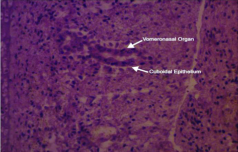
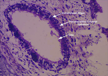
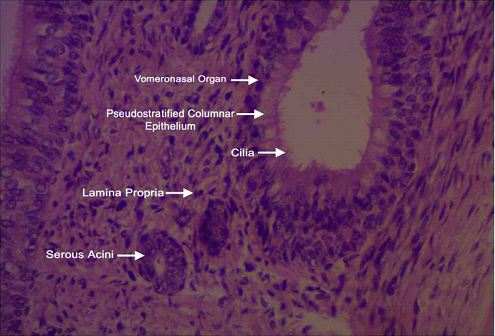
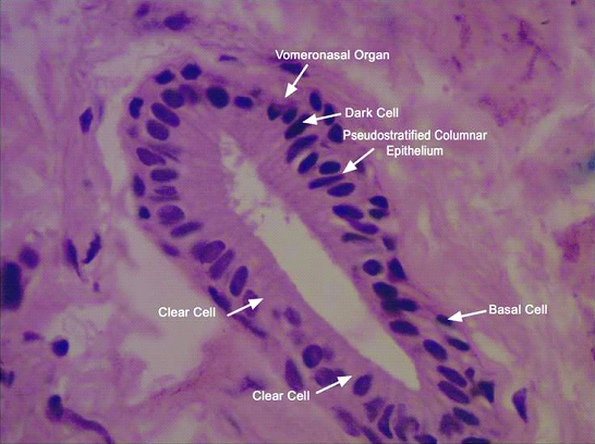
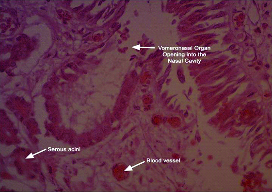
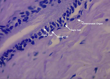
Initially the organ was lined by nonciliated cuboidal epithelium in 11 weeks fetus [Table/Fig-2]. Later from 16 weeks fetus, the organ was found to have Pseudostratified ciliated columnar epithelium [Table/Fig-3]. From 18 weeks fetus the Lamina propria was found which contained serous acini [Table/Fig-4]. In 28 weeks fetus, three types of cells were observed [Table/Fig-5] as dark cells, basal cells and clear cells. The duct opening into the nasal cavity was also observed [Table/Fig-6]. Blood vessels were observed in 28 weeks old fetus [Table/Fig-6]. This is represented systematically in the [Table/Fig-7]. Cell counting was not done because three types of cells were found only in 28 weeks fetus. The organ was not found to be disappearing. Each age group demonstrated appearance of a particular feature.
Special staining using Periodic acid Schiff stain [Table/Fig-8], Periodic acid Schiff stain with Alcian blue stains [Table/Fig-9] and Masson’s trichrome stain [Table/Fig-10] showed Vomeronasal organ. These special stains demonstrate acid mucin. They differentiated the nuclei from cytoplasm of a cell. Collagen demonstrated as blue in colour in Masson’s trichrome stain. Cell differentiation and Collagen identification was possible in these special stains.
Haematoxylin & Eosin staining of Section of Nasal septum of 11 weeks fetus (10x X 10). The organ was lined by Cuboidal epithelium without cilia.
Haematoxylin & Eosin staining of Section of Nasal septum of 16 weeks fetus (10x X 100). The cilia can be identified.
Haematoxylin & Eosin staining of Section of Nasal septum of 18 weeks fetus (10x X100). Lamina propria with its contained serous acini are identified.
Haematoxylin & Eosin staining of Section of Nasal septum of 28 weeks fetus (10x X40). Three types of cells – Dark cells, Light cells and Basal cells were observed
Haematoxylin & Eosin staining of Section of Nasal septum of 28 weeks fetus (10x X40). Vomeronasal duct opening into the nasal cavity and blood vessels are observed
Structure of vomeronasal organ in foetuses.
| Age in weeks | Lining Epithelium | Cilia | Lamina propria | Serous acini | Mucous Acini | Blood Vessel |
|---|
| 11 | Cuboidal and columnar | Absent | Absent | Absent | Absent | Absent |
| 16 | Pseudostratified columnar | Present | Absent | Absent | Absent | Absent |
| 18 | Pseudostratified columnar | Present | Present | Present | Absent | Absent |
| 22 | Pseudostratified columnar | Present | Present | Present | Absent | Absent |
| 28 | Pseudostratified columnar | Present | Present | Present | Absent | Present |
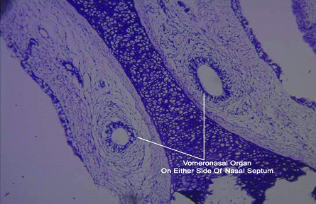
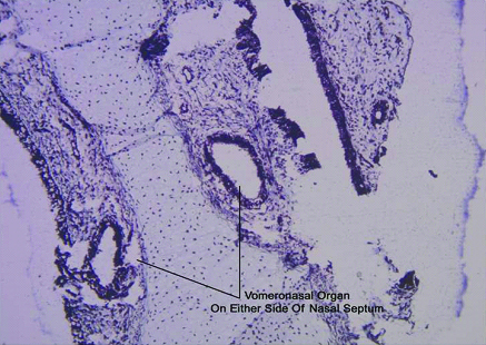
Periodic acid Schiff staining of Section of Nasal septum (10x X10) showing vomeronasal organ on either side of nasal septum.
Alcian blue with Periodic acid Schiff staining of Section of Nasal septum (10x X100).
Masson’s trichrome staining of Section of Nasal septum (10x X10).
Discussion
The first appearance of Vomeronasal organ was identified in the form of bilateral epithelial thickenings at 37 days of age which invaginated and formed tubular Vomeronasal organ between 37 and 43 days of development [4]. It was found that Vomeronasal organ grew prenatally and persisted in adults after examining 50 prenatal humans and two neonatal specimens and four lemur embryos. Three stages of Vomeronasal organ development have been observed. Formation of tubular Vomeronasal organ was found in 33 days-10 weeks as first stage. In second stage, gradual replacements of receptor population with ciliated cells were observed between 10-15 weeks. In third stage, volumetric growth of Vomeronasal epithelium and luminal expansion was observed from 14 weeks until birth. Vomeronasal organ is continuously present throughout embryonic and fetal development and persists beyond [4]. But in our study, we couldn’t stage the development of the organ.
Vomeronasal organ of Jacobson were observed as rudimentary diverticula which first appeared in 8 mm embryos as a pair of grooves on the medial wall of each nasal cavity. Grooves deepened and produced blind tubular sac which opened in front of the nasal septum. In late fetal life, Vomeronasal organ often degenerated because of disappearance of neuronal cells. This organ was not functional in man [7].
In lower animals a special part of olfactory nerve was distributed to Jacobson’s organ as Vomeronasal nerve. This small paired organ was located in the anterior portion of nasal septum. It reached its full development at the end of fifth month. The organ and its nerve might completely disappear in late fetal life but remnant had been found in the adult [8]. The axons of Vomeronasal nerves passing in bundles to an accessory olfactory bulb are seen in the Vomeronasal organs of rodents and other species. There is no evidence of the same structure in humans [9]. Structure and function of Vomeronasal organ was identified by Michael Meridith. He identified the presence of bipolar cells resembling Vomeronasal nerves found in other species and in early human embryos. The cells were positive for Neuron specific enolase. It was found in adult humans as a blind ending tube lined by pseudostratified epithelium and with associated sub mucosal glands. The size increased upto 30 weeks [9].
Vomeronasal organ is the peripheral sensory organ of accessory olfactory system in most amphibian, reptiles and mammals. In these animals, paired Vomeronasal organ are located either at the base of the nasal septum or in the mouth and are involved in chemical communication that often but not exclusively is mediated by Pheromones. In macro somatic animals the Vomeronasal organ consists of Vomeronasal duct which contains chemosensory cells and a Vomeronasal nerve which terminates in the accessory olfactory bulb in the central nervous system. Presence of Vomeronasal organ in adult humans is controversial. The duct may persist beyond embryonic life without neural connection [10].
The Vomeronasal organ was observed as bilateral epithelial thickenings between 37 and 43 days of development in the initial period. Greatest development was observed between 12-14 weeks. Then replacement of receptor population with patchy ciliated cells occurred [11].
On either side of anterior nasal septum, an invagination of ectoderm represents Vomeronasal organ. The Vomer ossified in the connective tissue covering residual posteroinferior cartilage from two centers which united below the cartilage creating deep groove in which Quadrangular cartilage lodges. As growth continued the bony lamellae fused and the quadrangular cartilage got absorbed during development and Vomeronasal organ disappeared [12]. In our study, we found the persistence of the organ in the fetal period. Vomeronasal organ increases its volume by second trimester [13].
Knect observed the Vomeronasal organ endoscopically [14]. Imaging of human Vomeronasal duct was observed by Nasreedin by endoscopic examination of nasal septum [15].
Vomeronasal organ does not function as a sensory organ in adult humans. It might have a function only during human fetal life. The presence of terminal nerve and other structures help in the migration of neurosecretory cells containing Leutinizing hormone releasing hormone to Hypothalamus in the brain. As in other mammals, the Vomeronasal system plays a role in the migration of Leutinizing hormone – releasing hormone neurons from the olfactory placode towards the brain during the development of embryo. Leutinizing hormone releasing hormone positive cells can be seen upto 19 weeks of fetuses [16].
The lemur Microcebus was studied since it belongs to a primate group (prosimii or Strepsirchini) showing complete development of Vomeronasal organ from peripheral receptor organ to accessory olfactory bulb. In humans, the fetal Vomeronasal organ develops initially as a sensory structure similar to Microcebus. It does not progress to a certain stage and loses some chemoreceptors. After birth, it transforms into a simple multilayered pseudostratified ciliated epithelial duct like structure. This structure continues to grow with the facial development and is retained in the adult throughout the life [13].
The bar shaped Paraseptal cartilages symbolize either an absent or nonchemosensory Vomeronasal organ [13]. The Vomeronasal organ in many vertebrates serves as a major duct for glandular complex. A neurosensory epithelium having chemosensory function might develop as a part of this ductal epithelium [13]. Immunomarkers like Neuron specific enolase can be studied in the future study to confirm this sensory role.
During surgical procedure of deviated nasal septum (Submucosal resection of nasal septum), the Vomeronasal organ may be damaged. Even though, it is a vestigial organ, the organ may present as a 2-8mm long putative pit posterior to anterior nasal spine. The pit may be damaged even by nasal endoscopy procedure. Hence the organ should be taken care during the surgery [15].
The study is still going on to identify the increase in frequency of presence of Vomeronasal organ in fetuses. In future, detection of cell types and counting of cells in all fetuses can be done with concomitant observation of same cells in Hypothalamus to detect their migration. Even in adult specimens obtained during nose surgeries, the identification of Vomeronasal organ to confirm the persistence of the organ in adults can be studied. Immunohistochemical studies using markers like Neuron specific enolase will be helpful for identification of sensory neurons and hence confirm its role as sensory organ.
Conclusion
Knowledge about the persistence of Vomeronasal organ in fetuses and its structure need to be known. The organ may be found as a putative pit posterior to anterior nasal spine. The organ may be damaged in nasal septal surgeries and nasal endoscopic procedures. This is only a preliminary study. Further studies are required to find out the time of appearance of the organ, development and upto which age it persists and the function of the lining cells.
[1]. Garcia Velasco J, Garcia–Casas S, The Vomeronasal organ & nose surgeryAesthetic Plas sur 1995 19(5):451-54. [Google Scholar]
[2]. Ruysch Jacobson or Kolliker with an English translation by Kunwar P Bhatnagar, Timothy D Smith. The human Vomeronasal organ V. An interpretation of its discoveryAnat Record part B New Anatomist 2003 270B:4-15. [Google Scholar]
[3]. Jacobson L, Description anatomique d’ un organe observe dans les mammiferesAnnals de museum d’ Histoire Naturellae (Paris)18(1811):412-14. [Google Scholar]
[4]. Timothy DS, Bhatnagar KP, Human VNO part II: Prenatal developmentJ Anat 2000 197:421-38. [Google Scholar]
[5]. Moren DT, Jafek BW, The Vomeronasal organ in man: Ultra structure and frequency of occurrenceJ Steroid Biochem Mol Biol 1997 39(4B):545-52. [Google Scholar]
[6]. Stensaas LJ, Lavker RM, Ultrastructure of human Vomeronasal organBiochem mol biol 1991 39(4B):553-60. [Google Scholar]
[7]. Leslie Brainerd Arey, Developmental Anatomy 1961 6th editionBombaySaunder’s company, Asia publishing house:527 [Google Scholar]
[8]. Hamilton Boyd and Mossman. Human embryology 1972 4th editionBaltimoreWilliams and Wilkin Company:501 [Google Scholar]
[9]. Meridith M, Human VNO function: A critical review of Best and Worst casesChem senses 2001 26:433-45. [Google Scholar]
[10]. Gray’s Anatomy. Anatomical basis of clinical practice 2008 40th editionSpainElsevier [Google Scholar]
[11]. Moore KT, Persaud TVN, Developing human, clinically oriented embryology 2008 8th editionPhiladelphiaElsevier publisher:185 [Google Scholar]
[12]. Hazarika P, Nayak DR, Ear, Nose and Throat and Head and Neck Surgery 2013 2nd editionNew delhiCBS publisher:240 [Google Scholar]
[13]. The human VNO III. Postnatal development from infancy to ninth decadeJ Anat 2001 199:289-302. [Google Scholar]
[14]. Knecht M, Nervenar ZT, The human Vomeronasal organNervenarzt 2003 74(10):858-62. [Google Scholar]
[15]. Abomaali ND, Kuhna D, Imaging of the Human Vomeronasal ductChemical senses 2001 26(1):35-39. [Google Scholar]
[16]. Doving KB, Trotier D, Structure and function of Vomeronasal organJ Exp Biol 1998 201:2913-25. [Google Scholar]