Gerbode Defect of Congenital Variety in an Infant: A Case Report
Ankur Singh1, Ravindra Kumar2, Abhishek Abhinay3, Rajniti Prasad4, Om Prakash Mishra5
1 Assistant Professor, Department of Pediatrics, Institute of Medical Sciences, Banaras Hindu University, Varanasi, Uttar Pradesh, India.
2 PG Student, Department of Pediatrics, Institute of Medical Sciences, Banaras Hindu University, Varanasi, Uttar Pradesh, India.
3 Senior Resident, Department of Pediatrics, Institute of Medical Sciences, Banaras Hindu University, Varanasi, Uttar Pradesh, India.
4 Professor, Department of Pediatrics, Institute of Medical Sciences, Banaras Hindu University, Varanasi, Uttar Pradesh, India.
5 Professor, Department of Pediatrics, Institute of Medical Sciences, Banaras Hindu University, Varanasi, Uttar Pradesh, India.
NAME, ADDRESS, E-MAIL ID OF THE CORRESPONDING AUTHOR: Dr. Ankur Singh, Assistant Professor, Department of Pediatrics, Institute of Medical Sciences, Banaras Hindu University, Varanasi-221005, India. E-mail : pediaankur@gmail.com
Gerbode defect is a rare communication from left ventricle to right atrium. It is of two types: congenital versus acquired OR Direct (type I) versus Indirect (type II). Acquired forms are more common and increasingly reported than congenital. We report a second Indian case of such a rare defect and highlight the salient points of all such previously reported cases to make aware the clinicians and paediatricians of need of early diagnosis and timely surgery/ referral for successful outcome.
Direct, Indirect, Ventriculo-atrial defect
Case Report
A two-month-old female baby presented with complains of fever and cough for 4 days and rapid breathing for 1 day. There was no history of cyanosis, repetitive chest infection. Baby was born as full term through normal vaginal delivery.
At the time of admission, baby was having respiratory rate of 82/minute with subcostal and intercostal retractions, flaring of alae nasi, heart rate 152/min. Respiratory system examination revealed bilateral extensive wheez and crepts. There was pansystolic murmur of grade 3/6 in left parasternal area with hepatomegaly of 2.5 cm. Anthropometric parameters revealed (Wt. 3.2 kg < - 3 z scores; and length 52 cm ~ 3 z score; and head circumference 34 cm < - 3 z scores).
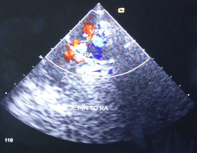
Blood biochemistry was normal. Chest radiograph showed apical lobe consolidation with irregular rt. heart border. 2D-ECHO showed acyanotic heart disease with two VSD of size 3.0 mm (perimembranous) and 3.3 mm (muscular) with small PFO 2.8 mm with mild pulmonary regurgitation with indirect Gerbode defect from left ventricle to right atrium. ECHO revealed cardinal findings suggestive of Gerbode Defect (Dilated right atrium and high pressure gradient of 144.7 mm of Hg (N: 65-144 mm of Hg) across the defect [Table/Fig-1]. TORCH profile was negative and blood culture was sterile.
Echocardiograph showing jet going from Left ventricle to right atrium.
Baby was managed with restricted and sodium free fluid, intravenous furosemide, digoxin, antibiotics. Condition improved over 5 days and baby started taking orally. Attendants were counselled about the disease and need of surgical management of defect.
Discussion
The present child presented to us in heart failure with underlying murmur consistent with diagnosis of VSD. Echocardiography revealed a communication between left ventricle and right atrium besides two 2 VSD (perimembranous 3.0 mm, muscular 3.3 mm) and patent foramen ovale (PFO ~ 2.8 mm). The dilated right atrium and high pressure gradient of 144.7 mm of Hg (N: 65-144 mm of Hg) across ventriculo-atrial defect confirmed the diagnosis of Gerbode defect in present case. The child was managed with conservative treatment and referred to surgical centre for correction of defect. LV-RA communication was a Gerbode defect (indirect type, congenital variety). The left ventricular to right atrium (LV-RA) shunt was first reported by Gerbode et al., in year 1958 [1]. It is of two types: direct (defect above tricuspid leaflet, Type I) and Indirect (defect below tricuspid leaflet, Type II) [2]. Gerbode defect could be congenital or acquired after trauma, infective endocarditis, post surgery and spontaneous closure of VSD [3–5]. Literature search revealed acquired defect are more common since advent of more surgical closure of VSD, both in paediatric and adults [6]. We found only 10 reports of congenital defects in English literature through search engines of Pubmed and Google scholar [2,7–10]. We summarised data of 11 cases (age < 18 years) including ours and reached to important findings [Table/Fig-2]. There were 5 males and 6 females with no sex predilection. Direct defect (7/10) was the most common anatomical location. Three cases were of indirect type including ours. Our patient was the youngest to be reported to have diagnosed at age of 2 months. Maximum number (9/11) of cases was symptomatic at presentation and defect was detected by Echocardiography. VSD was the most common additional cardiac shunt present in five of eleven cases. Surgery was the most important intervention done to save all children except one where child was kept in follow up as he was asymptomatic [7]. There have been many reports of acquired variety in adult population from Indian subcontinent. But, congenital forms are rarely picked and reported. H Swamy Rajesh reported in a 10-year-old male child in year 2012 [8].
Depicting clinical, cardiac and outcome profile of Congenital Gerbode Defect of eleven patients (including present case) [2,7–10].
| Serial Number | Parameter | Kelle et al.,(n=6) | Acar et al.(n=1) | Rajesh et al.,(n=1) | Panduranga et al.,(n=1) | Otaigbe et al.,(n=1) | Present Study (n=1) |
|---|
| 1 | Age | 1.6 years (median age at time of repair); range (0.4-19 years) | 14 years | 10 years | 13 years | 4 months | 2 months |
| 2 | Sex | 2 males. 4 females | Male | Male | Male | Female | Female |
| 3 | Clinical presentation | Symptomatic | Asymptomatic | Symptomatic | Asymptomatic | Symptomatic | Symptomatic |
| 4 | Type of defect | Congenital (direct) | Congenital (combined) | Congenital (indirect) | Congenital | Congenital(indirect) | Congenital(indirect) |
| 5 | Associated cardiac findings | Patent arterial duct & right sided aortic arch (1), left superior caval vein (3), anomalous left hepatic vein (2), | Perimembranous VSD | Small to medium sized VSD | 0.5 cm perimembranous subaortic VSD | Perimembranous VSD (7mm) | Two VSD; Perimembranous (3 mm); muscular (3.3 mm); PFO (2.8 mm) |
| 6 | Intervention | Surgery | None | Surgery | Surgery | Surgery | Surgery |
| 7 | Outcome | Alive | Alive (under follow up) | Alive | Alive at 6 months follow up | Alive at post operative period | Alive at follow up of 6 months |
Conclusion
This literature search highlights rarity of congenital variety of Gerbode defect. We report only second case of such rare defect from India. The review highlights salient parameters (no age predilection, direct more common congenital variety, symptomatic at presentation, required surgical correction in majority) from all reported cases so far to aware the treating clinicians and paediatricians of such rare defect that needs timely surgical correction/referral for better survival and outcome.
[1]. Gerbode F, Hultgren H, Melrose D, Osborn J, Syndrome of left ventricular-right atrial shunt; successful surgical repair of defect in five cases, with observation of bradycardia on closureAnn Surg 1958 148:433-46. [Google Scholar]
[2]. Kelle AM, Young L, Kaushal S, Duffy CE, Anderson RH, Backer CL, The Gerbode defect: the significance of a left ventricular to right atrial shuntCardiol Young 2009 19:96-99. [Google Scholar]
[3]. Cabalka AK, Hagler DJ, Mookadam F, Chandrasekaran K, Wright RS, Percutaneous closure of left ventricular-to-right atrial fistula after prosthetic mitral valve rereplacement using the Amplatzer duct occluderCatheter Cardiovasc Interv 2005 64:522-27. [Google Scholar]
[4]. Trehan V, Ramakrishnan S, Goyal NK, Successful device closure of an acquired Gerbode defectCatheter Cardiovasc Interv 2006 68:942-45. [Google Scholar]
[5]. Inoue H, Iguro Y, Kinjo T, Matsumoto H, Yotsumoto G, Sakata R, Acquired left ventricular-right atrial communication and severe aortic valve regurgitation caused by infective endocarditisThorac Cardiovasc Surg 2009 57:54-56. [Google Scholar]
[6]. Demirkol S, Gurkan Yesil F, Bozlar U, Balta S, Sahin MA, Guler A, Multimodality imaging of a congenital Gerbode defectKardiol Pol 2013 71:104 [Google Scholar]
[7]. Acar P, Séguela PE, Hascoet S, The Gerbode defect or left ventricular to right atrial shunt assessed by transthoracic 3D echocardiographyEchocardiography 2011 28:E140-42. [Google Scholar]
[8]. Rajesh HS, A case report of rare case VSD – Gerbode Defect (Lv to Ra Shunt)Pediat Therapeut 2012 2:77 [Google Scholar]
[9]. Panduranga P, Mukhaini M, A rare type of Gerbode defectEchocardiography 2011 28:E118-20. [Google Scholar]
[10]. Otaigbe BE, Orubide D, Rare presentation of gerbode defect in a 4-month-old nigerian and a review of the literatureCase Rep Cardiol 2013 2013:564786 [Google Scholar]