The Effects of Fetal Movement Counting on Pregnancy Outcomes
Masoumeh Delaram1, Lobat Jafarzadeh2
1 Assistant Professor, Faculty of Nursing and Midwifery, Shahrekord University of Medical Sciences, Shahrekord, Iran.
2 Department of Obstetrics and Gynecology, Shahrekord University of Medical Sciences, Shahrekord, Iran.
NAME, ADDRESS, E-MAIL ID OF THE CORRESPONDING AUTHOR: Dr Masoumeh Delaram, Assistant Professor, Faculty of Nursing and Midwifery Shahrekord University of Medical Sciences, Shahrekord, Iran.
E-mail: masoumehdelaram@yahoo.com
Introduction
Counting fetal movements may lead to timely assess fetal health and prevent the adverse effects of pregnancy.
Aim
The aim of this study was to determine the effect of fetal movement counting on pregnancy outcomes.
Materials and Methods
In a randomized controlled trial, 208 women with singleton pregnancy were randomly divided into two groups of fetal movement counting and control. Pregnancy outcomes were compared between the two groups. Data were analysed with SPSS and p<0.05 was considered significant.
Results
There was no significant difference in the mean maternal concern (p=0.36), admission to hospital due to the decreased fetal movements (p=0.99), birth weight (p=0.21), Apgar score (p=0.51), the mean of gestational age at the time of decreased fetal movements (p=0.49) and mode of delivery (p=0.69) between the two groups. There were no cases of premature labour, intrauterine growth retardation and fetal death in the two groups.
Conclusion
Pregnancy outcome was similar in the two groups of fetal movement counting and control. Further studies are needed to evaluate the effect of fetal movement counting on the major outcomes of pregnancy such as intrauterine fetal death.
Fetal activity, Nulliparous women, Outcomes of pregnancy
Introduction
Fetal movement counting by mother is a method used to assess the fetal well-being and this unstructured screening helps the mother to be reassured of the health of the fetus [1]. More than 99% of women who have given birth to a healthy baby say that it is important to feel the baby’s movements every day [2]. When the fetal momement is reduced, they are worried and visit their doctor or health care provider for further evaluations [3]. In women with decreased fetal movements, there is the risk of complications such as fetal growth restriction and stillbirth. More women notice to changes in fetal movement, its intensity and frequency [4].
Decrease in fetal movements concern the mother and she often sought unscheduled antenatal consultation [5]. Although in most cases with reduction of fetal movements, pregnancy continue without complications [6], the concern of mother should be taken seriously because the adverse outcomes, including intrauterine growth retardation and death may be associated with reduced fetal movements [7–10]. The counting of fetal movements by mother has been recommended as a instrument for boosting self-screening of mothers to reduce the fetal movements counting [9,11]. The daily fetal movement counting may increase the mother’s ability to recognise on time the warning signs and if the fetus is in danger, it will be properly intervened.
Although this method is simple, its usage is controversial. Gradual reduction of fetal movement and its perception by mother is an important sign of fetal damage that can demonstrate complication [12,13], preterm delivery [7], intrauterine growth retardation [14], stillbirth and emergency caesarean section [8]. Maternal perception of reduced fetal movements is the most important marker of decreased fetal activity [15]. If the mothers carefully control the fetal movement and report on time decrease in fetal movements to physician or health care providers, it is likely to prevent perinatal morbidity and mortality [5]. Although the health professionals do not have a same approach on the official count of fetal movements [16,17] and dispute that the counting of fetal movement probably trigger maternal psychological stress [9,16,18] and derive excess discrepancies and obstetric interposition [9], a current study has shown that the fetal movement counting reassured mothers, and result in to decrease in maternal concern [19]. Another study reported that the women who performed fetal movement counting from 28 to 37 weeks of gestation, reported less anxiety than those in the control group [20]. Because there is not enough evidence about the impact of fetal movement counting on pregnancy outcome, the current study was performed to detect the counting of fetal movement on pregnancy outcomes.
Materials and Methods
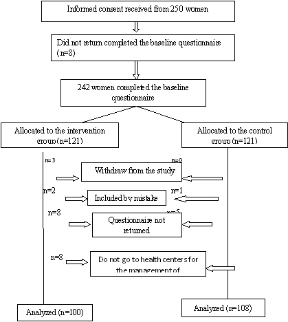
The study protocol was approved by the Ethics Committee of Shahrekord University of Medical Sciences and the study was registered in Iranian Registry of Clinical Trials as IRCT201207103078N9. In addition, informed consent was obtained from the participants. For ethical considerations of the research, the objectives of the study were explained to health care providers and participants. Intervention community of the study included all nulliparous women referred to health centers of Shahrekord. The sample included 208 cases of women who were selected through convenience sampling and were randomly assigned to the intervention group (n = 100) and control group (n = 108) [Table/Fig-1]. These women had diploma or higher education, singleton pregnancies, were not considered for the early termination of pregnancy and had not previously participated in any investigation of fetal movement counting. Exclusion criteria included oligohydramnios, multifetal pregnancy, fetal abnormalities and maternal smoking. The women who had information about the fetal movement counting were also excluded. At the beginning of the study, all subjects underwent ultrasound at 17-18 weeks of gestation for the detection of multiple pregnancies and fetal anomalies and then they completed the Personal Information Form at 28 weeks of gestation. Then, the women in the intervention group were instructed to count fetal movement and record it. Intervention group was asked to place in the left lateral position after breakfast every morning for half an hour and count and record the fetal movements. To ensure proper performance of this task, the subjects were telephoned once a week. They also were asked to show the fetal movement chart to the health care provider at each prenatal visit. Counting fetal movements continued for 28 to 37 weeks of pregnancy and the control group received the standard antenatal care. At postpartum, pregnancy outcomes (preterm delivery, intrauterine growth retardation, mode of delivery, birth weight and Apgar score, mothers concern about reduced fetal movements and hospitalization due to it) were compared in the two groups. Participants’ baseline characteristics and pregnancy outcome were evaluated through questionnaires and checklist.
Flowchart of participants in the study.
Statistical Analysis
Statistical analysis was performed by SPSS (version 16). We used the mean and standard deviation for quantitative variables and frequency and percentage for qualitative variables. P<0.05 was considered significant. The mothers in the control group were asked the question of whether they count fetal movements during pregnancy or not and if the answer was yes, they were excluded from the final analysis.
Results
Mean age of women was 26.35±4.34 years in the intervention group and 26.72±3.93 years in the control group with no significant difference. Also, no difference was seen in job, level of education, smoking and unplanned pregnancy between the two groups. Mean of body mass index (BMI) was 24.22±3.23 in the intervention group and 24.82±2.66 in the control group with no significant difference [Table/Fig-2].
Baseline characteristics of participants.
| Variable | Control Group (n=108)Mean (SD) or No. (%) | Intervention Group (n=100)Mean (SD) or No. (%) | p |
|---|
| Age (yr) | 26.72±3.93 | 26.35 (4.34) | 0.51 |
| BMI (kg/m2) | 24.82 (2.66) | 24.22 (3.23) | 0.14 |
| Education |
| High school graduate | 30 (27/7) | 29 (29) | 0.09 |
| >Colledge graduate | 78 (72/3) | 71 (71) | |
| Job |
| Employed | 14 (13.1) | 16 (16) | 0.56 |
| Un-Employed | 94 (86.9) | 84 (84) | |
| Unplanned pregnancy | 5 (4.7) | 4 (4) | 0.06 |
The p values refer to comparisons between the control and the intervention groups: chi-square test for categorical variables and t-test for continuous variables.
BMI; body mass index, SD; standard deviation
The comparison between pregnancy outcomes in the two groups was shown [Table/Fig-3]. As seen, there was no significant difference in the mean frequency maternal concern about the decreased fetal movements and admission to hospital due to it. Besides, no difference was found in birth weight and Apgar score of infants between the two groups. Mean gestational age at the time decreased the fetal movement, was 34.51±2.5 in the intervention group and 34.55±8.48 in the control group that there was no significant difference. No significant difference was found in mode of delivery between the two groups (p=0.09). There were no cases of premature labour, intra uterine growth retardation and fetal death in the two groups.
The comparison between pregnancy outcomes in the intervention and control groups.
| Pregnancy outcomes (Mean±SD) | Maternal concern | Admission to hospital | Birth weight | Apgar score |
|---|
| Intervention group | 1.80±1.7 | 1.95±0.21 | 3086±394 | 8.98±0.14 |
| Control group | 1.55±0.82 | 1.93±0.22 | 3153±380 | 8.96±0.23 |
| p | 0.36 | 0.99 | 0.21 | 0.51 |
SD; standard deviation
Discussion
The findings of this study showed that there was no significant difference in pregnancy outcome between the two groups of fetal movement counting and control. Saastad et al., reported that there was not a significant difference in the proportion of intrauterine growth restriction fetuses in both groups, but intrauterine growth restriction fetuses had been diagnosed earlier in fetal movement counting compared with controls and reduced the number of infants who were born with a low Apgar score. This study also reported that there was no fetal death [21]. Both of the above finding is in line with our findings. A similar situation existed in mean maternal concern due to reduced fetal movements and hospitalizations in the two groups. A study reported that the frequency of consultation with mother due to concerns about decreased fetal movement was not significantly different between the women who counted fetal movements and those who did not [21]. Another study reported that there was not a significant reduction in unexplained intrauterine fetal death rates in the two groups [22]. Saastad et al., reported that the fetal movement counting improved diagnosis of intrauterine fetal growth retardation, and reduced the number of children born with a low Agar score [2]. One of the factors that are associated with reduced fetal movements is low Apgar score and suggests that the fetus is in a precarious condition [21]. It is demonstrated that the fetus with agar score less than or equal to 3, in the first minutes after birth, is at a greater risk of disability later in life [13,23]. Small sample size in this prospective study may explain the differences between this study and the above-referenced study which was conducted on a much larger sample size.
In this study, the gestational age of the fetus at the time of decreased fetal movements was similar in the two groups. In Saastad et al., study, women who were in the fetal movements counting group, but not control group, were concerned about reduced fetal movements and had been early admitted to hospital [23]. In this study, there was no significant difference in the mode of delivery. In the fetal movement counting group, 53 women and in the control group, 58 women delivered by cesarean section. These findings are relevant to a study that reported no significant differences in the rate of emergency cesarean section between the groups [24]. The lack of significant differences in birth weight was another finding of that study [24], which is consistent with our study. The women participating in the study were mainly employed and educated. Therefore, the findings should be generalized to similar populations. The limitation of current study was that the most women participating in the study were more employed and educated. Therefore, the findings should be limited to the same population.
Conclusion
Pregnancy outcome was similar in the two groups of fetal movement counting and control. Further studies are needed to evaluate the effect of fetal movement counting on the major outcomes of pregnancy such as intrauterine fetal death.
Conflict of interest
Authors declared that there is no conflict of interest.
The p values refer to comparisons between the control and the intervention groups: chi-square test for categorical variables and t-test for continuous variables.
BMI; body mass index, SD; standard deviation
SD; standard deviation
[1]. Sheikh M, Hantoushzadeh S, Shariat M, Maternal perception of decreased fetal movements from maternal and fetal perspectives, a cohort studyBMC Pregnancy Childbirth 2014 14:286doi: 10.1186/1471-2393-14-286 [Google Scholar]
[2]. Mangesi L, Hofmeyr GJ, Smith V, Smyth RM, Fetal movement counting for assessment of fetal wellbeingCochrane Database Syst Rev 2015 10:CD004909doi: 10.1002/14651858.CD004909.pub3. Review [Google Scholar]
[3]. Saastad E, Tveit J, Flenady V, Stray-Pedersen B, Fretts R, Bordahl P, Implementation of uniform information on fetal movement in a Norwegian population reduced delayed reporting of decreased fetal movement and stillbirths in primiparous women - a clinical quality improvementBMC Research Notes 2010 3(2)doi:10.1186/1756-0500-3-2 [Google Scholar]
[4]. Froen JF, Saastad E, Tveit JV, Bordahl PE, Stray-Pedersen B, [Clinical practice variation in reduced fetal movements]Tidsskr Nor Laegeforen 2005 125(19):2631-34. [Google Scholar]
[5]. Tveit JV, Saastad E, Stray-Pedersen B, Bordahl PE, Flenady V, Fretts R, Reduction of late stillbirth with the introduction of fetal movement information and guidelines - a clinical quality improvementBMC Pregnancy Childbirth 2009 9(32):1-10. [Google Scholar]
[6]. Kamalifard M, Abbasalizadeh S, Ghojazadeh M, Ghatreh Samani F, Rabiei L, Diagnostic value of fetal movement counting by mother and the optimal recording durationJ Caring Sci 2013 2(2):89-95.doi: 10.5681/jcs.2013.011. eCollection 2013 Jun [Google Scholar]
[7]. Froen JF, Arnestad M, Frey K, Vege A, Saugstad OD, Stray-Pedersen B, Risk factors for sudden intrauterine unexplained death: epidemiologic characteristics of singleton cases in Oslo, Norway, 1986-1995Am J Obstet Gynecol 2001 184(4):694-702. [Google Scholar]
[8]. Raynes-Greenow CH, Gordon A, Li Q, Hyett JA, A cross-sectional study of maternal perception of fetal movements and antenatal advice in a general pregnant population, using a qualitative frameworkBMC Pregnancy Childbirth 2013 13:32doi: 10.1186/1471-2393-13-32 [Google Scholar]
[9]. Mangesi L, Hofmeyr GJ, Fetal movement counting for assessment of fetal wellbeingCochrane Database Syst Rev 2007 24(1):1-23. [Google Scholar]
[10]. Smith V, Begley C, Devane D, Detection and management of decreased fetal movements in Ireland: a national survey of midwives’ and obstetricians’ practicesMidwifery 2014 30(1):43-9.doi: 10.1016/j.midw.2013.02.006. Epub 2013 Mar 2 [Google Scholar]
[11]. Sharp GC, Stock SJ, Norman JE, Fetal assessment methods for improving neonatal and maternal outcomes in preterm prelabour rupture of membranesCochrane Database Syst Rev 2014 10:CD010209doi: 10.1002/14651858.CD010209.pub2. Review [Google Scholar]
[12]. Raynes-Greenow CH, Gordon A, Li Q, Hyett JA, A cross-sectional study of maternal perception of fetal movements and antenatal advice in a general pregnant population, using a qualitative frameworkBMC Pregnancy Childbirth 2013 13:32doi: 10.1186/1471-2393-13-32 [Google Scholar]
[13]. Nomura RM, Ferreira MV, Latif IO, Francisco RP, Zugaib M, Agreement between maternal perception of fetal movements and visualization by ultrasound]Rev Bras Ginecol Obstet 2013 35(2):55-9.Portuguese [Google Scholar]
[14]. Copel JA, Bahtiyar MO, A practical approach to fetal growth restrictionObstet Gynecol 2014 123(5):1057-69.doi: 10.1097/AOG.0000000000000232. Review [Google Scholar]
[15]. Mohr Sasson A, Tsur A, Kalter A, Weissmann Brenner A, Gindes L, Weisz B, Reduced fetal movement: factors affecting maternal perceptionJ Matern Fetal Neonatal Med 2015 :1-4.[Epub ahead of print] [Google Scholar]
[16]. Chauveau L, Raia-Barjat T, Noblot E, Fanget C, Gallot D, Chaleur C, [Maternal knowledge of fetal movements in late pregnancy]J Gynecol Obstet Biol Reprod (Paris) 2015 S0368-2315(15):00153-2.pii doi: 10.1016/j.jgyn.2015.06.006. [Epub ahead of print] French [Google Scholar]
[17]. Matsubara S, Kuwata T, Fukui S, Fetal movement count may prevent fetal death as early as 26 weeksActa Obstet Gynecol Scand 2013 92(12):1426doi: 10.1111/aogs.12200. Epub 2013 Jul 19 [Google Scholar]
[18]. Franks Z, Nightingale R, Decreased fetal movements: a practical approach in a primary care settingAust Fam Physician 2014 43(11):782-85. [Google Scholar]
[19]. Velazquez MD, Rayburn WF, Antenatal evaluation of the fetus using fetal movement monitoringClin Obstet Gynecol 2002 45(4):993-1004. [Google Scholar]
[20]. Grant A, Elbourne D, Valentin L, Alexander S, Routine formal fetal movement counting and risk of antepartum late death in normally formed singletonsLancet 1989 2(8659):345-49. [Google Scholar]
[21]. Delaram M, Shams S, The effect of foetal movement counting on maternal anxiety: A randomised, controlled trialJ Obstet Gynaecol 2015 :1-5.[Epub ahead of print] [Google Scholar]
[22]. Liston RM, Bloom K, Zimmer P, The psychological effects of counting fetal movementsBirth 1994 21(3):135-40. [Google Scholar]
[23]. Imdad A, Yakoob MY, Siddiqui S, Bhutta ZA, Screening and triage of intrauterine growth restriction (IUGR) in general population and high risk pregnancies: a systematic review with a focus on reduction of IUGR related stillbirthsBMC Public Health 2011 11(Suppl 3):S1doi: 10.1186/1471-2458-11-S3-S1. Review [Google Scholar]
[24]. Posthumus AG, Birnie E, van Veen MJ, Steegers EA, Bonsel GJ, An antenatal prediction model for adverse birth outcomes in an urban population: The contribution of medical and non-medical risksMidwifery 2015 S0266-6138(15):00289-2.pii doi: 10.1016/j.midw.2015.11.006. [Epub ahead of print] [Google Scholar]