The Extensor Indicis Brevis – A Rare Variation and its Significance
Rohini Arathala1, P.K. Sankaran2, Gunapriya Ragunath3, Soma Sri Harsha4, Tharani Sree Sugumar5
1 Student, Department of Anatomy, Saveetha Medical College and Hospital, Chennai, India.
2 Assistant Professor, Department of Anatomy, Saveetha Medical College and Hospital, Chennai, India.
3 Professor, Department of Anatomy, Saveetha Medical College and Hospital, Chennai, India.
4 Student, Saveetha Medical College, Chennai, India.
5 Student, Saveetha Medical College, Chennai, India.
NAME, ADDRESS, E-MAIL ID OF THE CORRESPONDING AUTHOR: Dr. P.K. Sankaran, 18 Pillaiyar Koil St, Thirumazhisai, Chennai - 600124, India.
E-mail: drpks@live.com
Variations in muscle attachments are valuable in interpreting rare clinical conditions. This case report discusses about the abnormal extensor indicis muscle attachment in left hand of male cadaver and its clinical relevance. The extensor indicis muscle was arising from lunate carpal bone and posterior radiocarpal ligament which further continued as short belly and had its distal attachment on the proximal phalanx of the index finger. This muscular variation has been described in earlier studies with 1.0% incidence. Abnormal origin of extensor indicis may be associated with pain and swelling at the back of the hand. This pain may be confused with other clinical conditions like synovial cyst and ganglion. Knowledge of such rare variations are useful to clinicians, surgeons, occupational and physical therapists for planning better surgical decisions.
Extensor muscles, Hand muscles, Tendon graft/surgeries, Wrist pain
Case Report
During routine dissection teaching for the first year MBBS students in Saveetha Medical College, we observed an anomalous origin of the extensor indicis muscle in an adult male cadaver of around 60 years of age on the left side.
The extensor indicis muscle originated as a small belly from the articular upper surface of lunate bone and dorsal radiocarpal ligament instead of arising from posterior surface of ulna and inserted in to base of distal phalanx of the index finger [Table/Fig-1,2]. The belly of this muscle is approximately 5.3 cm in length and its tendon was approximately 6 cm in length. After its origin the muscle passes along the ulnar side of extensor digitorum tendon to the index finger and unites with it opposite to second metacarpophalangeal joint then inserted in to the base of distal phalanx. This muscle was supplied by posterior interosseous nerve. No variation was observed in the right side of extensor indicis muscle.
Arrow showing the extensor indicis brevis.
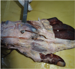
1. Origin of the extensor indicis brevis
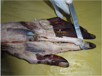
2. Tendon inserted in to the base of distal phalanx of the index finger along with tendon of Extensor digitorum.
Note: EIB- Extensor indicis brevis.
ED- Extensor digitorum
Discussion
The posterior compartment muscles of the forearm are known as the extensor group of muscles that produces extension at the wrist and fingers [1]. The extensor indicis is a narrow, elongated muscle located on the dorsum of the forearm, medial to extensor pollicis longus. This extensor muscle originates from the posterior surface of the ulna and posterior surface of the interosseous membrane. The tendon of the extensor indicis muscle usually passes under the fourth compartment of extensor retinaculum along with the tendon of the extensor digitorum for index finger [2]. Opposite the head of the second metacarpal the extensor indicis joins along with extensor digitorum tendon to index finger and extends the index finger independently, without affecting remaining fingers. Though the anomalous variation of Extensor Indicis (EI) tendon is very rare the anatomical knowledge of such variation is very important while performing tendon graft surgeries in the hand. In the present case we are reporting a unique and rare variation of the extensor indicis muscle originating from lunate bone and dorsal radiocarpal ligament. This muscle is inserted into the distal phalanx of the index finger on ulnar side of the extensor expansion. Earlier study on extensor indicis muscles were done on 263 specimens which showed abnormal origin of this muscle like two belly and one tendon in between and two origin one from ulna and other head from proximal carpal bones finally inserting into carpal bones [3]. Another study showed an anomalous origin of extensor indicis from dorsum of wrist covered by extensor retinaculum [4]. Murakami et al., observed an extensor indicis brevis with an unusual ganglion [4]. This kind of muscle variations can be confused with a ganglion or other tumor and overuse of this will produce pain [5].
The variations of muscles and tendons in hand are not uncommon however; this type of variation is relatively rare. The extensor indicis muscle is widely utilized in surgeries for tendon transfer designed to restore a variety of finger movements [6–8]. It is utilized in tendon graft surgeries in dysfunctions or functional loss of the abductor pollicis brevis, opponens pollicis muscles [7], and extensor pollicis longus [9]. The preference for using this extensor indicis tendon, for tendon grafts surgeries because index finger receives extensor digitorum tendon (to index finger) also [6]. Noorda et al., did not observe pain during mobility, or significant weakness in the donor finger after surgery although there was poor finger function in 11% of the evaluated cases [9]. On the other hand, Kitano et al., observed a functional reduction in the independent indicis extension in 26% of the cases [8]. Due to anomalous origin the extension of index finger may be weak as compared to the normal individuals. Also, this extensor indicis brevis lies more superficial in the dorsum of the hand can be easily damaged in minor injuries and any surgeries at this region.
Conclusion
To conclude, in this present case we found abnormal origin of extensor indicis muscle from lunate bone and dorsal radiocarpal ligament. Hence, it can be stated that attention should be paid to the individual variations in the pattern of distribution of extensor tendons. Accordingly, any surgical procedure in this particular region should be planned carefully in advance to encounter any variation. This may help clinicians in accurately managing suspected painful conditions in the back of the wrist and hand.
[1]. Romanes GJ, Cunningham manual of practical anatomy 1986 1(15)New YorkOxford University Press Inc:117 [Google Scholar]
[2]. Susan Standring, Gray’s Anatomy; The anatomical basis of clinical practice 2005 Churchill Livingstone:882 [Google Scholar]
[3]. Li J, Ren ZF, Bilateral extensor indicis brevis: a rare muscular variantRom J Morphol Embryol 2012 53:185-87. [Google Scholar]
[4]. Murakami Y, Todanik The extensor indicis brevis muscle with and unusual ganglionClin Orthop 1982 162:207-09. [Google Scholar]
[5]. Tountas CH, Bergman R, Anatomic variations of the upper extremity 1993 New YorkChurchill Livingstone Inc [Google Scholar]
[6]. Gonzalez MH, Weinzweig N, Kay T, Grindel S, Anatomy of the extensor tendons to the index fingerJ Hand Surg 1996 21A:988-91. [Google Scholar]
[7]. Batra S, Sakamuri R, Kanvinde RN, Sequential traumatic bilateral extensor pollicis brevis rupture: a case reportJ Hand Surg 2007 32:685-87. [Google Scholar]
[8]. Kitano K, Tada K, Shibata T, Yoshida T, Independent index extension after indicis proprius transfer excision of junctureetendinumJ Hand Sur 1996 21A:992-96. [Google Scholar]
[9]. Noorda RJP, Hage JJ, Groot PJM, Bloem JJAM, Index finger extension and strength after extensor indicis proprius transferJ Hand Surg 1994 19A:844-49. [Google Scholar]