Bladder Endometriosis Mimicking TCC – A Case Report
Asish Gupta1, Atul Bhatnagar2, B N Seth3, Arbinder Dang4, Vineeta Gupta5
1 Consultant, Department of Gen and Laproscopic Surgery, Sant Parmanand Hospital, Civil Lines Delhi, India.
2 Consultant, Department of Gen and Laproscopic Surgery, Sant Parmanand Hospital, Civil Lines Delhi, India.
3 Head of Department, Department of Anesthesia, Sant Parmanand Hospital, Civil Lines Delhi, India.
4 Consultant, Department of Gynecology, Sant Parmanand Hospital, Civil Lines Delhi, India.
5 Professor and Head, Department of Oral Pathology, IDSTModinagar, Uttar Pradesh, India.
NAME, ADDRESS, E-MAIL ID OF THE CORRESPONDING AUTHOR: Dr. Vineeta Gupta, 18/23, First Floor, Shakti Nagar, Delhi 110007, India. E-mail : vineeta9999@gmail.com
Endometriosis is the ectopic presence of endometrial tissue outside the uterus. Though on its own endometriosis is not a rare lesion, the involvement of the urinary tract is rare but with the bladder being the most commonly affected organ. Endometriosis is usually seen in females between the ages of 30-40 years and may occur due to fluctuating levels of oestrogen and progesterone. Clinically the patient maybe asymptomatic or show symptoms of dysmenorrhea, irregular or heavy periods, pain in the pelvic area, lower abdomen or in the back. It has been suggested that ultrasonography should be done either before or during menstruation as the lesion becomes more evident and a biopsy taken during this period is a strong aid in reaching a final diagnosis. We report here an unusual case of bladder endometriosis where the patient came with severe pelvic pain and an endoluminal mass seen on the ultrasonographic report. Based on these findings a differential of transitional cell carcinoma was given which was ruled out based on the cystoscopic findings.
Cystoscopy, Endometrial tissue, Transitional cell carcinoma, Urinary system
Case Report
A female patient age 32-year-old reported to the surgical OPD to consult regarding her ultra -sonography report which showed a lobulated endoluminal vesicle mass lesion measuring 3x2 cm in the trigonal area of the urinary bladder and with her left ovary showing PCOD configuration. Her history revealed no urinary symptoms like burning micturition; haematuria etc. There was no loss of appetite or weight though she did complain of severe recurrent pelvic pain. She was recently diagnosed with diabetes and she was G3P1L1A1. In 2010 she conceived and in the 35th week ultrasonographic report the foetus was then diagnosed with hydrocephalus and with breech presentation. Elective C-section was carried out but the child was still born. Following this episode she developed secondary infertility and was started on hormonal therapy but had not conceived since then. The present ultra sound was done to evaluate the follicles and ovaries on day-10 of the cycle on suggestion from her gynaecologist. She came to our OPD for advice regarding the lobulated endoluminal vesicle mass lesion.
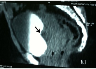
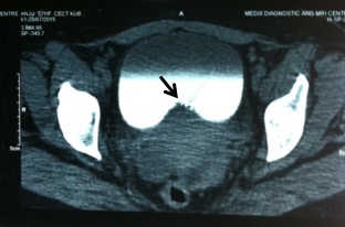
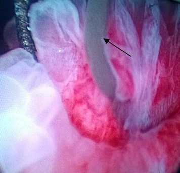
We advised a contrast enhanced CT scan to evaluate the bladder mass. Contrast enhanced chromatography (CeCT) revealed an enplaque soft tissue mass arising from the posterior wall of the urinary bladder which showed moderate post contrast enhancement with associated thickening of the adjacent urinary bladder wall. No extra vesicle extension was seen. The mass measured 4x 1.7 cm. Impression on the scan was likely Urothelial Neoplasm (Transitional Cell Carcinoma) [Table/Fig-1,2]. Patient was advised cystoscopic evaluation and biopsy to rule out malignancy. Patient was worked up with routine blood and urine examination which were within the normal parameters. Patient was subjected to cystoscopy under anaesthesia. The cystoscopy revealed an elevated lesion in the posterior wall of the bladder with a blue-black cystic base [Table/Fig-3]. On taking an incisional biopsy there was extravasation of jelly like material from the cyst.
CeCT image showing an enplaque soft tissue mass arising from the posterior wall of the urinary bladder (arrow), sagittal section.
CeCT image showing an enplaque soft tissue mass arising from the posterior wall of the urinary bladder (arrow), transverse section.
Elevated lesion in the posterior wall of the bladder with a blue-black cystic base. With extravasation of jelly like material (arrow) from the cyst.
The biopsy specimen was sent for routine histopathological examination and was reported as Non-Specific Cystitis.
Evaluating the entire history of secondary infertility, treatment with hormonal therapy, severe pelvic pain every two weeks and per-op findings of blue dome like cyst which oozed out jelly like material we came to the final diagnosis of Bladder Endometriosis.
A gynaecological opinion was sought and the patient was prescribed progestin hormonal therapy. In the two months postoperative follow up period she was advised a follow up CeCT, but she refused as she was symptom free.
Discussion
Endometriosis is the ectopic presence of endometrial tissue outside the uterus. Per-se endometriosis is not rare but the involvement of the urinary tract is rare (1-2%). In the urinary tract the bladder is the most commonly affected organ (84%) [1]. Endometriosis is usually seen in females between the age of 30-40 years. and may occur due to fluctuating levels of oestrogen and progesterone. Here usually the patient shows primary infertility or may have developed secondary infertility due to endometriosis [2]. This also can explain the secondary infertility seen in our patient.
Various theories have been put forth regarding the cause of endometriosis, like, retrograde menstruation, metaplasia, hormones, oxidative stress and inflammation, immune dysfunction, apoptosis suppression, genetic and stem cells [3]. The patient developed secondary infertility post her third pregnancy and was started on hormone therapy for her IVF treatment.
Various authors have suggested that ultrasonography should be done either before or during menstruation as the lesion becomes more evident. Biopsy taken via cystoscopy on the 17th day of the cycle will aid in reaching a final diagnosis and may show the characteristic appearance of endometrial stroma with surrounding glandular spaces, but even if these features are not present and a nonspecific picture is seen the diagnosis of endometriosis cannot be ruled out [1,4]. This was seen in our case also where the histopathological report of nonspecific cystitis was given. Taking deeper biopsy samples is not advised in case of bladder involvement as there is a fear of bladder perforation [5]. Detailed history and analysis of the clinical features should be done and only then should treatment be decided. Treatment in such cases may range from conservative hormonal therapy to the more radical approach of surgical resection [6].
Literature advises the use of hormonal therapy as the first line of approach in cases where the symptoms are not severe and the lesion is small and/or localized. This approach is also more suitable in cases where the patient wishes for future pregnancies [2]. We also placed the patient on hormonal therapy under the guidance of the gynaecological department who placed her on progestins. We did not consider surgical approach as the symptoms were not severe, she had no urinary symptoms and she also had the desire of a future pregnancy. Considering the CeCT findings we had suspected bladder CA and to rule that out a cystoscopy was done.
Bladder Carcinoma (TCC) though is more commonly seen in males has also been reported in females. The most common symptom reported is Haematuria, but literature also shows that bladder carcinoma may also be seen along with non-visible i.e. microscopic haematuria [7]. Our patient did not show any urinary signs like macro or microscopic haematuria etc., 80-90%. TCC patients present with gross (macro) haematuria. The bleeding in the bladder may be episodic in which case microscopic haematuria will be absent. Irritative bladder symptoms are presented in 20-30% TCC patients [7]. While our patient did not show any of the above we gave a differential diagnosis of TCC based on the CeCT report.
On receiving the histopathological report of the cystoscopy specimen we could, with complete certainty rule out TCC or Bladder Carcinoma and so we proceeded with the medical management of the patient under the guidance of the Department of Gynaecology. The patient was kept under six month follow up and though advised with a repeat CeCT during this period, she refused and showed no recurrence of any symptoms.
Conclusion
We present here a case showing an asymptomatic mass lesion in the urinary bladder which was visible on both the ultrasonography and CeCT. The patient presented with a gynaecological history of G3P1L1A1, showing secondary infertility after her third pregnancy and on IVF for the same. Due to the ultrasonography and CeCT findings a differential diagnosis of TCC and bladder endometriosis was given. Histological analysis of the cystoscopically retrieved tissue revealed a nonspecific diagnosis of cystitis which thus ruled out TCC. Bladder endometriosis is rare but can be cured by medication as we observed in our case.
[1]. Fedele L, Bianchi S, Raffaelli R, Portuese A, Pre-operative assessment of bladder endometriosisHum Reprod 1997 12:2519-22. [Google Scholar]
[2]. Kumar S, Tiwari P, Sharma P, Goel A, Singh JP, Vijay MK, Urinary tract endometriosis: Review of 19 casesUrol Ann 2012 4(1):6-12. [Google Scholar]
[3]. Sourial S, Tempest N, Hapangama DK, Theories on the pathogenesis of endometriosisInt J Reprod Med 2014 :1-7. [Google Scholar]
[4]. Chapron C, Dubuisson JB, Jacob S, Fauconnier A, Da Costa Vieira M, Laparoscopy and bladder endometriosisGynecol Obstet Fértil 2000 28:232-37. [Google Scholar]
[5]. Aldridge KW, Burns JR, Singh B, Vesical endometriosis: A review and two case reportsJ Urol 1985 134:539-41. [Google Scholar]
[6]. Nezhat CR, Nezhat FR, Laparoscopic segmental bladder resection for endometriosisObstet Gynecol 1993 81:882-84. [Google Scholar]
[7]. Price SJ, Shephard EA, Stapley SA, Barraclough K, Hamilton WT, Non – visible versus visible haematuria and bladder cancer risk: a study of electronic records in primary careBr J Gen Pract 2014 64(626):e584-89. [Google Scholar]