A Rare Case of Mandibular Exostoses and its Review
Sunil S Nayak1, Vanishri S Nayak2
1 Assistant Professor, Department of Oral and Maxillofacial Surgery, Srinivas Institute of Dental Sciences, Mukka, Mangalore, India.
2 Senior Grade Lecturer, Department of Anatomy, Kasturba Medical College, Manipal University, Manipal, India.
NAME, ADDRESS, E-MAIL ID OF THE CORRESPONDING AUTHOR: Dr. Vanishri S Nayak, Senior Grade Lecturer, Department of Anatomy, Kasturba Medical College, Manipal University, Manipal-576104, India. E-mail : vanishri.nayak@manipal.edu
Mandibular exostosis is a type of bony prominence caused due to hyperostosis in the mandibular bone. They are benign, broad-based surface masses on the outer or facial aspect of the jaw bones; slowly enlarge over the years to form the bulky masses. During the period between the 10th to 13th week of intrauterine life, changes in the structure of the Meckel’s cartilage and the protrusion of the medial lamina of the mandible onto the cartilage can result in the formation of such exostosis. We discuss here a very rare case of a 49-year-old male, in which the buccal exostoses formed underwent changes in size and shape due to certain factors, resulting in a bony bar formation in the mandibular anterior region.
Exostosis, Meckel’s cartilage
A 49-year-old male patient, who suffered a road traffic accident, reported to our casualty center with facial injuries. He was in a conscious state, responsive and well oriented. On examination he had a deep laceration on the left side of the face, extending from the medial canthus of the left eye to the left nostril region traversing the lateral aspect of the nose.
He had bilateral circumorbital ecchymosis with associated facial edema and bleeding through both the nostrils. An intraoral examination showed a normal occlusal relationship and his mouth opening was found to be adequate with no mandibular deviation. After the primary care of the injured site, a 3-D CT scan was carried out in view of suspected fractures of the maxillofacial bones.
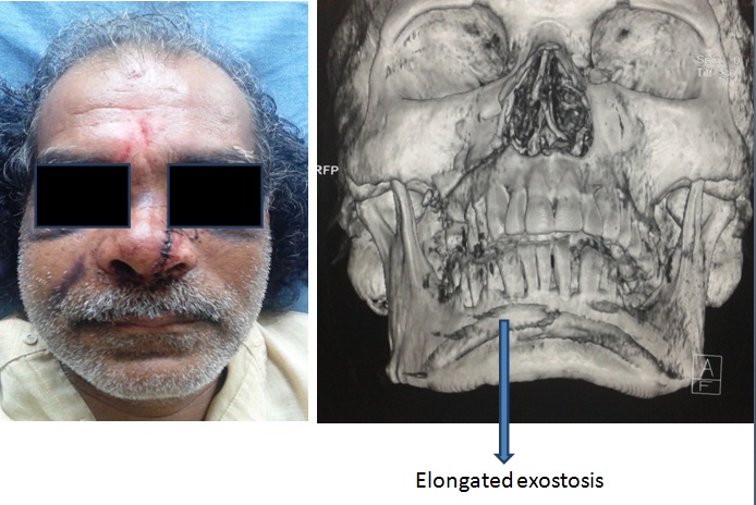
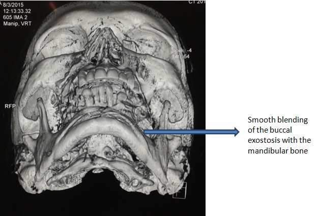
The 3-D CT scan showed a partial lefort 1 maxillary fracture (only the right side was involved). Surprisingly, in the mandibular anterior region, we noted a thin bar of bone extending from one mental foramen region to the other across the symphysis. This additional bar of bone was a distinct variation from the normal anatomic structure of the mandible. This additional anterior bar of mandibular bone was found to be smoothly merging with the underlying mandibular body at the mental foramen region on both sides [Table/Fig-1,2]. The partial lefort 1 fracture of the maxilla was not associated with any displacement or movement of the maxillary segment and the occlusion was stable. The patient was not willing to undergo any surgical procedure to fix the facial bone fracture as he had no functional discomfort. He was managed conservatively and later discharged after one week.
Frontal view of patient with 3-D CT Scan
3-D CT Scan of the patient- Inferior view
The formation of the mandible begins by the membranous ossification lateral to the Meckel’s cartilage in the sixth to seventh week of intrauterine life [1,2]. The bony prominence comprising the bending Meckel’s cartilage and the protruding medial lamina of the intramembranous bone comprises the origin of the torus, which is a type of bony hyperostosis in the mandible [3–5]. Surgical removal of the bony buccal exostoses is indicated if they cause pain and tenderness in the overlying mucosa or interfere with normal occlusal function.
A single ossification center for each half of the mandible arises in the region of the bifurcation of the inferior alveolar nerve into its mental and incisive branches [1]. Ossification spreads medially below the incisive nerve and then upwards to form a trough of bone formed by lateral and medial plates which are united beneath the nerve [6,7].
The bony prominence comprising the bending of Meckel’s cartilage and the protruding medial lamina is the origin of the exostosis. Although, it was topographically found to be located usually above the mylohyoid muscle attachment, another aspect clearly suggests its location corresponding to the mental foramen region on the lateral lamina of the mandibular bone [3].
Certain factors such as occlusal stress and bruxism could increase the size of the exostosis or affect the growth of the entire mandible [5]. Sometimes these tori or exostoses change in size and shape varying from small to large and bulky structures usually during the fourth decade of life. The bony bar seen in this particular individual, could well be the extension of similar such exostoses which underwent changes in and size and shape to form such a variation.
A protruding medial lamina of the intramembranous bone and the bending of the Meckel’s cartilage resulting in the formation of a bony prominence are considered to be the fetal origin of the mandibular buccal exostoses. The bony exostoses have to be surgically removed or smoothened if they cause pain and tenderness in the overlying mucosa or interfere with normal occlusal function.
[1]. Lipski M, Tomaszewska IM, Lipska W, Lis GJ, Tomaszewski KA, The mandible and its foramen: anatomy, anthropology, embryology and resulting clinical implicationsFolia Morphol 2013 71(4):285-92. [Google Scholar]
[2]. Diewert VM, Growth movements during prenatal development of human facial morphologyProg Clin Biol Res 1985 187:57-66. [Google Scholar]
[3]. Rodriguez-Vazquez JF, Sakiyama K, Verdugo-Lopez S, Amano O, Murakami G, Abe S, Origin of the Torus Mandibularis: An embryological hypothesisClin Anat 2013 26(8):944-52. [Google Scholar]
[4]. Basha S, Dutt S, Buccal-sided mandibular angle exostosis – A rare case reportContemp Clin Dent 2011 2(3):237-39. [Google Scholar]
[5]. Johnson OM, The tori and masticatory stressJ Prosthet Dent 1959 9:975-77. [Google Scholar]
[6]. Lee SK, Kim YS, Oh HS, Yang KH, Kim EC, Chi JG, Prenatal development of the human mandibleAnat Rec 2001 263:314-25. [Google Scholar]
[7]. Wyganowska-S’wiatkowska M, Przystanska A, The Meckel’s cartilage in human embryonic and early fetal periodsAnat Sci Int 2011 86:98-107. [Google Scholar]