Hypersensitivity Pneumonitis (HP) or Extrinsic Allergic Alveolitis (EAA) is a disease resulting from immunologically induced inflammation in response to inhalation of a wide variety of airborne allergens. The condition develops mainly in non atopic individuals sensitized to organic dust due to repeated exposures. It is a relatively rare disease constituting upto 2% of interstitial lung diseases. Knowledge of classical High Resolution Computed Tomography (HRCT) of lung findings aid in early diagnosis. We report a case of subacute hypersensitivity pneumonitis in a housewife who despite being symptomatic remained undiagnosed for two years. She showed a good response to therapy, but soon relapsed. Visit to her home revealed that she lived in a damp house full of moldy walls.
Centrilobular nodules, Extrinsic allergic alveolitis, Ground glass opacities, Radiography
Case Report
A 43-year-old housewife presented with chief complaints of dry cough, breathlessness and chest discomfort for over two years. Prior to presenting to us, she had received antibiotics, bronchodilators and steroids on multiple occasions but with temporary relief. There was no history of haemoptysis, chest pain, fever, night sweats or weight loss. She did not have significant past or family history or any addictions.
Her respiratory rate was 20 breaths/ min, pulse 90/min, blood pressure 120/80mmHg and oxygen saturation 97% on room air.
Physical examination was normal. Auscultation of lung revealed bilateral crepitations and rhonchi. Cardiac, per abdomen and CNS examination were normal. Investigations showed normal haematology, renal and liver function and a negative collagen profile.
Chest X-ray showed diffuse haziness in bilateral lung fields. Pulmonary function test (spirometry) was normal. HRCT thorax [Table/Fig-1a,b] showed areas of ground glass opacities, poorly defined small centrilobular nodules and areas of mosaic perfusion, findings classical of hypersensitivity pneumonitis. Patient was always a housewife and never had pets. We could not identify any allergens from her history and in view of diagnosis of Hypersensitivity Pneumonitis bronchoalveolar lavage was planned. However, she did not give consent. She was started on prednisolone at 1mg/kg/day and tapered over six months. Patient responded well to the treatment [Table/Fig-2a,b].
(a) HRCT scan obtained through upper lungs shows ground glass opacities and poorly defined small nodules; (b) Areas of mosaic perfusion, ground glass opacities and patchy nodules in lower lung fields.

Resolution of lesions seen in both lung fields, few areas of mosaic perfusion persists.

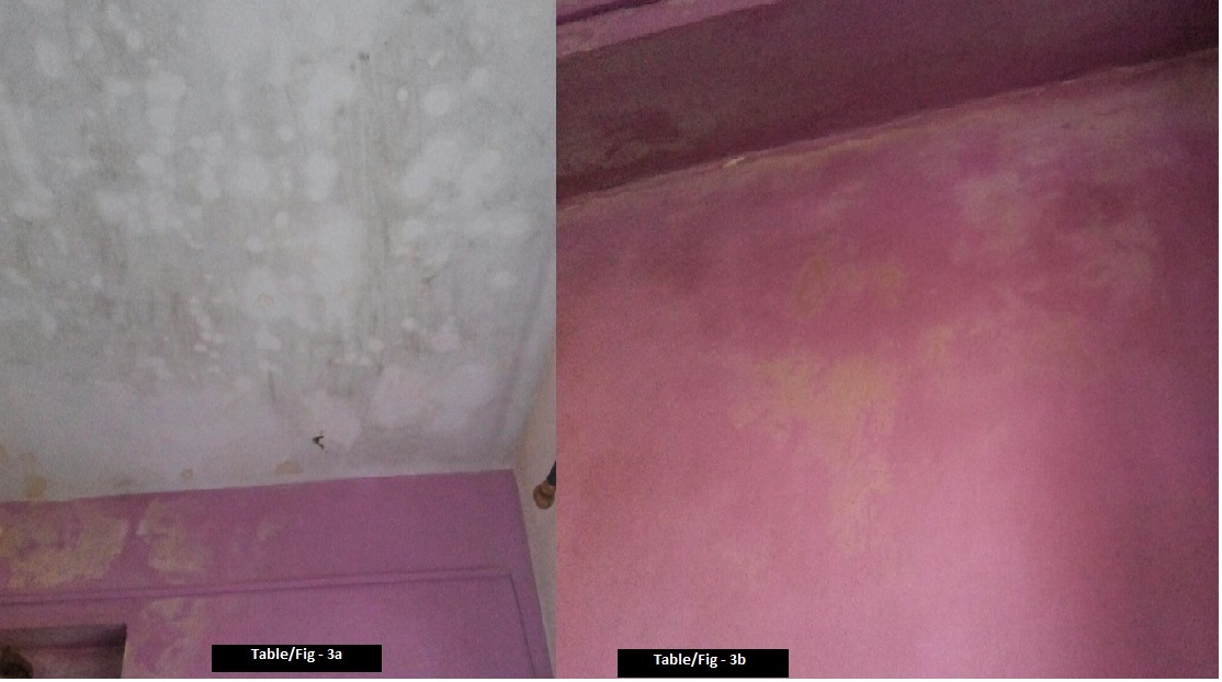
Within two months patient relapsed. As history could not reveal any offending agent and she being a housewife, it was decided to inspect her home for any possible risk factor. Her house had a musty odour. She was living in a three room house with moist walls and roofs, covered with molds [Table/Fig-3a,b]. Scrapings were taken from multiple sites from the walls. Examination on 10% KOH showed hyaline septate hyphae, 3-4μ with dichotomous branching. Growth on Sabouraud’s dextrose agar from all sites was identified as Aspergillus flavus [Table/Fig-4]. The growth was velvety, green and brown on reverse. Microscopically conidiophores were rough, pitted and spiny, phialides single and double covering entire vesicle, pointing in all directions suggestive of Aspergillus flavus. Repeated demonstration in direct smear and culture established the diagnosis [1]. Skin prick test for Aspergillus species as well as specific IgE was negative.
Damp roof and walls showing fungal growth
Growth of Aspergillus flavus. Conidiophores are long, rough and spiny. Spherical, smooth conidia are derived from double row of phialides covering entire circumference of spherical vesicles, pointing out in all directions

Patient had commercial home assessment and house waterproofing was done. Patient responded well to treatment following removal of risk factor.
Discussion
Hypersensitivity Pneumonitis (HP) results from repeated exposure to inhaled organic particles or low molecular weight chemicals. The prevalence varies depending on disease definition, diagnostic methods, exposure, geographical conditions and host risk factors [2]. In large population based study in UK, the incidence was 1 per 100,000 [3].
Clinical findings mimic other diseases, hence a high degree of clinical suspicion and a thorough occupational and environmental history are essential for accurate diagnosis [4].
Fungi cause hypersensitivity pneumonitis, asthma, aspergilloma, allergic bronchopulmonary aspergillosis, fungal sinusitis and constitutional symptoms like fatigue, nausea, cognitive dysfunction, immune dysfunction and toxic mold syndrome [5,6]. A meta-analysis showed that building dampness and molds are associated with 30-50% increase in respiratory and asthma related health outcomes [7]. In some locales, Aspergillus flavus is more common in air than Aspergillus fumigatus for unclear reasons [8].
In 1932, the first clinical presentation of HP was described in Maple bark strippers due to Cryptostroma corticale, a fungus found under the bark [9]. In the same year Farmer’s lung was described [10].
The offending antigens are ubiquitous but individuals with genetic susceptibility and environmental factors contribute to the risk. Antigen acts as inducing factor and genetic and environmental factors as promoting risk factors [2], leading to Type III and Type IV hypersensitivity reaction. The inflammation involves alveoli, terminal bronchioles and interstitium. Histologically, noncaseating granulomas and areas of fibrosis are seen while in chronic HP, alveolar destruction in the form of honeycombing is present. Centrilobular, peribronchiolar and bridging fibrosis are important hallmarks [11,12].
Acute stage manifests with fever, chills, malaise, cough, dyspnea, chest tightness, headache and arthralgia which resolves in few days. In the early stage lung function can be normal. In subacute stage, there is cough, dyspnea and fatigue and HRCT shows ground glass opacities signifying airspace disease and poorly defined centrilobular nodules. In chronic stage, the presentation is insidious with HRCT showing reticular-nodular opacities, ground glass shadows, interlobular septal thickening, loss of volume, traction bronchiectasis and honeycombing which spares the lung bases.
HP presents a diagnostic challenge. Sensitivity of chest radiograph to detect HP is low [13], however HRCT can be very specific. Absence of offending agent, negative serum precipitins and normal lung function can lead to a diagnostic dilemma. Early diagnosis and removal of the offending antigen is important, hence examination of living and working environment is recommended. In subacute disease, 3-6 months of prednisolone leads to remission. In chronic disease steroids are required for a prolonged period. Some patients progress to irreversible pulmonary fibrosis and die within few years [14]. Chronic HP with pulmonary hypertension is associated with greater risk of death [15].
Our patient responded well after the risk factor was eliminated. This case emphasizes the importance of home visit as an investigative tool. Relapse should raise a strong suspicion of the antigenic source still existing. In difficult to manage cases, all efforts should be made to identify the offending antigen so as to prevent the development of Chronic HP and death.
Conclusion
Indoor fungi and human health have gained a lot of significance. Living in moldy conditions can lead to hypersensitivity pneumonitis. Clinicians must be aware of such non occupational sources of antigens.
[1]. Winn W, Allen S, Janda W, Koneman E, Procop G, Schrekenberger P, Woods G, Mycology. In: Elmer W Koneman, editorKoneman’s Color Atlas & Textbook of Diagnostic Microbiology 2006 6th edPhiladelphiaLippincott Williams & Wilkins:1151-1243. [Google Scholar]
[2]. Selman M, Pardo A, King TE, Hypersensitivity pneumonitis: insights in diagnosis and pathobiologyAm J Respir Crit Care Med 2012 186:314-24. [Google Scholar]
[3]. Solaymani-Dodaran M, West J, Smith C, Hubbard R, Extrinsic allergic alveolitis: incidence and mortality in the general populationQJM 2007 100:233-37. [Google Scholar]
[4]. Glazer CS, Rose CS, Lynch DA, Clinical and radiologic manifestations of hypersensitivity pneumonitisJ Thorac Imaging 2002 17:261-72. [Google Scholar]
[5]. Bush RK, Portnoy JM, Saxon A, Terr AI, Wood RA, The medical effects of mold exposureJ Allergy Clin Immunol 2006 117:326-33. [Google Scholar]
[6]. Pettigrew HD, Selmi CF, Teuber SS, Gershwin ME, Mold and human health: separating the wheat from the chaffClin Rev Allergy Immunol 2010 38:148-55. [Google Scholar]
[7]. Fisk WJ, Lei-Gomez Q, Mendell MJ, Meta-analyses of the associations of respiratory health effects with dampness and mold in homesIndoor Air 2007 17:284-96. [Google Scholar]
[8]. Hedayati MT, Pasqualotto AC, Warn PA, Bowyer P, Denning DW, Aspergillus flavus: human pathogen, allergen and mycotoxin producerMicrobiology 2007 153:1677-92. [Google Scholar]
[9]. Towey JW, Sweany HC, Huron WH, Severe bronchial asthma apparently due to fungal spores found in Maple barkJAMA 1932 99:453-59. [Google Scholar]
[10]. Campbell JM, Acute symptoms following work with hayBMJ 1932 2:1143-44. [Google Scholar]
[11]. Takemura T, Akashi T, Ohtani Y, Inase N, Yoshizawa Y, Pathology of hypersensitivity pneumonitisCurr Opin Pulm Med 2008 14:440-54. [Google Scholar]
[12]. Katzenstein AL, Mukhopadhyay S, Myers JL, Diagnosis of usual interstitial pneumonia and distinction from other fibrosing interstitial lung diseasesHum Pathol 2008 39:1275-94. [Google Scholar]
[13]. Lynch DA, Rose CS, Way D, King TE, Hypersensitivity pneumonitis: sensitivity of high-resolution CT in a population-based studyAJR Am J Roentgenol 1992 159:469-72. [Google Scholar]
[14]. Pérez-Padilla R, Salas J, Chapela R, Sánchez M, Carrillo G, Pérez R, Mortality in Mexican patients with chronic pigeon breeder’s lung compared with those with usual interstitial pneumoniaAm Rev Respir Dis 1993 148:49-53. [Google Scholar]
[15]. Koschel DS, Cardoso C, Wiedemann B, Höffken G, Halank M, Pulmonary hypertension in chronic hypersensitivity pneumonitisLung 2012 190:295-302. [Google Scholar]