A Comparison Between Phorbol 12 Myristate 13 Acetate and Phorbol 12, 13 Dibutyrate in Human Melanocyte Culture
Divya Padma1, Kumar M.R. Bhat2
1 PhD Scholar, Department of Anatomy, Kasturba Medical College, Manipal University, Manipal, India.
2 Additional Professor, Department of Anatomy, Kasturba Medical College, Manipal University, Manipal, India.
NAME, ADDRESS, E-MAIL ID OF THE CORRESPONDING AUTHOR: Dr. Kumar M.R. Bhat, Additional Professor, Department of Anatomy, Kasturba Medical College, Manipal University, Manipal-576104 Karnataka, India.
E-mail: kumar.mr@manipal.edu
Introduction
Melanocyte culture is an integral part of the studies of skin biology and cosmetic applications. After the introduction of selective medium for the culture of human melanocyte using Phorbol 12-myristate13-acetate (PMA) in 1982, a lot of methods of culturing were tried but till date PMA is a preferred mitogen because of its cost effectiveness compared to growth factors. We have tried to preliminarily evaluate the efficacy of another phorbol ester, Phorbol 12, 13-dibutyrate (PDBu) in melanocyte culture because of its less hydrophobic nature compared to PMA. This property minimizes the trace amount of mitogen in cell culture after washing off and hence does not interfere in other biological assays.
Aim
To evaluate the differences in the melanocyte survival rate, morphology and mitotic index when grown in media supplemented with PMA and PDBu.
Materials and Methods
Foreskins were collected from children undergoing circumcision. Epidermal cells were isolated from foreskin and cultured using PMA and PDBu. Melanocytes in culture were monitored for the better establishment and documented. In proliferative assay, melanocytes were treated with PMA and PDBu for 24, 48 and 72 hours and proliferation was measured using 3-(4,5-Dimethylthiazol-2-yl)-2, 5-diphenyltetrazolium bromide (MTT) assay method.
Results
When cultured, melanocytes acquired proliferative status and bipolar morphology quicker in PDBu medium than in PMA medium. Keratinocytes survived as contamination in PMA medium whereas PDBu medium had minimal keratinocytes. MTT assay showed that PDBu has higher proliferative induction capacity than PMA. In even lower concentration of PDBu in medium, melanocytes survived till 72 hours without significant cell loss in compared to PMA medium.
Conclusion
PDBu can be a valuable replacement for PMA in human melanocyte culture. Higher proliferation induction, unfavourable to keratinocyte survival and less hydrophobicity make PDBu a promising alternative for quicker establishment of pure human melanocyte cultures especially in cosmetic in vitro experimental dermatology.
Cell culture, Melanocyte, Phorbol esters
Introduction
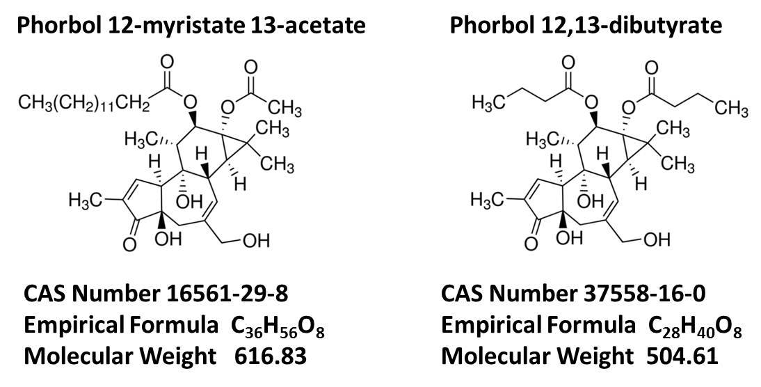
Culturing melanocyte isolated from human skin is an important aspect of pigmentation studies in human skin biology and cosmetic applications [1]. Although a number of cell lines originated from melanocytes or melanoma are used in pigmentation studies, the primary monolayer culture of epidermal melanocytes remains as a more reliable model system for the studies [2,3]. Isolation and establishment of melanocyte culture is difficult because of not dividing nature of melanocytes and hence, need potent mitogens to proliferate. It also includes the risk of contamination of keratinocytes and other cells. Decades of studies has introduced selective media for isolation and culture of melanocytes including usage of mitogens like Phorbol myristate acetate (PMA), cholera toxin, alpha-melanotropin, endothelin-1, and basic fibroblast growth factor [4,5]. PMA a protein kinase C activator, is largely used as a mitogen in melanocyte culture [6]. It is reported as a tumour promoter [7,8] but the usage of it has not been reduced as it is cheaper and efficient mitogen for melanocyte culture than other expensive growth factors like bFGF. Phorbol myristate acetate and Phorbol 12, 13 dibutyrate are structurally [Table/Fig-1] and functionally similar [9]. PDBu is reported as a less potent activator of protein kinase C than PMA. PDBu is less hydrophobic than PMA and hence can be easily washed off in cell culture to prevent the interference in other biological assays. The usage of PDBu in melanocyte culture is not common. PDBu is reported as probable replacement but exact quantification of the difference in efficacy is not reported.
Schematic structures of Phorbol 12 myristate 13 acetate and Phorbol 12,13 dibutyrate
As PDBu is less hydrophobic and hence preferred in cell culture we wanted to evaluate the ability of PDBu to replace PMA in melanocyte culture. In this study we have evaluated the differences between PMA and PDBu in melanocyte culture in the context of cell survival, cell morphology and cell proliferation induction efficacy.
Materials and Methods
In vitro study was conducted in School of Life Sciences and Kasturba Medical College, Manipal University, Manipal for the duration of 4 months (from August 2013 to November 2013), two foreskin samples were collected for the study and used for different experimental purposes (from August 2013 to November 2013).
Chemicals
PMA and PDBu both were purchased by Sigma Aldrich. Catalogue numbers were P8139 and P1269 respectively. Both were dissolved in ultrapure Dimethyl sulphoxide (DMSO) at the concentration of 10mM. These chemicals’ stock solutions were further diluted in melanocyte medium to get desired concentration.
Isolation of melanocytes: Foreskin pieces were collected from 2 children below 10 years who had undergone circumcision, after obtaining the institutional ethical clearance and consent from the parents/guardian of the subjects. Two samples were used in this study to compare the proliferative efficiency of PMA and PDBu In vitro for 10 days. All experiments were done separately for melanocytes obtained from two different individuals and average representative value is expressed in the results. Skin pieces were dipped in ethanol for 30 seconds and washed with PBS repeatedly. The skin sample was cut into small pieces and incubated in dispase (BD Bio sciences) for 16-18 hours at 40C. After the incubation, epidermis was peeled off the dermis. The separated epidermis was incubated with 0.25% trypsin having 0.5mM Ethylene diamine tetraacetic acid (EDTA) for 5-10 minutes. Trypsin digestion and gentle shaking was done to get the cell suspension. Trypsin activity was inhibited by adding Phosphate Buffer saline (PBS) having 10% Fetal bovine serum (FBS). The cell suspension was centrifuged at 1000rpm for 5 minutes to get pellet. The pellet was disturbed and PBS was added. The cell suspension was spun again to get the cell pellet. The washed cell pellet was reconstituted in fresh medium and was plated onto culture flasks with selective complete medium for melanocytes (Nutrient mixture Ham F10 supplemented with 5% FBS, 85nM PMA, 0.1mM Iso butyl methyl xanthine (IBMX) and 0.25nM cholera toxin). For the comparative study, cells were isolated from foreskin, processed and at the end split and seeded into two T25 flasks having same amount of cells and different media having either 85nM PMA or 85nM PDBu. Cells were regularly observed under microscope and photographed in different magnification.
MTT Assay
Melanocytes were plated onto tissue culture 96 well plates at the concentration of 1X10⁴ cells/well. Next day melanocyte were added with 100μl of different concentration of PMA and PDBu in F10 medium alone or with 5% of FBS, cholera toxin and IBMX as a complete medium. Cells were incubated for 24, 48 and 72 hours separately at 370C temperature and 5% of CO2. After the incubation 20 μl of 5mg/ml MTT was added and incubated for 4 hours. Medium was discarded and 100μl of DMSO was added to dissolve the formazan crystals formed. The optical density (OD) was read at 570nm with reference at 650nm. The readings were used as it was or calculated as percent of control, considering nutrient mixture Ham F10 alone as control. The experiments were repeated to confirm the results.
Statistical Analysis
Data was presented as mean ± standard deviation. One-way analysis of variance (ANOVA) followed by Dunnett’s test was used to compare the data and determine the statistical significance of difference using Graphpad prism 6 software. Difference was considered significant at p< 0.05.
Results
Isolation and culture of melanocytes
Melanocytes were isolated and cultured in melanocyte growth media having either PMA or PDBu at the concentration of 85nM. Cells were attached and continued to grow in both media. But the striking differences observed microscopically between these two media were, morphology of melanocytes and presence of keratinocytes [Table/Fig-2]. As depicted in [Table/Fig-2], in PDBu medium, number of melanocytes having proliferative bipolar shape were comparatively more than in PMA medium. In PMA medium, dendritic cells were more and the culture took more time to establish bipolar, proliferative, pure melanocyte culture. Another observed factor was, keratinocytes could not attach and grow properly in PDBu medium compared to PMA medium. The presence of keratinocytes in PMA medium made multiple differential trypsinizations necessary to establish pure culture whereas PDBu medium had minimum keratinocytes and could attain pure culture in minimum passages.
Micrographs of cultured human epidermal cells. a, c, e and g - Melanocytes isolated and cultured in complete medium with 85nM of PMA on 2nd, 5th, 5th and 10th day post isolation respectively. b, d, f and g- Melanocytes isolated and cultured in complete medium with 85nM of PDBu on 2nd, 5th, 5th and 10th day post isolation respectively. a, b, e and f white arrows indicate presence of keratinocytes in culture. PMA medium showed increased amount of keratinocytes in culture where as PDBu medium had minimum keratinocytes. c, d, g and h white arrows indicate the morphology of melanocytes. PMA medium had more polydendritic cells whereas PDBu medium had more bipolar shaped cells.

Proliferation Assay
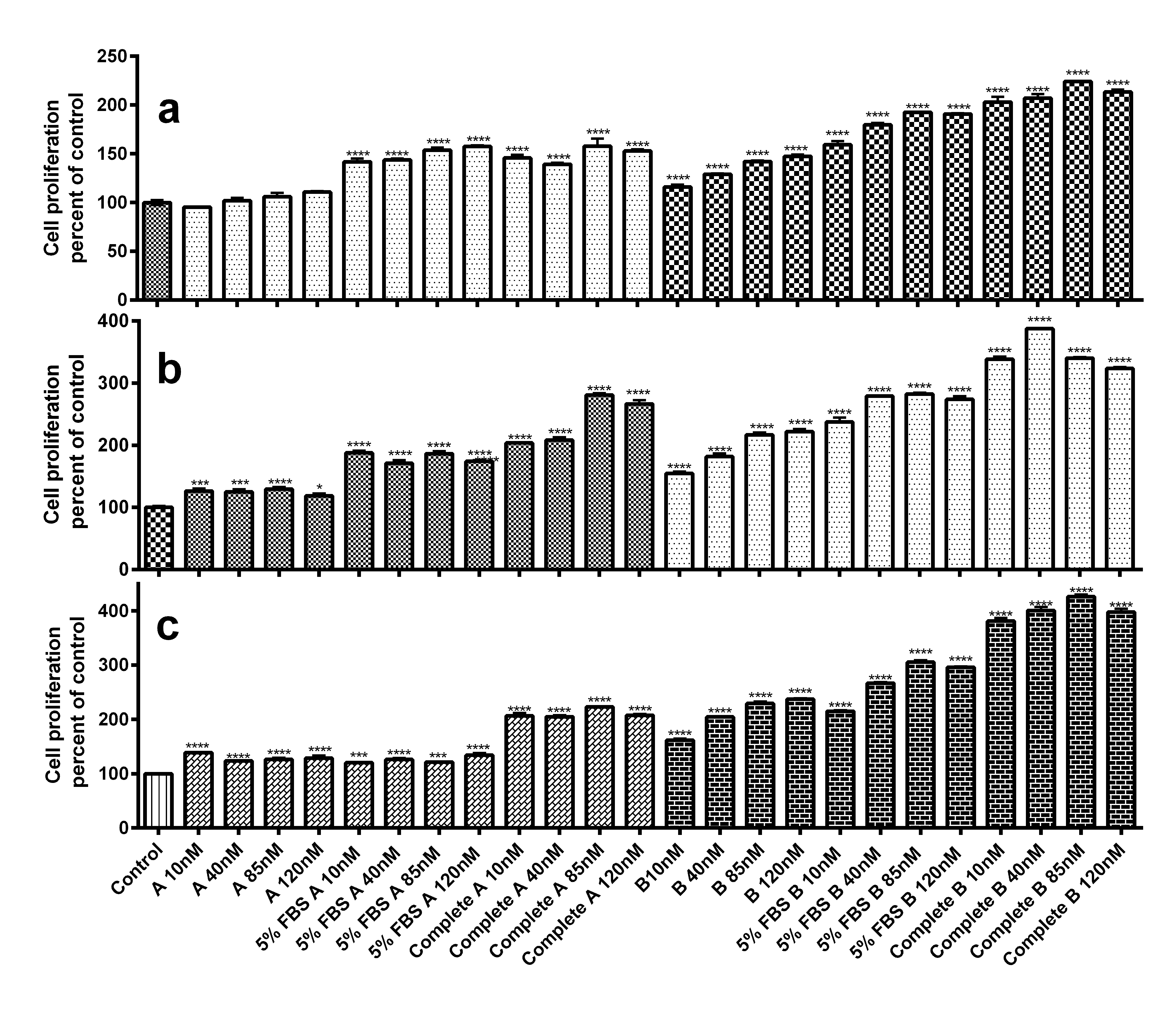
Melanocytes isolated using conventional medium having 85nM of PMA, were treated with PMA and PDBu separately for 24, 48 and 72 hours. The cells proliferated in the presence of PMA and PDBu both to statistically significant amount [Table/Fig-3]. In nutrient mixture F10 alone set, the amount of cells were not significantly more than control at 24 hours in PMA set whereas PDBu set showed higher amount of cells. All other combinations of both PMA and PDBu showed high amount of cells compared to control confirming the proliferation induction. In all respective sets of experiments, PDBu showed statistically highly significant proliferative capacity than PMA. It is important to know that PDBu with 5% FBS, induced and maintained the cells better than complete medium with PMA. This indicates that in the presence of PDBu, cholera toxin and IBMX are not necessary for cell survival. This reduces the usage of biohazard components like cholera toxin in cell culture.
Proliferation assay for human melanocytes. a, b and c - Melanocyte incubated for 24, 48 and 72 hours with difference concentration of PMA and PDBu respectively. In X axis A corresponds to Phorbol 12 myristate 13 acetate and B to Phorbol 12, 13 dibutyrate. PDBu showed highly significant proliferative capacity compared to PMA. The p-value is represented as non-significant (ns) p > 0.05, *P ≤ 0.05, **p ≤ 0.01, *** p ≤ 0.001, **** p ≤ 0.0001.
Analysis of OD of the Proliferation Assay
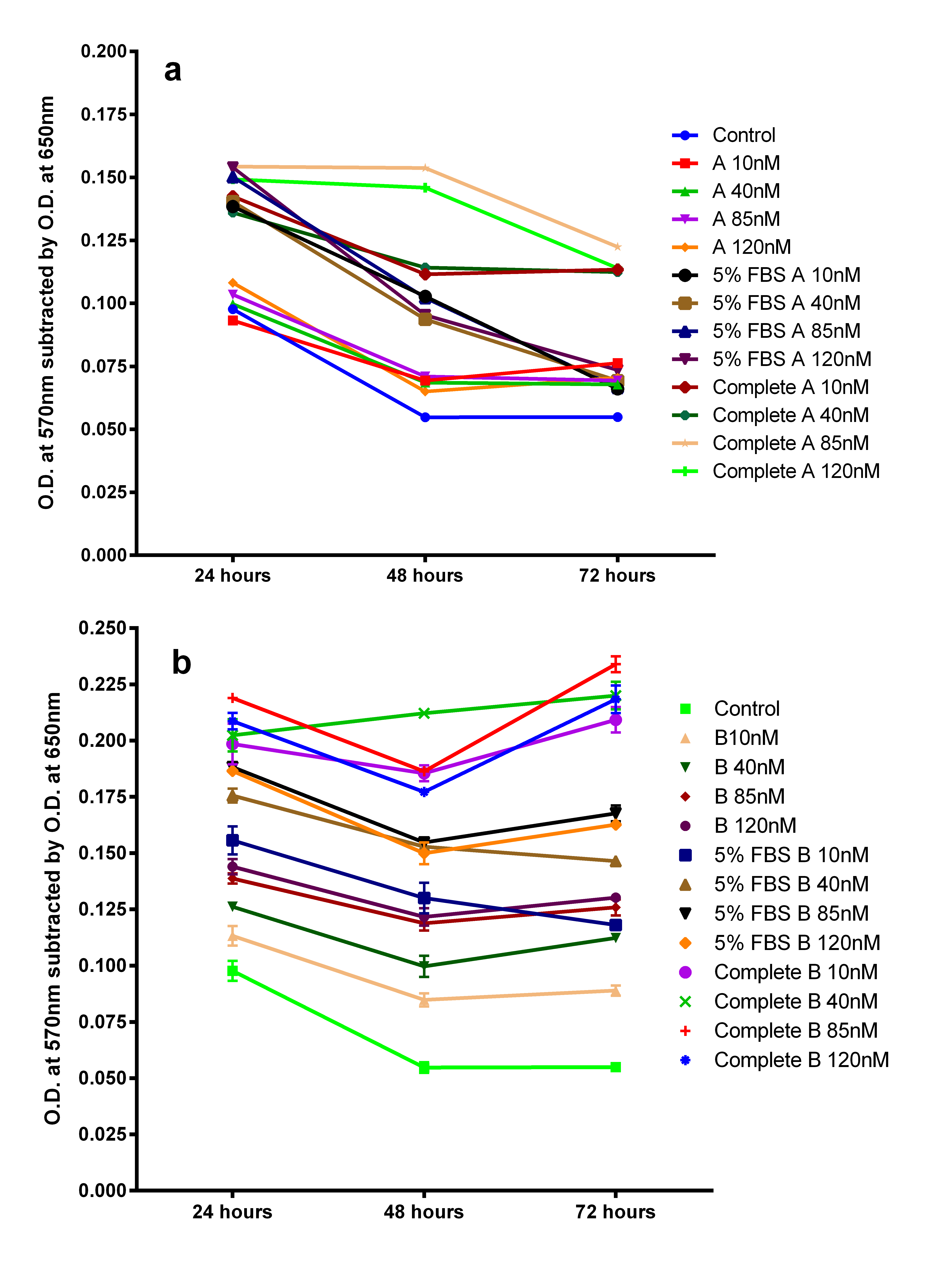
As the control was only nutrient mixture F10 medium devoid of any supplements, melanocytes were doubted to be dying in prolonged exposure. So to avoid the false interpretation of the data we analysed the OD of all sets to get a clear picture of the trend. The analysis of OD showed that melanocytes divided within 24 hours and till 72 hours continued to be in the same amount in some sets whereas in control, cells decreased in number [Table/Fig-4]. In PMA set, cells in complete medium 85nM and 120nM PMA showed highest proliferative capacity at 24 hours and survived till 48 hours. In all other PMA sets, cells decreased after 24 hours. Comparatively in PDBu sets, cells showed higher proliferative capacity at 24 hours and survived till 72 hours without significant cell loss. It confirms the capacity of PDBu to maintain the cells to longer duration whereas PMA quickly loses its efficacy.
Optical density readings of proliferative assay: a) PMA medium proliferation assay showing reduction of OD after 24 hours; b) PDBu medium proliferation assay showing higher OD and maintaining the OD till 72 hours.
Discussion
In our study for the first time, we have shown that PDBu is a better replacement for PMA in the context of proliferation induction to melanocytes and unfavourable condition for keratinocytes. The time duration for establishing a pure culture was significantly reduced in the presence of PDBu than PMA because of these properties. PDBu is also less hydrophobic than PMA and thus can be washed off when not necessary [10]. This will minimize the unwanted reactions between the trace phorbol esters and cell components in other biological assays. Morphologically PMA induced dendritic melanocytes more than bipolar shaped cells. This could be because of differentiation property of PMA [11]. PMA is not recommended in regenerative medicine because of tumour promoting properties, so as PDBu [12]. There are no reports suggesting that PDBu is less tumourogenic than PMA and long term usage of PDBu to culture the melanocytes may also alter the gene expression similar to the effects of PMA [4]. But the use of PDBu could be very useful in research, especially in cosmetic sciences where primary melanocytes were cultured in large number for different assays using melanocytes in vitro pigmentation models. As primary melanocytes culture has short life span, a better and potent mitogen would shorten time duration needed to establish the pure culture [13]. A small change in medium, using a similar mitogen PDBu in lower concentration enhanced the efficacy of the melanocyte culture significantly compared to PMA. Hence, PDBu can be used as a better replacement with minimal usage of itself and other mitogens in melanocyte culture medium. However, further study is required to evaluate the tumourogenic properties of these two mitogens and detailed molecular analysis is required to understand genetic alteration when used long term. Additionally, the results of this study is only of melanocytes derived from the foreskin samples of children under the age of 10 years, to assess the efficacy of the PMA and PDBu. Further, the effects on the adult skin melanocyte culture would confirm the significance of PDBu in melanocyte culture. Thus may be useful in cosmetic useful in cosmetic application.
Conclusion
In melanocyte culture, PDBu is a promising alternative mitogen to PMA because of its higher survival, stronger proliferative induction and less hydrophobic nature. PDBu is less favourable than PMA for keratinocytes to survive. This shortens the time duration to establish pure cutures of melanocytes. Our study has qualitatively as well as quantitatively, evaluated the differences between PMA and PDBu and concluded that PDBu is better than PMA in primary culture, to get quicker and more homogenous pure cultures of melanocytes which can be used in different assays in pigmentation studies.
[1]. Isabelle G, Laetitia A, Jessica J, Roxane P, Pigmented Skin Models: Understand the Mechanisms of Melanocytes 2013 [Google Scholar]
[2]. Smit N, Vicanova J, Pavel S, The hunt for natural skin whitening agentsInt J Mol Sci 2009 10(12):5326-49. [Google Scholar]
[3]. Duval C, Smit NP, Kolb AM, Regnier M, Pavel S, Schmidt R, Keratinocytes control the pheo/eumelanin ratio in cultured normal human melanocytesPigment Cell Res 2002 15(6):440-46. [Google Scholar]
[4]. Eisinger M, Marko O, Selective proliferation of normal human melanocytes In vitro in the presence of phorbol ester and cholera toxinProc Natl Acad Sci U S A 1982 79(6):2018-22. [Google Scholar]
[5]. Swope VB, Medrano EE, Smalara D, Abdel-Malek ZA, Long-term proliferation of human melanocytes is supported by the physiologic mitogens alpha-melanotropin, endothelin-1, and basic fibroblast growth factorExp Cell Res 1995 217(2):453-59. [Google Scholar]
[6]. Arita Y, O’Driscoll KR, Weinstein IB, Growth of human melanocyte cultures supported by 12-O-tetradecanoylphorbol-13-acetate is mediated through protein kinase C activationCancer Res 1992 52(16):4514-21. [Google Scholar]
[7]. Blumberg PM, Delclos KB, Dunn JA, Jaken S, Leach KL, Yeh E, Phorbol ester receptors and the In vitro effects of tumour promotersAnn N Y Acad Sci 1983 407:303-15. [Google Scholar]
[8]. Griner EM, Kazanietz MG, Protein kinase C and other diacylglycerol effectors in cancerNat Rev Cancer 2007 7(4):281-94. [Google Scholar]
[9]. Bell RM, Burns DJ, Lipid activation of protein kinase CJ Biol Chem 1991 266(8):4661-64. [Google Scholar]
[10]. Wang QJ, Fang TW, Fenick D, Garfield S, Bienfait B, Marquez VE, The lipophilicity of phorbol esters as a critical factor in determining the pattern of translocation of protein kinase C delta fused to green fluorescent proteinJ Biol Chem 2000 275(16):12136-46. [Google Scholar]
[11]. Chao-Hsing KA, Hsin-Su YU, A study of the effects of phorbol 12-myristate-13-acetate on cell differentiation of pure human melanocytes In vitroArch Dermatol Res 1991 283(2):119-24. [Google Scholar]
[12]. Driedger PE, Blumberg PM, Specific binding of phorbol ester tumour promotersProc Natl Acad Sci U S A 1980 77(1):567-71. [Google Scholar]
[13]. Im S, Hann SK, Park YK, Kim HI, Culture of melanocytes obtained from normal and vitiligo subjectsYonsei Med J 1992 33(4):344-50. [Google Scholar]