Conventional methods of tooth replacements are slowly and steadily being replaced by newer modes of tooth replacements like implants. Thus the introduction of dental implants has changed the face of dentistry over the last 50 years [1]. Dental implants should be considered as an option for replacing missing teeth and they often provide more predictable results than conventional treatments like RPD, FPD and CD [2,3].
The use of titanium and its alloys as an implantation material: In 1969 Branemark, an orthopedic surgeon solidly embedded titanium in the bone. He called this phenomenon as “Osseointegration”. He hypothesized that titanium might be capable of withstanding occlusal forces and published long term success of titanium as an artificial implantation material [4].
Inspite of the high success rate, occurrence of implant failures have also been reported. The aetiology of implant failures can be broadly classified into three categories- infection impaired healing and occlusal overloading. Peri-implantitis is the most common cause for implant failures. Peri-implantitis is referred to as an inflammatory reaction with the loss of supporting bone in the soft tissues surrounding the implants [5,6]. Numerous microbes and bacteria are involved in the pathogenesis of peri-implantitis. Thus to improve the antimicrobial potential of the implants, titanium alloys were used followed by mechanical blasting, acid etching, bioactive coating, anodized LASER modified surfaces [7].
HA nano coated dental implants could induce a chemical bond with bone and achieve biological fixation. Zirconium and aluminium nano composites are well known for their high flexural strength and biocompatibility and are classified as a bioinert material [8]. Titanium nano particles are said to be the most biocompatible material with more osteogenic potential and offers an effective avenue for the development of implant surfaces with better bone generative and regenerative potential [9]. The centers for disease control recommended 37oC for 0.5,1,6,12,48,72 and 96 hours incubation or more (until the colonies were seen on the plates) to determine the numbers of CFU/mL. Hence this study was done to evaluate the anticandidal effect of titanium, zirconium and aluminium nanoparticles against C. albicans at 24 at hours,72 hours and one week time interval [10].
Materials and Methods
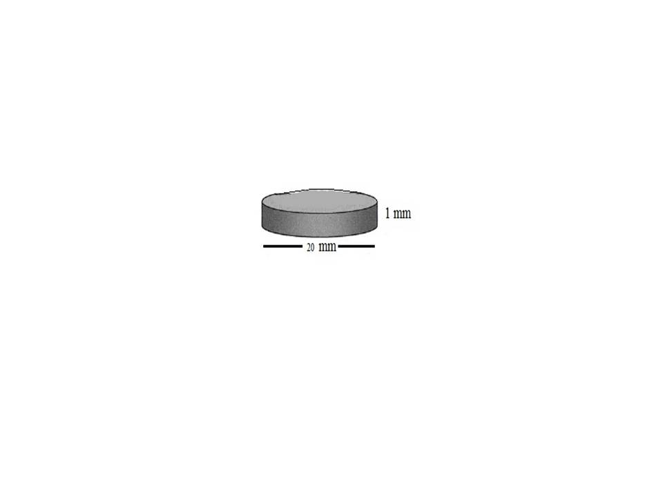
According to ISO/TR 11175:1993, the samples were prepared with the dimension of 20mm diameter and 1mm thickness. Commercially available pure grade IV titanium (Orotec titec) was made into sheets of 1mm thickness. The master press was used to prepare the individual discs measuring 20mm in diameter and 1mm in thickness [Table/Fig-1]. A total of 40 samples were prepared by using the master press.
Line diagram of master die.
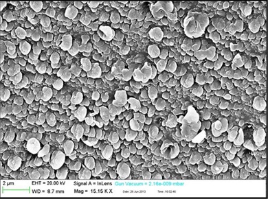
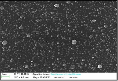
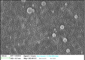
The samples were decontaminated by treating it in acetone and subsequently in water. The decontamination procedure was repeated 3 times for 15 minutes. The samples were then coated with titanium (TiO2) (Alfa Aesar, Bredent), zirconium (ZrO2) (Aldrich) and aluminium (Al2O3) (SRL chem) nanoparticles by a method called Pulsed laser deposition (PLD) [Table/Fig-2,3 and 4].
SEM of Titanium nanoparticle coating.
SEM of Zirconium nanoparticle coating.
SEM of Aluminium nanoparticle coating.
Initially titanium, zirconium and aluminium nanoparticles were made into pallots to act as targets. The nanoparticles were made in the form of discs about 16mm in diameter and four mm in thickness by compressing the nanoparticles in the pallotizer at 300N for five minutes. Then the pallots were retrieved carefully from the pallotizer and sintered at a temperature of 1600°C in a box furnace (VB ceramics pvt ltd) for 8 hours and was allowed to cool down to room temperature. The sintered pallots were then retrieved from the furnace after 24 hours.
The samples without any surface coating were considered as Group A (control), samples coated with titanium nano particles were Group B, samples coated with zirconium nano particles were Group C and samples coated with aluminium nano particles were Group D. Sabouraud Dextrose Agar (SDA) was used as the culture media for C.albicans. SDA is a combination of 5% of enzymatic digest of Casein and animal tissue, 40% of dextrose and 15% of agar. The colonies were counted under digital colony count tester (Toshiba) in 24 hours, 72 hours and 1 week interval [Table/Fig-5].
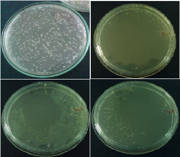
Growths of C.albicans at 24,72 hours and 1 week interval.
Results
One way Analysis of variance (ANOVA) was used for comparison within the groups and Tukey’s HSD Post Hoc test for multiple group comparison [Table/Fig-6,7,8 and 9]. Showed the mean colony forming units of Group A, B, C and D against C.Albicans in 24 hours were 283.60, 72, 365.55 and 444.33. The Significance p-value was <.001, which showed significant difference in C.F.U among the groups. The statistical analysis showed the p-value <0.001, hence it was statistically significant between group A & B and insignificant for group C & D. [Table/Fig-10,11 and 12] showed the mean colony forming units of Group A,B,C and D against C. albicans in 72 hours were 332.70, 97.20, 402.36 and 558. The significance p-value was <.001 that showed significant difference in C.F.U among the groups. Multiple group comparison showed the p-value is <0.001, hence it was statistically significant between group A & B and insignificant for group C&D. [Table/Fig-13,14 and 15] showed the mean colony forming units of Group A, B, C and D against C.Albicans at one week time interval were 359.60,93.70,388.09 and 557.11. The significance p-value was <.001 that showed significant difference in C.F.U among the groups. Multiple group comparison showed the p-value <0.001, hence it was statistically significant between group A & B and insignificant for group C & D.
Mean and SD of CFU of group A,B,C & D for C.albicans in 24 hours.
| Groups | Mean | Std. Dev | Min | Max |
|---|
| Group A | 283.60 | 10.844 | 269 | 300 |
| Group B | 72.00 | 7.165 | 64 | 86 |
| Group C | 365.55 | 50.097 | 301 | 461 |
| Group D | 444.33 | 32.265 | 389 | 476 |
| Total | 289.40 | 142.120 | 64 | 476 |
One-way ANOVA for group A,B,C & D on C. albicans in 24 hours.
| Sum of Squares | df | Mean Square | f-value | p-value |
|---|
| Between Groups | 752782.473 | 3 | 250927.491 | 258.502 | <0.001 |
| Within Groups | 34945.127 | 36 | 970.698 | | |
| Total | 787727.600 | 39 | | | |
Tukey’s HSD test between groups for C. albicans at 24 hours.
| Pairs | Mean Difference | p-value |
|---|
| Control | TiO2 nanoparticle | 211.60 | <0.001 |
| ZrO2 nanoparticle | -81.95 | <0.001 |
| Al2O3 nanoparticle | -160.73 | <0.001 |
| TiO2 nanoparticle | ZrO2 nanoparticle | -293.55 | <0.001 |
| Al2O3 nanoparticle | -372.33 | <0.001 |
| ZrO2 nanoparticle | Al2O3 nanoparticle | -78.79 | <0.001 |
Mean and SD of CFU for group A,B,C & D on C. albicans in 72 hours.
| Groups | Mean | Std. Dev | Min | Max |
|---|
| Group A | 332.70 | 6.865 | 321 | 341 |
| Group B | 97.20 | 9.390 | 83 | 111 |
| Group C | 402.36 | 78.139 | 311 | 560 |
| Group D | 558.00 | 23.611 | 512 | 589 |
| Total | 343.68 | 169.973 | 83 | 589 |
One-way ANOVA for group A,B,C & D on C. albicans in 72 hours.
| Sum of Squares | df | Mean Square | f-value | p-value |
|---|
| Between Groups | 1060008.530 | 3 | 353336.177 | 190.608 | <0.001 |
| Within Groups | 66734.245 | 36 | 1853.729 | | |
| Total | 1126742.775 | 39 | | | |
Tukey’s HSD test between the Groups for C. albicans in 72 hours.
| Pairs | Mean Difference | p-value |
|---|
| Control | TiO2 nanoparticle | 235.50 | <0.001 |
| ZrO2 nanoparticle | -69.66 | 0.004 |
| Al2O3 nanoparticle | -225.30 | <0.001 |
| TiO2 nanoparticle | ZrO2 nanoparticle | -305.16 | <0.001 |
| Al2O3 nanoparticle | -460.80 | <0.001 |
| ZrO2 nanoparticle | Al2O3 nanoparticle | -155.64 | <0.001 |
Mean and SD of CFU for group A,B,C & D on C. albicans in 1 week.
| Groups | Mean | Std. Dev | Min | Max |
|---|
| Group A | 359.60 | 29.029 | 278 | 374 |
| Group B | 93.70 | 9.286 | 79 | 104 |
| Group C | 388.09 | 64.074 | 311 | 560 |
| Group D | 557.11 | 33.569 | 512 | 618 |
| Total | 345.40 | 169.268 | 79 | 618 |
One-way ANOVA for group A,B,C & D on C. albicans in 1 week.
| Sum of Squares | df | Mean Square | F-Value | P-Value |
|---|
| Between Groups | 1058987.302 | 3 | 352995.767 | 217.487 | <0.001 |
| Within Groups | 58430.298 | 36 | 1623.064 | | |
| Total | 1117417.600 | 39 | | | |
Tukey’s HSD test between the Groups for C. albicans in 1 week.
| Pairs | Mean Difference | P-Value |
|---|
| Control | TiO2 nanoparticle | 265.90 | <0.001 |
| ZrO2 nanoparticle | -28.49 | 0.381 |
| Al2O3 nanoparticle | -197.51 | <0.001 |
| TiO2 nanoparticle | ZrO2 nanoparticle | -294.39 | <0.001 |
| Al2O3 nanoparticle | -463.41 | <0.001 |
| ZrO2 nanoparticle | Al2O3 nanoparticle | -169.02 | <0.001 |
Comparison of the anti-candidal effect of TiO2, ZrO2, Al2O3 nanoparticle coated titanium plates at 24 hours, 72 hours and 1 week time interval.
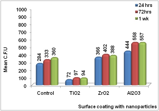
[Table/Fig-15] showed the Bar diagram of the values obtained for colony formation in Group A, B, C and D against C. albicans at 24 hours, 72 hours and one week time interval.
Discussion
The goal of modern dentistry is to restore aesthetics to natural form, function, comfort, speech and health regardless of the atrophy, disease or injury of the stomatognathic system. As a result of continued research in materials and techniques, the predictable success is now a reality for the rehabilitation of many challenging situations. The documented high survival rate of osseointegrated root form dental implants has led to their acceptance as a realistic alternative treatment in modern dentistry [1–3].
Titanium and its alloys have been used as the gold standard material in implant dentistry because of the osseointegration phenomenon [4]. Inspite of the high success rate, implant failures have also been reported [11] [Table/Fig-16]. The reasons for implant failures are infection impaired healing and occlusal overloading. The previous studies result showed that peri-implantitis was the most common cause for implant failures [5,6]. Peri-implantitis is an inflammatory reaction with the loss of supporting bone and the soft tissues surrounding the implants [5].
Factors related to the failure of dental implants.
| Factors | Comment |
|---|
| Implant | Previous failureSurface roughnessSurface purity and sterilityFit discrepanciesIntra-oral exposure time |
| Mechanical | Premature loading |
| Overloading (Late failure) | Traumatic occlusion due to inadequaterestorations |
| Patient (local factors) | Oral hygieneGingivitisBone quantity/qualityAdjacent infection/inflammation (periimplantitis)Presence of natural teethPeriodontal status of natural teethImpaction of foreign bodies (includingdebris from surgical procedure) in theimplant pocketSoft tissue viability |
| Patient (systemic factors) | Vascular integritySmokingAlcoholismPredisposition to infection, e.g. age,obesity, steroid therapy, malnutrition,metabolic disease (diabetes)Systemic illnessChemotherapy/radiotherapyHypersensitivity to implantcomponents |
| Surgical technique/ environment | Surgical traumaOverheating (use of handpiece)Perioperative bacterialcontamination, e.g. via saliva, perioralskin, instruments, gloves, operatingroom air or air expired by patient |
The most commonly involved microbes in peri-implantitis are Streptococcus mutans, Staphylococcus aureus, Porphyromonas gingivalis, Tannerella forsythia, Candida albicans, Actinomycetum concomitans, prevotella intermedia and fusobacterium [12,13]. Many studies are there related to bacteria. But no other studies are there related to Candida albicans.
In the previous studies, vector ultrasound system and carbon fiber curettes were used to treat peri-implantitis. There were no significant differences between the two techniques to treat peri-implantitis [14]. Earlier Er: YAG laser was used to treat perimplantitis but it could only stop bleeding on probing [15]. Also, the treatments with local antibiotics improved clinical parameters such as pocket depth and bleeding [16].
For the success of the implants, antimicrobial property of the implant surface is mandatory so that the initial attachment and colonization of the micro organisms can be prevented. When the initial attachment is prevented it obviously prevents the proliferation of the organisms. It had been shown in literature that coating of nanoparticles like titanium, zirconium and aluminium nanoparticles over titanium surfaces have likely to change not only the physical properties but also increased the osteogenic potential and the antimicrobial efficacy of the titanium implants [9,17].
Pulsed laser deposition is an advanced and trusted method of coating when compared to sputtering, plasma spraying, Chemical Vapour Deposition (CVD) and Powder Vapour Deposition (PVD) because a relatively thin, homogenous and uniform coating can be obtained through PLD. The particles will be evenly distributed along the entire surface area of the samples when the coating is done through PLD [18,19].
In this study, statistical analysis was done using one-way analysis of variance (ANOVA) among the groups and multiple group comparisons were done using Tukey’s HSD Post Hoc test for the antimicrobial effects of the titanium, zirconium and aluminium nanoparticles against C. albicans in 24 hours, 72 hours & one week time interval. The calculated p-value is < 0.001, hence the results were significant. The antifungal effect of titanium nanoparticles coated implant was significant when compared to zirconium and aluminium nanoparticles coated implant against C. albicans at 24 hours, 72 hours and one week time interval.
SDA is used for culturing pathogenic & commensal fungi and yeasts. The high dextrose concentration and acidic pH of the media permits selectivity of fungi. According to RP Allaker, the antimicrobial efficacy of titanium nanoparticle was due to the ion release by the particles. When the Biotrophic Interfacial Complex (BIC) was invaded by the microbes, the titanium nanoparticles started releasing positively charged titanium ions. These positively charged ions were attracted towards the microbes which carry negative charge. When the ions bind to the cell wall, they slowly started dissolving the cell wall and destroyed the cellular components of the organism [20]. So titanium nanoparticles have been proven to have a potent bactericidal as well as fungicidal potential.
Limitation
The limitation of the study is that the uniformity of the nanoparticle is unpredictable.
Clinical significance: The necessity of preventing periimplantitis is mandatory for success of implants. Coating the implant surface with titanium nanoparticles might be an efficient method to prevent the fungal adhesion, thereby preventing periimplantitis.
Conclusion
Within the limitations of the study, following conclusions were made. The antifungal effect of titanium nanoparticles coated implants against C. albicans was significant at 24 hours, 72 hours and one week time interval, it was significant at 24 hours and insignificant at 72 hours and one week time interval for zirconium nanoparticle coating and insignificant for aluminium nanoparticles coating at all intervals.