Birefringent substances can be detected by using a pair of crossed polarizers as they shine out in a dark background. A polarizing microscope consists of two crossed linear polarizers, one below and one above the specimen. An additional component, a first order red retardation plate, is needed to evaluate the sign of birefringence (positive ornegative) especially for crystal evaluation [1]. A polarizing microscope with retardation plate is very expensive and hence not widely used. There are several models of polarizing microscope available in the market ranging from $1000- $7000 [2]. The cost of a pair of crossed polarizers is minimum $40 (around 2500/- Indian rupees). On adding the third component, red retardation plate, the cost is minimum $120 (approx. 7200/- Indian rupees) [3]. Moreover, the knowledge that these polarizers can be easily fitted in an existing microscope is not widely known amongst pathologist. In developing and underdeveloped countries even if an institution has a polarizing microscope, it is often out of reach of most of the junior pathologists/residents. Thus, an affordable yet effective substitute would enable widespread use of the polarizing system. The modern cinemas use 3D Polaroid glasses for efficient stereoscopic viewing [4]. The same 3D glasses can be used in a bright field microscope to detect birefringence instead of an expensive polarizing microscope. We demonstrate a brief description of such a simplified, easy to use and affordable system.
Materials and Methods
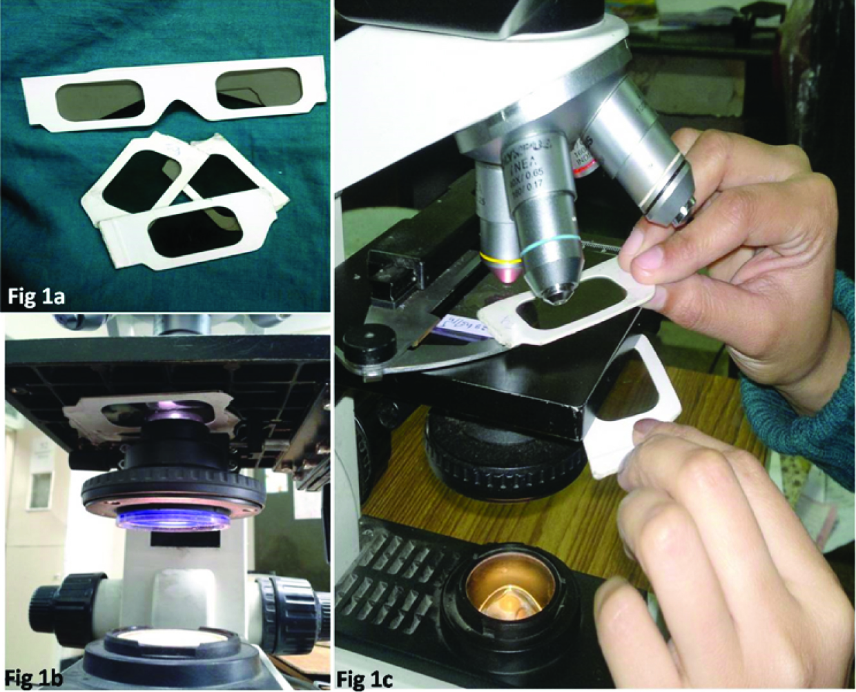
Passive 3D Polaroid glasses with a paper frame, easily available at cinema halls were procured and arranged in specific manner in light microscopes of various models and different companies. The glasses were cut in two halves [Table/Fig-1a]. One of the half “polarizer” was placed above the sub stage condenser [Table/Fig-1b] and just below the stage. Another half “analyser” was placed over the slide [Table/Fig-1c]. The side alignment of glasses was specific in order to attain the desired birefringence.
a) The 3D Polaroid glasses from cinema halls were cut into halves and aligned in specific manner. b) One half was placed above the field diaphragm and just below the stage called as polarizer. c) Another half placed over the slide called as analyser.
Using a certain alignment, polarizing effect without red retardation function was obtained. Using the same pair of glasses, but when placed after flipping them upside down (without changing their location), polarizing effect with red retardation plate was obtained. Thus, the same pair could be used as polarizer without and with red retardation plate function depending on the desire of the user. For microscopes using reflected light as the source of illumination, the “polarizer’ can simply be kept above the field diaphragm. Either of the Polarizer or the Analyser can be rotated to detect birefringent substance. Various birefringent substances diagnosed by commercial polarizing microscope were analysed using the new system with adequate negative controls like hyalinization which may resemble amyloid on routine staining. The comparison was based on subjective interpretation of intensity and quality of birefringence. The various models of microscope available in the department that were used to study the new system includes Nikon Optiphot 2, Nikon Eclipse 80i (Nikon Corporation, Tokyo, Japan), Olympus CH20i, and Olympus BX 51 (Olympus Corporation, Tokyo, Japan).
All the images using the system were clicked on Olympus BX51. The commercial polarization was seen and compared on the available polarizing microscope Nikon Eclipse E 600 (Nikon Corporation, Tokyo, Japan). Red retardation plate was not available in the department and the comparison was done with the images published in various standard textbooks and internet.
Results
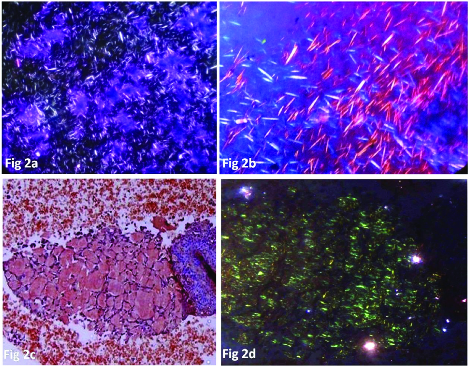
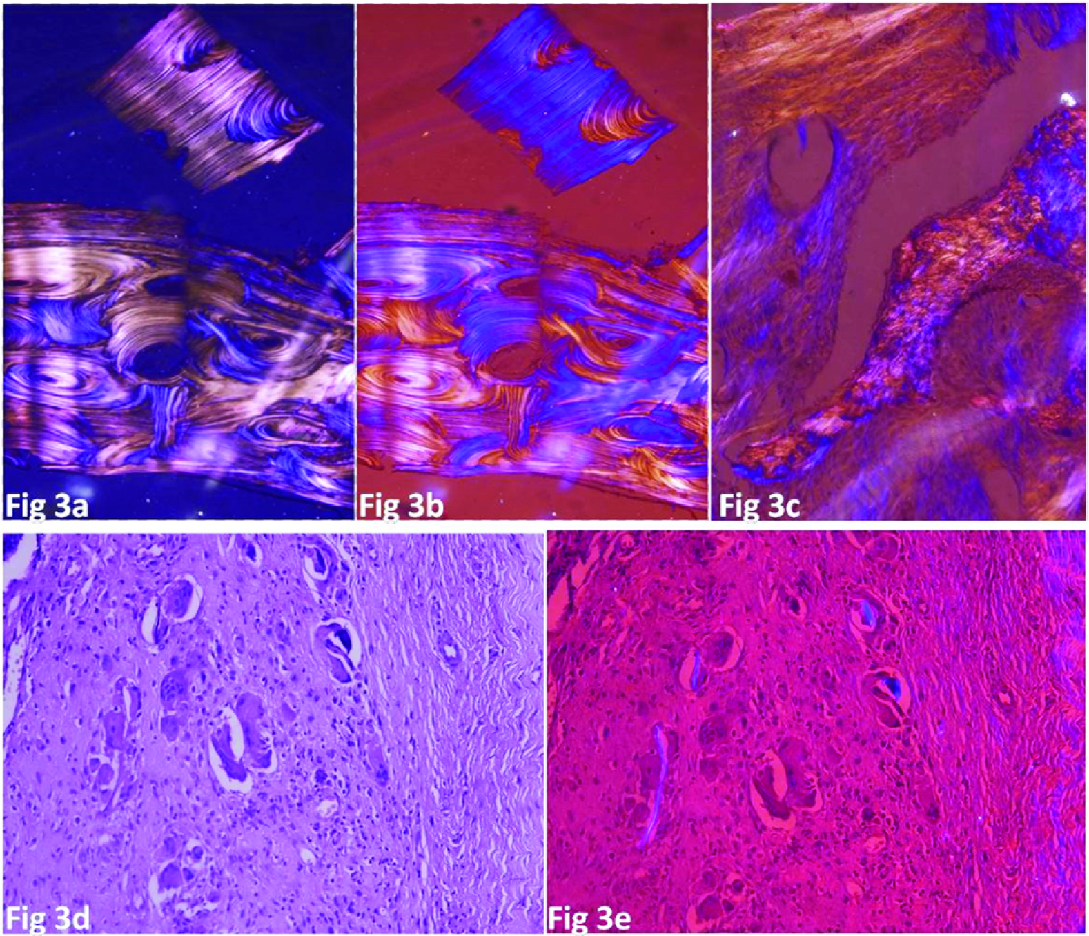
The birefringence observed by our system was comparable to the commercially available polarizing system with respect to intensity and quality. Also, there were no false positive/negative results when compared with the commercial Polarizing microscope. Moreover, the system had an inbuilt red retardation plate function to determine sign of birefringence. The system had an added advantage of being used in any light microscope model as well as in multi-header microscopes. The portability of both Analyser and Polarizer was an added distinct advantage. Various specimens that were observed were mono sodium urate crystals in synovial fluid cytology and tophus biopsy specimens ([Table/Fig-2a] from a case of gout synovial fluid cytology) which were negatively birefringent using red retardation plate function [Table/Fig-2b], amyloid deposits ([Table/Fig-2c] from a case of medullary carcinoma of thyroid) showing apple green birefringence [Table/Fig-2d] on polarization, lamellar bone. [Table/Fig-3a&b] showing long well arranged collagen lamellae as compared to woven bone showing haphazard short stretched collagen arrangement ([Table/Fig-3c] from a case of fibrous dysplasia), foreign body granulomas [Table/Fig-3d&e]. A case of a finger swelling showed numerous giant cells [Table/Fig-3d] which showed the presence of birefringent foreign body on cross polarization using our indigenous cross polarizing system [Table/Fig-3e]. Various other birefringent substances were observed without and with the red retardation plate function. These substances included skeletal muscle striations, fibrosis, nerve myelination, meglumine crystals in urine and glove powder (Maltese cross appearance).
a) Urate crystals without and b) with red retardation plate function. (Unstained under cross polarized light with red retardation plate function, 100X and 400X respectively) showing long needle shaped crystals with negative sign of birefringence. c) Amorphous substance deposit in a case of medullary carcinoma thyroid (H&E, 200X) and d) same substance showing apple green birefringence. (Congo Red under cross polarized light, 200X)
a&b) Lamellar bone showing reversal of colours on rotating the analyser/polarizer. (H&E under cross polarized light with red retardation plate function 100X). c) Woven bone from a case of fibrous dysplasia showing haphazard osteoid arrangement. (H&E under cross polarized light with red retardation plate function, 100X). d) Many giant cells with granulomas in a biopsy from a finger tip nodule (H&E, 100X). e) Birefringent foreign body seen clearly (H&E under cross polarized light with red retardation plate function, 100X)
Discussion
Polarized light is a beam of light whose electric field vectors vibrate only in one direction. Birefringence is property of a substance to split a beam of light, incident on it, into two polarized rays that are mutually perpendicular and experience different refractive indices [5].
Polaroid films were made by Edwin H Land in 1930 [6]. The 3D glasses used in cinema hall are made from Polaroid sheets that use Iodoquinine sulfate crystals aligned in aspecific direction in a polymeric sheet. Polaroid films have also been extensively used in 3D image reconstruction, in sunglasses, liquid crystal displays (LCD) & various other media facets [4]. The polarizing microscopes use glass prism systems or pure linear polaroid materials. They thus do not have retardation plate function for which another requipment called full wave plate is needed. The full wave plate/first order retardation plate is designed to introduce a relative retardation of exactly one wave length in the green or 550 nanometer region between the two light beams (produced due to the birefringence of the specimen) passing through it. As a result, green wavelengths emerge from the retardation plate still linearly polarized and having the same orientation as when they entered the retardation material i.e. parallel to the polarizer. These wavelengths are perpendicular to the analyser, thus are absorbed and do not pass through. So, from the original white light, the green wavelengths are subtracted giving rise to a magenta-red background, hence the name ‘Red Retardation plate’ [7].
The 3D Polaroid glasses are different since they are a combination of circular and linear polarized glasses in different axes to achieve stereoscopy [8]. Thus, these 3D Polaroid glasses can also be modified to detect birefringence and its sign with an added advantage of being affordable and easily available. So, they are an excellent alternative to commercial polarizing microscopes. Any simple light microscope irrespective of model or manufacturer/company can be used to detect birefringence of various known pathologic substances using this indigenous system.
This system using 3D Polaroid glasses has some limitations that were encountered. First, the amount of light entering the two eye pieces is slightly different even after optimum rotation. This creates a little eye strain which makes prolonged viewing difficult. However, in most of the cases, prolong viewing is not needed. Second, it was observed that in rare (one instance) cases, the quality of glasses procured from the cinema hall was of slightly inferior quality. Thus, it was felt necessary that glasses procured from different cinema halls should be tested and the best one should be carefully preserved for further use. Another caveat in using this system is that the system does not have a compensator in it. Exact quantification of birefringence is not possible due to lack of compensators, though it is seldom needed in diagnostic pathology. RJ Maude et al., have described the use of Polaroid sunglasses for screening of smears for malarial parasite as hemozoin pigment is birefringent [2]. However; we found no study describing indigenous methods for a polarizing system that can be used in all the situations, especially with the use of 3D viewing glasses.
Conclusion
The new Analyser- Polarizer system using 3D Polaroid glasses are comparable to costly commercially available Polarizing microscopes. Their specific placement in a light microscope of any model can convert the latter into a polarized microscopy unit with aretardation plate function without virtually any added expense. The system is efficient, easily accessible, portable and compatible with all models of light microscopes. We hope that this will enable the use of polarizing system by every pathologist, overcoming the economic constraint. With its widespread use, we also hope that the list of birefringent substances and their diagnostic utility grows further. All of this requires just a visit to the nearest 3D movie theatre and enjoying the show!