Granulocytic Sarcoma in an Adult with Relapsed Acute Myeloid Leukaemia
Neethu Ann Kurien1, Deepa John2, Geeta Chacko3, Pushpa Jacob4
1 Resident, Department of Ophthalmology, Christian Medical College, Vellore, Tamil Nadu, India.
2 Associate Surgeon, Department of Ophthalmology, Christian Medical College, Vellore, Tamil Nadu, India.
3 Professor, Department of Pathology, Christian Medical College, Vellore, Tamil Nadu, India.
4 Professor, Department of Ophthalmology, Christian Medical College, Vellore, Tamil Nadu, India.
NAME, ADDRESS, E-MAIL ID OF THE CORRESPONDING AUTHOR: Dr. Deepa John, Associate Surgeon, Department of Ophthalmology, Schell Eye Hospital, Arni Road, Christian Medical College, Vellore – 632001, Tamil Nadu, India.
E-mail: deeparebeccajohn@gmail.com
Granulocytic sarcoma is an extramedullary tumour consisting of malignant granulocytic precursor cells that is common among children with acute myeloid leukaemia (AML). We report a case of orbital granulocytic sarcoma in an adult with relapsed undifferentiated AML–M0. It presented as bilateral medial canthal swellings. An incisional biopsy confirmed the diagnosis of granulocytic sarcoma. The swelling resolved with re-induction chemotherapy.
AML-M0 subtype, Extramedullary tumour, Myeloid sarcoma, Orbital mass
Case Report
A 40-year-old male with a past history of undifferentiated acute myeloid leukaemia (AML-M0) presented with gradually increasing bilateral medial canthal swellings for one month [Table/Fig-1].
Swelling along medial canthal region
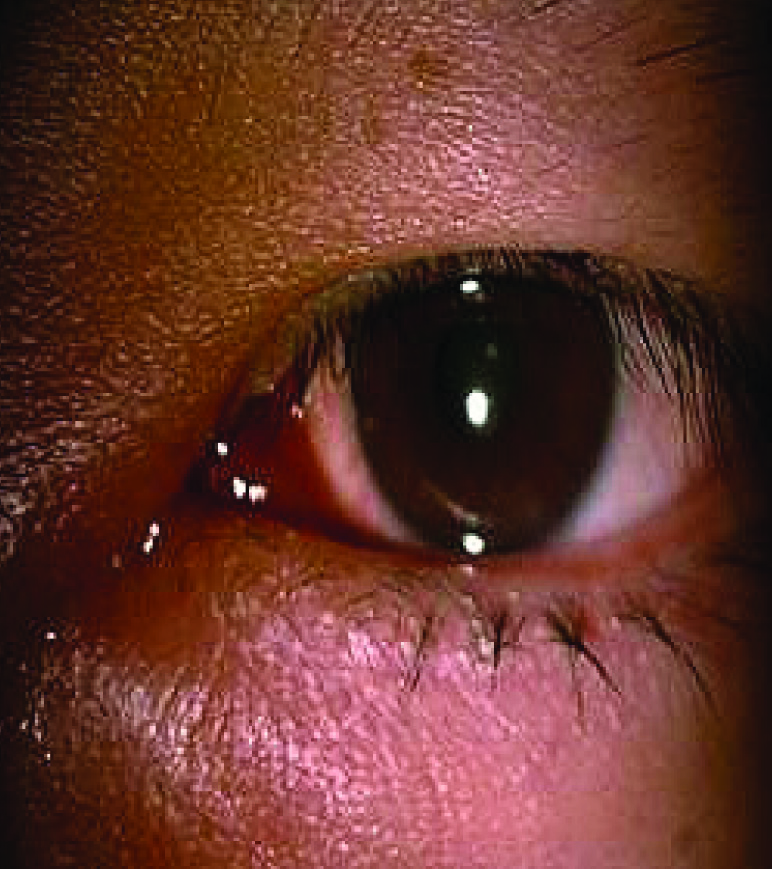
On examination, his best corrected visual acuity was 6/6, J1 in both eyes. The medial canthal swelling was bilateral, firm, erythaematous and non-tender measuring 10x5 mm in the right and 13x6 mm in the left eye. The anterior and posterior segment eye examination was otherwise unremarkable. Systemic examination was normal.
Computerised tomography scan of the orbit showed bilateral, symmetrical, enhancing soft tissue mass measuring 24x17x30 mm on the left and 21x17x26 mm on the right in the antero-medial aspect of the orbit [Table/Fig-2].
CT orbit axial view showing bilateral, symmetrical, enhancing soft tissue mass in antero-medial aspect of the orbit (white arrows)

The biopsy sample consisted of a circumscribed greyish white firm nodule. Histopathological examination showed conjunctival epithelium with a subepithelial circumscribed cellular tumour composed of closely packed sheets of mitotically active medium-sized polygonal cells with moderately pleomorphic oval to irregular nuclei with dispersed chromatin, small nucleoli and scanty to vacuolated cytoplasm. There were no areas of necrosis or haemorrhage.
The tumour cells were immunopositive for CD34 and CD43 and negative for myeloperoxidase (MPO) [Table/Fig-3], consistent with granulocytic sarcoma (GS). Cerebrospinal fluid analysis was normal, but bone marrow biopsy showed 20% blasts signifying concurrent medullary relapse.
Photomicrograph illustrating: a) circumscribed tumor beneath the epithelium (H&E x100); b) high power view of the same with sheets of mitotically active medium sized polygonal cells with moderately pleomorphic oval to irregular nuclei with dispersed chromatin, small nucleoli and scanty to vacuolated cytoplasm (H&E x400); c)Immunopositivity for CD34 [x400]; d) Immunopositivity for CD43 [x400].

Patient was referred to haematology department and re-induction chemotherapy was started with cytosine and idarubicin for six days followed by high dose cytosine arabinoside. Stem cell transplantation was considered due to the extramedullary relapse of AML. With re-induction chemotherapy, he attained complete remission of AML along with regression of the swelling. However, after 3 months of treatment, while awaiting stem cell transplantation, patient died of a cardiac arrest.
Discussion
Granulocytic sarcoma was first described by Allen Burns in 1881, and its association with AML in 1893 [1]. It occurs in 3-9% of patients with AML, either co-existent or prior to the onset of bone marrow leukaemia [2], and is common among children and adolescents. Myeloid sarcomas are most common in certain subtypes of AML, viz. M5a (monoblastic), M5b (monocytic), M4 (myelomonocytic), and M2 (myeloblastic with maturation) [3]. The orbit is one among the various locations where GS can initially manifest [4]. Other ocular manifestations are ptosis, lacrimal gland involvement, conjunctival mass or diffuse uveal involvement [5]. Orbital involvement may occur many years after remission, either unilaterally or bilaterally [6].
GS or chloroma represents an extramedullary mass of myeloblasts in the myelodysplastic syndromes [1]. The greenish coloration of this lesion is attributed to the MPO in the cells of granulocytic lineage. In these lesions the normal haematopoietic progenitor cells bind to bone marrow stroma and begin to proliferate and differentiate, while in vitro they bind to skin fibroblasts. Binding of these cells to the fibroblasts localized in non-haematopoietic tissues may result in the formation of extramedullary myeloid metaplasia [5]. Myeloid leukaemia associated with GS has a worse prognosis than leukaemia presenting alone [7]. Though our patient was in remission, he had bilateral medial canthal swelling initially which on biopsy proved to be extramedullary relapse. Bone marrow tests showed a concurrent medullary relapse.
The diagnosis of AML is established by morphologic examination of the bone marrow aspirate which shows 30% or more blasts relative to all nucleated marrow cells and cytochemical stains which are positive for MPO or alpha naphthyl butyrate esterase activity [4]. The most common marker expressed is CD68-KP1 [8]. Other typical immunochemistry markers seen are CD43, CD20 MPO, CD68, CD117, CD3 and lysozyme [1]. Cells that contain lysozyme are identified by immunohistochemical stain which utilizes antilysozyme antibody [4]. In our case CD34 and CD43 was positive but was negative for MPO.
GS is seen in certain subtypes of AML, in particular M5a, M5b, M4, and M2 of AML [9]. M4 and M5 subtypes are associated with a poor prognosis [10]. Minimally differentiated AML (AML-M0) is one among the eight recognized French-American-British subtypes accounting for less than 3% of AML. Even though GS in the oral cavity has been reported in AML-M0 subtype [11], GS has not been reported earlier with orbital involvement. We report a rare case of GS in AML-M0 with extramedullary relapse.
Conclusion
Granulocytic sarcoma should be considered in the differential diagnosis of adults with AML with orbital or conjunctival swelling, even if in remission. An early biopsy to confirm the diagnosis and referral to the haematologist for re-induction chemotherapy is warranted.
[1]. Mangla D, Dewan M, Meyer DR, Adult orbital myeloid sarcoma (granulocytic sarcoma): two cases and review of the literatureOrbit 2012 31(6):438-40. [Google Scholar]
[2]. Neiman RS, Barcos M, Berard C, Granulocytic sarcoma: A clinicopathologic study of 61 biopsied casesCancer 1981 48:1426-37. [Google Scholar]
[3]. Byrd JC, Edenfield WJ, Shields DJ, Dawson NA, Extramedullary myeloid cell tumors in acute nonlymphocytic leukaemia: a clinical reviewJ Clin Oncol 1995 13:1800-16. [Google Scholar]
[4]. Stockl FA, Dolmetsch AM, Saornil MA, Font RL, Burnier MN, Orbital granulocytic sarcomaBr J Ophthalmol 1997 81:1084-88. [Google Scholar]
[5]. Davis JL, Park DW, Font RL, Granulocytic sarcoma of the orbit. A histopathologic studyOphthalmology 1985 92:1758-62. [Google Scholar]
[6]. Banna M, Aur R, Akkad S, Orbital granulocytic sarcomaAm J Neuroradiol 1991 12:255-58. [Google Scholar]
[7]. Patil P, Bharambe BM, Binayke RS, Grace DC, Myeloid Sarcoma of Orbit in a Case of Acute Myeloid LeukaemiaJ Cytol Histol 2012 3:157doi:10.4172/2157-7099.1000157 [Google Scholar]
[8]. Campidelli C, Agostinelli C, Stitson R, Pileri SA, Myeloid Sarcoma: xtramedullary Manifestation of Myeloid DisordersAm J Clin Pathol 2009 132:426-37. [Google Scholar]
[9]. Shields JA, Stopyra GA, Marr BP, Bilateral orbital myeloid sarcoma as initial sign of acute myeloid leukaemia: case report and review of the literatureArch Ophthalmol 2003 121:138-42. [Google Scholar]
[10]. Lin CH, Wu KH, Lin WC, Tsai JD, Peng CT, Chen AC, Granulocytic sarcoma of the colon in a child with acute myeloid leukaemia presenting as haematocheziaJ Pediatr Haematol Oncol 2008 30:981-83. [Google Scholar]
[11]. Amin KS, Ehsan A, McGuff HS, Albright SC, Minimally differentiated acute myelogenous leukaemia (AML-M0) granulocytic sarcoma presenting in the oral cavityOral Oncol 2002 38:516-19. [Google Scholar]