Pleomorphic Adenoma in Retromolar Area: A Very Rare Case Report and Review of Literature
MD Yousuf Qureshi1, Tahseen Ali Khan2, Venkata Naga Nalini Dhurjati3, Kavitha Gaddikeri4, MD Zainuddin E. Khany5
1 Assistant Professor, Department of Oral & Maxillofacial Surgery, Malla Reddy Institute of Dental Sciences, Hyderabad, India.
2 Assistant Professor, Department of Oral & Maxillofacial Surgery, Sri Sai College of Dental Surgery, Vikarabad, Telangana, India.
3 Graduate Student, Ragas Dental College, Chennai, Tamilnadu, India.
4 Reader, Department of Oral & Maxillofacial Pathology, S.B Patil Dental College and Hospital, Bidar, Karnataka, India.
5 Post Graduate Student, Department of Oral & Maxillofacial Surgery, MNR Dental College & Hospital, Sanga Reddy, Telangana, India.
NAME, ADDRESS, E-MAIL ID OF THE CORRESPONDING AUTHOR:Dr. MD Yousuf Qureshi, Assistant Professor, Department of Oral & Maxillofacial Surgery, Malla Reddy Institute of Dental Sciences, Hyderabad-500055, India.
E-mail: jksaheb4@gmail.com
Among all neoplasms affecting head and neck region, salivary gland neoplasms are rare. Pleomorphic adenomas are the most common benign salivary gland tumours making up to 50% of major and minor salivary gland tumours. Intraorally pleomorphic adenoma is mostly found on palate and lips and very rarely in retromolar area. Here we are reporting a rare case of pleomorphic adenoma in right lower retromolar area in a 31-year-old female, the lesion was excised in toto with safety margins under local anaesthesia and postoperative follow up after six months didn’t showed any recurrence.
Enclavoma, Minor salivary glands, Myoepithelial cells, Salivary gland neoplasm
Case Report
A 31-year-old female patient came to the Department of Oral Medicine and Radiology, with a complaint of painless swelling in the right mandibular posterior region since six months. She went to a local doctor who prescribed her medicines. History revealed that the patient noticed a small swelling six months back, which slowly increased to the present size. She did not had any pain, tenderness or discharge associated with the swelling. She did not had any difficulty in mouth opening, but had slight discomfort while mastication and the swelling did not subsided on taking medications. On clinical examination, a solitary oval painless swelling measuring about 2cm x 3cm, was found in right retromaolar area [Table/Fig-1].
Intraoral view showing a swelling in lower right retromolar area.

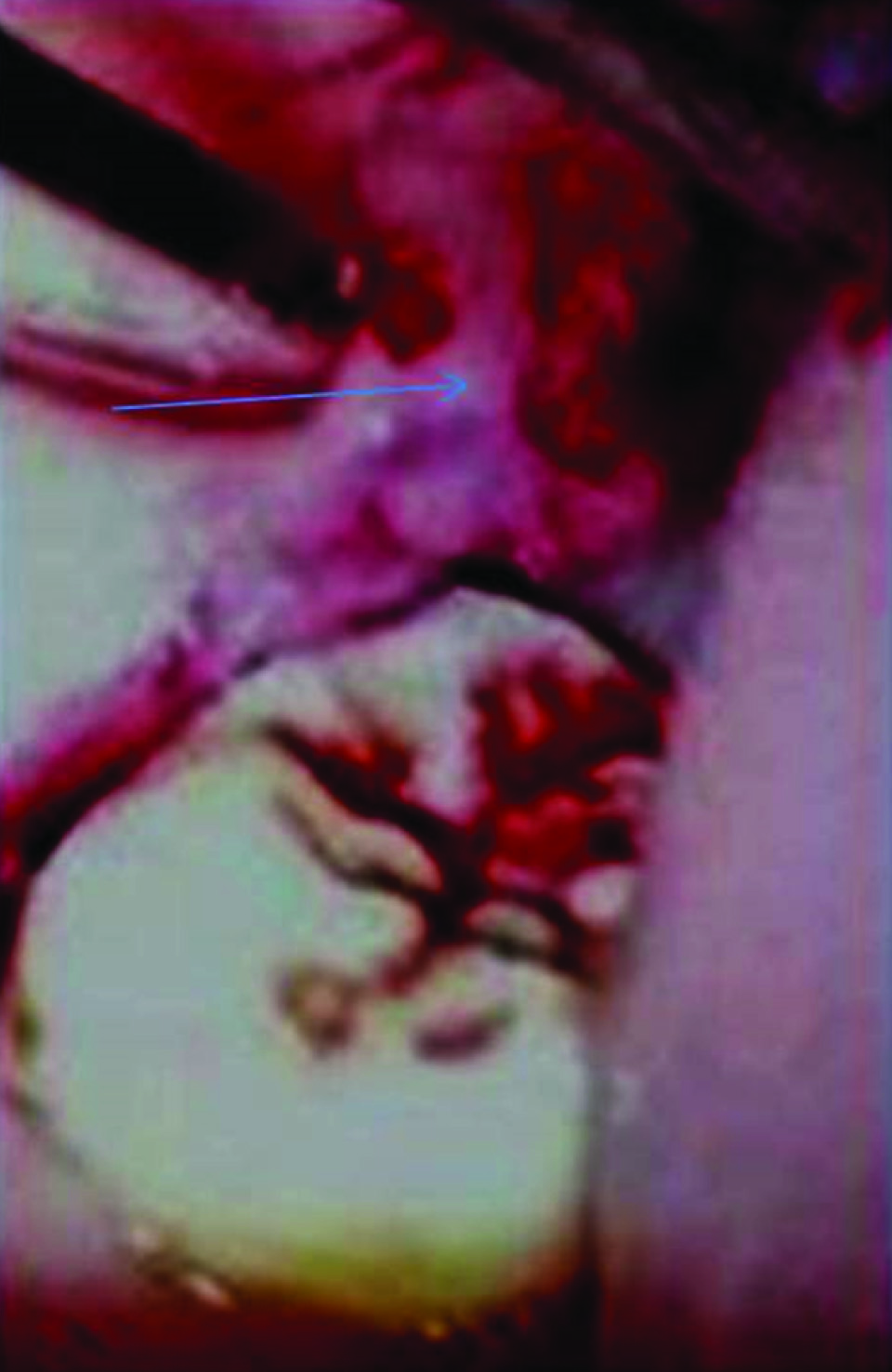
The growth extended from occlusal surface of 18 region to the occlusal surface of 48 region superio-inferiorly and from mesial of 48 to the anterior faucial piller posteriorly. No visible pulsations were seen and the mucosa over the growth appeared to be normal. There was no discharge from the swelling. On palpation the growth was movable, firm in consistency and non tender. It was not fixed to the underlying structures, non reducable and no pulsations were felt. Teeth in the vicinity of the swelling were not mobile. The regional lymph nodes were non palpable. There was no history of trauma, fever, past and family history was not significant. Radiographs and all the haematological investigations revealed normal values. Based on the history and correlating with the clinical findings, a provisional diagnosis of benign minor salivary gland tumour was given and differential diagnosis of fibroma, lipoma, neurilemmoma, mucocele and malignant salivary gland lesions (mucoepidermoid carcinoma) was made. Treatment of the lesion was planned in the surgery department as surgical excision. After explaining the procedure to the patient and his relatives consent was taken and, the lesion was totally excised with safety margins under local anaesthesia. After administering inferior alveolar nerve block an intraoral vertical incision was given in retromolar trigone area extending from retro molar area to 47. With help of dissecting scissors the mucosal tissue was undermined. The tumour was visualized and the surface of the tumour was firm in consistency. The complete tumour along with capsule was excised cautiously by holding it with curved artery mosquito forceps and surgical site was irrigated with 10% betadine mixed with normal saline in 1:1 ratio. After excision haemostasis was achieved [Table/Fig-2]. Closure was done by giving interrupted sutures with 3-0 black braided silk sutures. After surgery, antibiotics {augmentin 625 mg (500 mg amoxicillin and 125 mg clavulanic acid)} were prescribed twice a day for five days, and the sutures were removed after one week. The excised tumour mass was sent for histopathological evaluation.
Intraoperative photograph showing excision being done.
Macroscopic examination revealed a single large soft tissue specimen which was creamish in color, oval in shape, measuring about 2cm x 3cm in size [Table/Fig-3]. On cutting the inner aspect, the specimen appeared gelatinous and showed cystic degeneration. It was cut into two halves, one half was preserved and the other was taken for processing.
Gross specimen of the excised lesion.

Microscopic examination of haematoxylin and eosin stained sections revealed a highly cellular lesional tissue encompassing many myoepithelial cells separated by hyalinized, osseous and myxoid areas. Myoepithelial cells were spindle, round and epitheliod in shape with hyperchromatic nucleus. The myoepithelial cells showed proliferation in the form of sheets. In many areas duct like areas were scattered and small to large cyst formation containing eosinophilic coagulum could be appreciated [Table/Fig-4,5 and 6].
Photomicrograph (5x) showing epithelium and duct like areas, cystic areas with eosinophilic coagulum.

Photomicrograph (10x) showing duct like areas and cystic spaces.

Photomicrograph (40x) showing cystic areas and myoepithelial cells.

A diagnosis of pleomorphic adenoma was made. The patient was followed postoperatively for six months and there was uneventful healing [Table/Fig-7].
Postoperative photograph.

Discussion
Salivary gland neoplasms constitute about 1-4% of all neoplasms and 3-5% of neoplasms affecting head and neck regions [1,2]. Generally they affect major salivary glands and rarely about in 10-15 % cases affect minor salivary glands. Among neoplasms of minor salivary glands, pleomorphic adenoma is the most common benign neoplasm [3,4].
Studies have shown that females are more prone than males, as in indexed patient [5,6]. Pleomorphic adenoma cases show a mean age of 52 years with most of them in 4th to 6th decades, where as indexed patient was a young 31 year female [7,8].
Most familiar sites were palate (42.8-68.8%), upper lip (10%). Few cases have been reported at rare intraoral sites like throat (2.5%) and retromolar area (0.7 %) [4]. In the indexed patient it was found at a very rare site of right retromolar area. Most of the cases found in this site were malignant in nature, the present one was a benign neoplasm. Round, slow growing, painless, mobile, firm swelling without any overlying ulceration is the commonest finding of intraoral pleomorphic adenomas. Cervical lymph node involvement is uncommon. Mean duration was more than two years but the history revealed the lesion was of six months duration. Rare signs and symptoms are ulceration, speech difficulties, taste abnormalities and ill fitting dentures [1,9].
Very few cases of pleomorphic adenoma have been reported in retromolar area in the literature. Ajit Singh Rathore et al., have reported pleomorphic adenoma in a 30-year-old male patient in left lower retromolar area [1]. Rao PK et al., also reported pleomorphic adenoma in a 58-year-old male patient in left lower retromolar area. Whereas, Hirabayashi S et al., reported pleomorphic adenoma in a 41-year-old female in upper right retromolar area [10,11].
Pleomorphic adenoma cases have also been reported in palate in many cases [12–14]. Two cases of pleomorphic adenoma have been reported in parapharyngeal spaces [14].
The reported case is unique in that it is reported at a very rare site, retromolar area, whereas palate is the commonest site. The patient was much younger than the mean age of intraoral pleomorphic adenomas.
Histopathologically we see cells with both epithelial and mesenchymal differentiation. Recent studies have proven that it arises from a single myoepithelial cell or a ductal reserve cell. This cell differentiates in to epithelial ductal cells and mesenchymal chondroid, osseous and myxoid areas, hence called as mixed tumour, enclavoma, endothelioma, branchioma, endochondroma etc. Minor salivary gland lesions are more cellular with less chondroid and myxoid areas and do not have a fibrotic capsule [12].
Generally treatment includes wide excision with 1mm safety margins around the lesion. It should be diagnosed early as it causes disfigurement and loss of function. The lesion is non encapsulated and hence there are chances of recurrence, which was not seen in indexed patient after six months follow up. Most of minor salivary gland neoplasms are malignant with only 18% showing benign nature, the present case was a benign lesion. Malignant potential was found to be 2-9% [13–15].
Pleomorphic adenoma in retromolar area is a very rare neoplasm. It is rarely found in a 31-year-old patient, of lesser duration of six months and at a rare site of lower right retromolar area and its benign nature. Due to its high recurrence rate and malignant potential, it should be carefully excised with normal margins. A long term follow up is recommended and it should be kept in differential diagnosis of intraoral lesions as wrong diagnosis may result in wrong treatment and may affect its prognosis [14,15].
Conclusion
A rare case report of pleomorphic adenoma in lower right retromolar area in a 31-year-old female is reported, which was surgically excised and it is suggested to keep this lesion in differential diagnosis of swellings reported in this area.
[1]. Rathore AS, Chhina S, Garg K, Pleomorphic adenoma in retromolar area: a rare case report and review of literatureAmerican Journal of Medical Case Reports 2014 2(4):90-93. [Google Scholar]
[2]. Vijaya Kumar M, pleomorphic adenoma of soft Palate - A rare presentationInternational Journal of Biomedical Research 2015 6(06):439-41. [Google Scholar]
[3]. Yih WY, Kratochvil FJ, Stwart JL, Intraoral minor salivary gland neoplasms: review of 213 casesJ. Oral Maxillofac Surg 2005 63(6):805-10. [Google Scholar]
[4]. Moghe S, Pillai AK, Prabhu S, Nahar S, Kartika UK, Pleomorphic adenoma of the palate: Report of a caseInt J Sci Study 2014 2:54-56. [Google Scholar]
[5]. Prajapati N, Aggarwal A, Kapoor A, Pleomorphic adenoma of soft palate, a rare lesionThe Health Agenda 2013 1(2):42-44. [Google Scholar]
[6]. Pillai AK, Satpathy M, Nahar S, Moghe S, Pleomorphic Adenoma in Cheek: An Uncommon FindingIJSS Case Reports & Reviews 2014 1(1):19-22. [Google Scholar]
[7]. Wang D, Intraoral minor salivary gland tumours in a Chinese population: a retrospective study on 737 casesOral Surg Oral Med Oral Pathol Oral Radiol endod 2007 104:94-100. [Google Scholar]
[8]. Sunil S, Gopakumar D, Pleomorphic adenoma. A case report and review of literatureInt J Odontostomat 2013 7(2):171-74. [Google Scholar]
[9]. Dhillon M, Agnihotri PG, Raju SM, Lakhanpal M, Pleomorphic Adenoma of the Palate: Clinico-radiological Case ReportJournal of Indian Academy of Oral Medicine and Radiology 2011 23(3):286-88. [Google Scholar]
[10]. Rao PK, Shetty SR, Hegde D, Ectopic pleomorphic adenomaNorth American Journal of Medical Sciences 2012 4(4):190-92. [Google Scholar]
[11]. Hirabayashi S, Yanai A, Muraishi Y, Huge pleomorphic adenoma of the upper retromolar areaAnn Plast Surg 1997 38(2):184-86. [Google Scholar]
[12]. Gothwal AK, Pleomorphic Adenoma of the PalateJournal of Clinical and Diagnostic Research 2012 6(6):1109-111. [Google Scholar]
[13]. Sharma Y, Maria A, Chhabria A, Pleomorphic adenoma of the palateNational Journal of Maxillofacial Surgery 2011 2(2):169-71. [Google Scholar]
[14]. Verma P, Sachdeva SK, Verma KG, Sachdeva K, Pleomorphic adenoma of cheek: A rare case report and review of literatureIndian J Dent Res 2014 25:122-24. [Google Scholar]
[15]. Basavaraj KF, Karthikeyan Ramalingam, Vegf Expression and its Role in Recurrence of Intraoral Pleomorphic Adenomas – A Study on 10 CasesUJP 2013 02(03):118-26. [Google Scholar]