Comparison of the Perioperative Outcomes of a Vessel Sealing Instrument-Assisted Technique with a Conventional Technique in Abdominal Myomectomy
Mustafa Ulubay1, Mustafa Öztürk2, Fahri Burçin Firatligil3, Ulas Fidan4, Ugur Keskin5, Murat Dede6, Müfit Cemal Yenen7
1 Assistant Professor, Department of Obstetrics and Gynecology, Gulhane Military Medical Academy, Etlik, Ankara, Turkey.
2 Specialist, Department of Obstetrics and Gynecology, Gulhane Military Medical Academy, Etlik, Ankara, Turkey.
3 Specialist, Department of Obstetrics and Gynecology, Gulhane Military Medical Academy, Etlik, Ankara, Turkey.
4 Assistant Professor, Department of Obstetrics and Gynecology, Gulhane Military Medical Academy, Etlik, Ankara, Turkey.
5 Assistant Professor, Department of Obstetrics and Gynecology, Gulhane Military Medical Academy, Etlik, Ankara, Turkey.
6 Associate Professor, Department of Obstetrics and Gynecology, Gulhane Military Medical Academy, Etlik, Ankara, Turkey.
7 Professor, Department of Obstetrics and Gynecology, Gulhane Military Medical Academy, Etlik, Ankara, Turkey.
NAME, ADDRESS, E-MAIL ID OF THE CORRESPONDING AUTHOR: Mustafa Ulubay, M.D., Gulhane Military Medical Academy, Obstetrics and Gynecology Department, Ankara, Turkey.
E-mail: mulubay@gata.edu.tr
Introduction
In gynaecologic practice, LigaSure PreciseTM is generally used in endoscopic and open surgeries, such as hysterectomy, adnexectomy, and cancer surgery. However, there is no case report or main research article where LigaSure PreciseTM has been used for myomectomy. We want to compare a technique using a vessel sealing instrument with a conventional technique in abdominal myomectomy.
Materials and Methods
Fifty-five women who underwent abdominal myomectomy were divided two groups: (1) a vessel sealing instrument-assisted technique (24 patients); and (2) a conventional technique (31 patients) between January 2011 and December 2014 at the Department of Gynaecology and Obstetrics, Gulhane Military Medical Academy, Ankara, Turkey. The data for the operation times, the occurrence of perioperative complications, the hospitalization times, and changes in haemaglobin and haematocrit levels for the two techniques were collected and compared.
Results
The mean operation time was 48 minutes for the vessel sealing instrument-assisted technique and 54 minutes for the conventional technique. No statistically significant differences were determined for haemoglobin and haematocrit changes, hospital stay and perioperative complications.
Conclusion
We did not find any difference in the occurrence of complications, changes in haemoglobin or haematocrit levels, or hospital stay. The vessel sealing instrument-assisted technique is feasible and effective in reducing operation times.
Blood loss, Conventional myomectomy, Operation time
Introduction
The uterus is a thick-walled muscular organ comprised of smooth muscle cells. The monoclonal proliferation of these smooth muscle cells results in the occurrence of benign lesions called fibroids. Fibroids are the most common benign neoplasm of the uterus [1,2]. Although the pathogenesis of leiomyomas remains obscure, it is thought that hormonal mechanisms play a critical role in the development of fibroids, which explains the presence of fibroids in 20–25% of women who are of reproductive age [3].
Many fibroids are asymptomatic and are usually detected incidentally during routine gynaecological examinations [4]. However, 20–50% of fibroids become symptomatic and requires surgery [5]. These symptoms are: menorrhagia, pelvic pain, pelvic or urinary obstructive symptoms, infertility, and loss of pregnancy [6].
A fair proportion of women with uterine fibroids do not require treatment. A woman’s age and the severity of her symptoms are important factors in considering treatment options. There are three main treatment options for uterine fibroids: watchful waiting, medical therapy, and surgery [6].
In the surgical option, the standard treatment of symptomatic uterine fibroids is hysterectomy for women who do not desire to preserve their childbearing abilities, or are near or past menopause, while myomectomy is offered to women who wish to preserve their fertility [7]. Hysterectomy, the surgical removal of the uterus, eliminates fibroid symptoms and the chance of recurrence. However, many women who suffer from myomas wish to have children in the future or want to preserve the uterus. For these women, the first-choice surgery is myomectomy. Myomectomy is the surgical removal of myomas with restoration of the normal anatomy and preservation of the uterus [7]. Conventional, Laparoscopic and robotic myomectomy can be used for treatment. Laparoscopic and Robotic myomectomy are expensive treatment and need experience.
The fibroids are usually hypervascular in most areas with an increased number of arterioles and venules, so myomectomy may involve significant blood loss [8]. Several techniques have been developed to limit excessive blood loss. Laparoscopic preventive uterine artery occlusion, Osada method of uterus banding, laparoscopic bipolar coagulation vs. was described for reducing blood loss [9–11]. In this study, a vessel sealing instrument was used to limit blood loss.
We aimed to evaluate the use of a vessel sealing instrument compared with a conventional technique during the abdominal removal of the uterine fibroids.
Materials and Methods
The local ethics committee approval was obtained for this retrospective study. Written consent was received from all the patients. This Retrospective study included a total of 55 patients with subserous or intramural uterine fibroid(s) who underwent abdominal myomectomy between January 2011 and December 2014.
The principle reasons for seeking treatment were menometrorrhagia or pelvic pain. The majority of our patients wished to have no more children or did not wish to preserve their fertility.
Two groups were constituted: Group 1 included patients who underwent vessel sealing instrument-assisted abdominal myomectomy (n=24) while Group 2 included patients who underwent conventional myomectomy (n=31). Two experienced consultant gynaecologists performed the operations. The patients for each of the two operators were block-randomized.
The fibroid(s) were evaluated pre-operatively using abdominal and transvaginal ultrasonography (General Electric Logiq S6®, 1.5–4.5 MHz probe, Waukesha, WI USA).
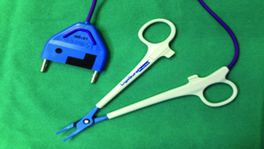
In Group 1, all patients underwent general anaesthesia and endotracheal intubation. A Foley catheter was inserted into the bladder as a standard procedure. The incision to access the abdomen was a Pfannenstiel incision. A transverse incision was made through the wall of the uterus above the fibroid. First, the cleavage between the myometrium and the fibroid had to be defined. The fibroid was then dissected bluntly and sharply, and vessel sealing was done using LigaSure PreciseTM (Covidien, Boulder Co, USA) [Table/Fig-1]. The entire fibroid was removed. After the fibroid had been removed, the defect was sutured in layers. To minimize adhesion, the uterine serosa was sutured in a running baseball fashion with 2/0 VicrylTM (Polyglactin 910 Suture, Ethicon Co).
LigaSureTM precise is a curved-jaw vessel sealing instrument that provides fine-grasping and effectively dissects tissue planes in open procedures.
In Group 2, all patients underwent general anaesthesia and endotracheal intubation. A Foley catheter was inserted into the bladder as a standard procedure. The incision to access the abdomen was a Pfannenstiel incision. A transverse incision was made through the wall of the uterus above the fibroid. First, the cleavage between the myometrium and the fibroid was defined. The fibroid was then dissected bluntly using curved Rochester® pean forceps (14 cm) and sharply using curved Metzenbaum Fino® scissors (18 cm) until the entire fibroid had been removed. After the fibroid(s) had been removed, the defect was sutured in layers, and the uterine serosa was sutured in a running baseball fashion with 2/0 VicrylTM (Polyglactin 910 Suture, Ethicon Co, USA) to minimize adhesion.
All patients underwent intraoperative antibiotic prophylaxis with CefamezinTM 1000 mg IV flacons (Cefazolin) and did not receive additional antibiotics postoperatively. The Foley catheter was extracted at the postoperative sixth hour once the patient had been mobilized.
Both groups were compared in terms of their operation times, perioperative complications, and changes in haemoglobin (Hb) and haematocrit (Htc) levels. The operation time was considered as the time elapsing from anaesthesia induction to awakening. All perioperative complications during the first month following surgery were recorded. Changes in Hb (Δ-Hb) and Htc (Δ-Hb) before and 36 hours after the operation made it possible to evaluate blood loss. Δ-Hb was calculated by comparing preoperative Hb vs. postoperative Hb formulas, while Δ-Htc was established by comparing preoperative Htc with postoperative Htc formulas.
Statistical Analysis
SPSS software for Windows (version 15.0) was used for data analysis. The Mann–Whitney U test was used to compare the discrete data between the groups. A p-value of less than 0.05 was considered to be statistically significant.
Results
The data of the patients’ demographics, myomatous disease, surgical interventions, changes in Hb and Htc levels, and postoperative complications are presented in [Table/Fig-2].
The data of the patients’ demographics, myomatous disease, surgical interventions, changes in Hb levels, and postoperative complications.
| Group 1(n:24) | Group 2(n:31) | p-value |
|---|
| Age (year) | 38 (29-45) | 39 (27-48) | 0.632 |
| Marital status |
| Married | 20 (83.3%) | 29 (93.5%) | |
| Single | 0 (0%) | 0 (0%) | |
| Divorced | 4 (16.7%) | 2 (6.5%) | |
| Delivery status |
| Caserean | 4 (16.7%) | 6 (%19.4) | |
| Vaginal delivery | 20 (83.3%) | 25 (%80.6) | |
| Nulligravida / nullipara | 0 (%0) | 0 (%0) | |
| Preoperative topography |
| Number of myomas | | | |
| Mean (min-max) | 2.29 (1-7) | 3.03 (1-21) | 0.889 |
| Size of the largest myoma | | | |
| Mean (size in mm) | 68.3 (30-120) | 70.6 (20-130) | 0.993 |
| Duration of the operation in minutes. Mean (min–max) | 48 | 54 | 0.006 |
| Delta Hb in g/dl Mean (min–max) | 1,75 (0,3-4,2) | 1,77 (0,3-4,5) | 0.980 |
| Duration of hospitalization in days. Mean (min–max) | 2.12 (2-3) | 2.09 (2-3) | 0.741 |
| Postoperative complications |
| Wound infection | 1 | 1 | |
| Intraoperative haemorrhage | 1 | 2 | |
| Weight (kg) Mean (min-max) | 59.9 (49-76) | 64.0 (50-90) | 0.101 |
| Height (cm) Mean (min-max) | 161.1 (152-168) | 162.5 (154-172) | 0.341 |
The mean age was not statistically different between the two groups (38 vs. 39 years); however, all other background factors that could have potentially affected the results were well balanced between the two groups. The mean operation time in Group 1 was 48 minutes compared with 54 minutes in Group 2. The reduction of the operation time in favor of Group 1 was statistically significant (p= 0.006). There were a few postoperative complications in the two groups. In Group 1, one patient required a blood transfusion for intraoperative haemorrhaging, and a wound infection was treated in another. In Group 2, two patients required blood transfusions for intraoperative haemorrhaging while one patient was treated for a wound infection.
The mean length of hospital stay was the same for both groups (2.12 vs. 2.09 days). There was no statistically significant difference in the Δ-Hb and Δ-Htc levels between the two groups (p > 0.05).
Discussion
To our knowledge, our study is the first research article designed to examine perioperative outcomes using the vessel sealing instrument-assisted technique in abdominal myomectomy. LigaSure PreciseTM is a vessel sealing instrument that is used in various types of operations, such as haemorrhoidectomy, appendectomy, thyroidectomy, etc [12–14]. In gynaecologic practice, LigaSure PreciseTM is generally used in endoscopic and open surgeries, such as hysterectomy, adnexectomy, and cancer surgery [15,16]. However, in English medical research literature, there is no case report or main research article where LigaSure PreciseTM has been used for myomectomy.
Myomectomy is the surgical removal of uterine fibroids. This surgery can be associated with significant blood loss because fibroids are usually hypervascular in most areas with an increased number of arterioles and venules [7,8]. This life-threatening bleeding can necessitate emergency blood transfusions. Globally, various interventions have been used to reduce bleeding during myomectomy, but it is unclear whether or not the interventions have been effective [7]. It is therefore essential to enable evidence-based clinical decisions.
In clinical practice, there are a few methods that have successfully reduced bleeding during the surgical removal of uterine fibroids. In the Cochrane Collaboration Reviews, misoprostol, vasopressin, bupivacaine plus epinephrine, tranexamic acid, gelatin-thrombin matrix, peri-cervical tourniquet, mesna, ascorbic acid, dinoprostone, loop ligation of the myoma pseudocapsule, and fibrin sealant patches (collagen sponges with thrombin and fibrinogen) have been found to significantly reduce bleeding during myomectomy [7].
We collected data regarding operation times, perioperative complications, and length of hospital stays. Differences in Δ-Hb and Δ-Htc levels, hospital stay, and postoperative complications were found to be insignificant in both groups in our study; however, a statistically signification reduction in operation time in favor of Group 1 was found (p < 0.05).
In some studies, the electrosurgical instruments used in myomectomy were found to have a thermal effect on uterine tissues [17,18]. Therefore, it was thought that using LigaSure PreciseTM in myomectomy may result in uterine rupture due to the thermal effect. In our study however, adverse effects caused by the LigaSure PreciseTM-assisted myomectomy technique were lacking, especially in patients wishing to have children.
Conclusion
In conclusion, given the limitation of sample sizes in the groups in our study, we did not find any differences in the occurrence of complications, changes in haemoglobin or haematocrit levels, and length of hospital stay. As our study shows, LigaSure PreciseTM is an effective and reliable instrument that shortens the duration of operation times in abdominal myomectomy. Nevertheless, due to the lack of adverse effects, well-designed randomized controlled trials using LigaSure PreciseTM in myomectomy are required.
[1]. Wallach EE, Vlahos NF, Uterine myomas: an overview of development, clinical features, and managementObstet Gynaecol 2004 104(2):393-406. [Google Scholar]
[2]. Buttram VC, Reiter RC, Uterine leiomyomata: aetiology, symptomatology, and managementFertil Steril 1981 36(4):433-45. [Google Scholar]
[3]. Baird DD, Dunson DB, Hill MC, Cousins D, Schectman JM, High cumulative incidence of uterine leiomyoma in black and white women: ultrasound evidenceAm J Obstet Gynaecol 2003 188(1):100-07. [Google Scholar]
[4]. Vollenhoven BJ, Lawrence AS, Healey DL, Uterine fibroids. A clinical reviewBJOG 1990 97:285-98. [Google Scholar]
[5]. Vercellini P, Vendola N, Ragni G, Trespidi L, Oldani S, Crosignani PG, Abnormal uterine bleeding associated with iron-deficiency anaemia: Aetiology and role of hysteroscopyJ Reprod Med 1993 38:502-04. [Google Scholar]
[6]. Evans P, Brunsell S, Uterine fibroid tumors: diagnosis and treatmentAm Fam Physician 2007 75(10):1503-08. [Google Scholar]
[7]. Kongnyuy EJ, Wiysonge CS, Interventions to reduce haemorrhage during myomectomy for fibroidsCochrane Database Syst Rev 2014 8:CD005355 [Google Scholar]
[8]. Sampson JA, The blood supply of uterine myomataSurg Gynaecol Obstet 1912 14:215 [Google Scholar]
[9]. Dubuisson J, Popescu S, Dubuisson JB, Petignat P, Preventive Uterine Artery Occlusion: Benefits of the Laparoscopic Posterior ApproachJ Minim Invasive Gynaecol 2015 :31 [Google Scholar]
[10]. Raba G, Kotarski J, Szczupak K, Obloza B, Fudali-Walczak M, Uterus banding with the Osada method effectively reduces intraoperative blood loss during myomectomyMinim Invasive Ther Allied Technol 2015 2:1-5.[Epub ahead of print] [Google Scholar]
[11]. Liu WM, Laparoscopic bipolar coagulation of uterine vessels to treat symptomatic leiomyomasJ Am Assoc Gynaecol Laparosc 2000 7:125-29. [Google Scholar]
[12]. Yeh CC, Jan CI, Yang HR, Huang PH, Jeng LB, Su WP, Comparison and Efficacy of LigaSure and Rubber Band Ligature in Closing the Inflamed Cecal Stump in a Rat Model of Acute AppendicitisBiomed Res Int 2015 2015:260312 [Google Scholar]
[13]. Molnar C, Voidazan S, Rad CC, Neagoe VI, Rosca C, Barna L, Total thyroidectomy with LigaSure Small Jaw versus conventional thyroidectomy – a clinical studyChirurgia (Bucur) 2014 109(5):608-12. [Google Scholar]
[14]. Chen CW, Lai CW, Chang YJ, Hsiao KH, Modified LigaSure haemorrhoidectomy for the treatment of haemorrhoidal crisisSurg Today 2014 44(6):1056-62. [Google Scholar]
[15]. Demir RH, Marchand GJ, Adnexal Masses Suspected to Be Benign Treated with LaparoscopyJSLS 2012 16(1):71-84. [Google Scholar]
[16]. Hagen B, Eriksson N, Sundset M, Randomised controlled trial of LigaSure versus conventional suture ligature for abdominal hysterectomyBJOG 2005 112(7):968-70. [Google Scholar]
[17]. Zhu Q, Ruan J, Zhang L, Jiang W, Liu H, Shi G, The study of laparoscopic electrosurgical instruments on thermal effect of uterine tissuesArch Gynaecol Obstet 2012 285(6):1637-41. [Google Scholar]
[18]. Sentilhes L, Sergent F, Verspyck E, Gravier A, Roman H, Marpeau L, Laparoscopic myomectomy during pregnancy resulting in septic necrosis of the myometriumBJOG 2003 110(9):876-78. [Google Scholar]