HSV Encephalitis with Normal CSF — A Case Report with Review of Literature
Gautam Rawal1, Sankalp Yadav2, Umar Rasool Wani3, Alok Kumar Ambastha4
1 Attending Consultant, Department of Critical Care, Rockland Hospital, Qutab Institutional Area, New Delhi, India.
2 General Duty Medical Officer-II, Chest Clinic, Moti Nagar, North MCD, New Delhi, India.
3 Junior Consultant, Department of Critical Care, Rockland Hospital, Qutab Institutional Area, New Delhi, India.
4 Senior Resident, Department of Critical Care, Rockland Hospital, Qutab Institutional Area, New Delhi, India.
NAME, ADDRESS, E-MAIL ID OF THE CORRESPONDING AUTHOR: Dr. Gautam Rawal, Flat No. 417, Dhruva Apartments, Plot No. 4, I P Extension, Patparganj, Delhi-110092, India. E-mail : drgautamrawal@hotmail.com
Herpes simplex virus encephalitis (HSVE) is one of the most potentially fatal infectious disease that should be detected as early as possible. The combination of clinical history and examination, brain computed tomography or magnetic resonance imaging (MRI) and lumbar puncture have been used to establish a diagnosis. The authors present a case of HSVE with normal CSF analysis, but typical MRI findings consistent with HSE and CSF PCR positive for Herpes simplex virus1 DNA, who responded to Acyclovir therapy with complete recovery.
Acyclovir, Cerebrospinal fluid, Herpes simplex virus, Polymerase chain reaction
Case Report
A 60-year-old Indian man presented to our institution, during the summer months, following referral by a local hospital. The patient was in a good health until five days before admission to our hospital, when he developed moderate grade fever followed by an episode of seizure the next day of fever. His past medical history was insignificant except that he was a chronic smoker. He was admitted to the local hospital where his neurological symptoms worsened during the next 72 hours. The patient started experiencing aggressive behaviour and also had multiple episodes of seizure. His Computed Tomography (CT) scan of the brain was inconclusive. The patient was transferred to our hospital for further management.
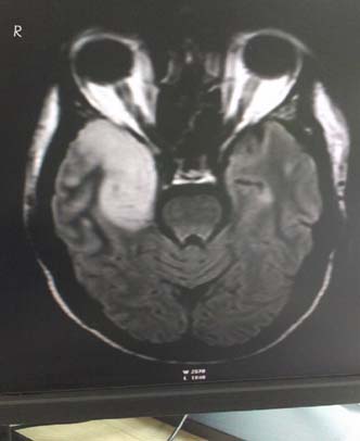
On admission, the patient was febrile (axillary temperature of 38°C), normotensive with mild tachycardia. His physical examination showed slow mentation along with generalized slowing of his responses to verbal commands and also generalized weakness. A chest X-ray showed bilateral infiltrations. Urine and blood cultures were taken. He was started on antibiotic-Cefepime, anti-epileptic, corticosteroid and empirical acyclovir due to suspected viral encephalitis along with supportive measures. A lumbar puncture was performed immediately and cerebrospinal fluid (CSF) analysis revealed a white blood cell (WBC) count of 0, a red blood cell (RBC) count of 0, a protein level of 18 mg/dL, and a glucose level of 57mg/dL (the synchronous serum value was 113mg/dL). A CSF polymerase chain reaction (PCR) for herpes simplex virus (HSV) was ordered, the result of which was received 72 hours later after the lumbar puncture had been performed. The CSF PCR was reported to be positive for HSV-1 DNA. A viral culture was not performed and the bacterial culture of the CSF was sterile. His contrast enhanced magnetic resonance imaging (MRI) of the brain was done which showed bilateral temporal enhancement (right more than left) [Table/Fig-1,2]. He improved neurologically and was continued on oral Acyclovir for a total of 21days. He was discharged in a stable condition without any neurological deficit. A written and informed consent was obtained from the patient for using the clinical images and the details of the case.
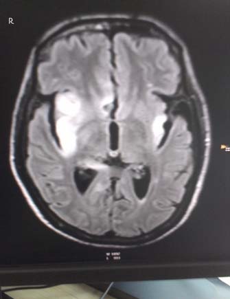
Flair image of CE MRI brain-showing bilateral temporal hyper intensities (Right > Left) involving the medial temporal lobes.
Discussion
Encephalitis refers to an acute or sub-acute, usually diffuse inflammation of the brain. The most common cause of sporadic encephalitis is due viral infection with herpes simplex type 1 (HSV1). It is characterized usually by the acute onset of fever, headache, seizures, focal neurologic signs, and impaired consciousness [1]. Herpes simplex viral encephalitis (HSVE), if not treated, is associated with a high mortality rate of up to 70% within 7-14 days and high morbidity of up to 90% (mostly neurological sequelae) among the survivors therefore making its early and accurate diagnosis of critical importance [1–4]. Prompt and early treatment with Acyclovir has been shown to decrease the mortality to approximately 20% [2,5].
The combination of a clinical history, a suggestive CT scan or MRI of the brain and the examination of the CSF by microscopy, biochemical analysis and DNA PCR for the presence of HSV DNA is needed for diagnosis of HSVE. The DNA PCR for HSV of the CSF is considered the gold standard test with sensitivity of 94-98% and specificity of 98-100% [6]. The presence of a totally normal CSF analysis in HSVE is rare, especially in an immunocompetent patient. In a study by Fodor et al., 1998, in patients with atypical HSVE presentation, only one out of the total 24 patients had normal CSF on the first day of admission [7]. Jakob et al., 2011 reported five cases of HSVE with a normal CSF in Germany, but in immunosuppressed patients who received whole brain irradiation for malignoma [8]. Avkan Oguz et al., 2006, documented the same in two immunocompetent patients in Turkey [9]. In 2009, the incidence of HSVE with normal CSF was reported to be 5–10% initially in patients who represented with encephalitis [10].
The pathogenesis of HSVE is not clearly understood in humans. It has been postulated that both mechanisms, first direct by virus-mediated and secondly indirect immune-mediated, may play a role in producing neuronal damage in the central nervous system which is distributed in an asymmetric fashion involving the medial temporal and inferior frontal lobes [11]. Temporal lobe involvement has been documented in about 60% of the patients [12].
The clinical presentation varies, with the typical presentation being a prodrome of malaise, fever (90%), headache (81%) and nausea, followed by acute or subacute onset of encephalopathy which manifests as lethargy, confusion, and delirium. Other clinical presentations include psychiatric symptoms (71%), seizures (67%), vomiting (46%), focal neurological deficit (33%) and memory loss (24%) [2,4].
This case of HSVE illustrated the scenario in a patient with clinical symptoms and typical findings on brain imaging but with a normal CSF examination. Assumptive early treatment with Acyclovir helped in complete neurological recovery of the patient in an otherwise highly fatal disease.
Conclusion
This is a rare case of HSVE with normal CSF findings, but typical MRI findings, which was confirmed by the CSF PCR and responded well to treatment with Acyclovir and steroids. A high degree of suspicion must be kept in mind of the clinician with patients presenting with impaired consciousness, seizure, or new onset temporal lobe symptoms and having a normal CSF. The potential diagnosis of HSVE must not be overlooked, especially in a tropical country like India, where there are multiple causes of infections affecting the brain, so that antiviral therapy with Acyclovir can be started without delay thus decreasing the associated mortality and morbidity. CSF for HSV PCR should be used as the method of choice to diagnose HSVE even in the presence of normal CSF examination.
[1]. Kennedy PG, Chaudhuri A, Herpes simplex encephalitisJ Neurol Neurosurg Psychiatry 2002 73(3):237-38. [Google Scholar]
[2]. Skelly MJ, Burger AA, Adekola O, Herpes simplex virus-1 encephalitis: a review of current disease management with three case reportsAntivir Chem Chemother 2012 23(1):13-18. [Google Scholar]
[3]. Domingues RB, Lakeman FD, Mayo MS, Whitley RJ, Application of competitive PCR to cerebrospinal fluid samples from patients with herpes simplex encephalitisJ Clin Microbiol 1998 36:2229-34. [Google Scholar]
[4]. Panagariya A, Jain RS, Gupta S, Garg A, Sureka RK, Mathur V, Herpes simplex encephalitis in North West IndiaNeurol India 2001 49(4):360-65. [Google Scholar]
[5]. Tyler KL, Herpes simplex virus infections of the central nervous system: Encephalitis and meningitis, including Mollaret’sHerpes 2004 11(Suppl 2):57A-64A. [Google Scholar]
[6]. Ladapo TA, Oyenusi E, Lesi F, Herpes simplex encephalitisNiger J Clin Pract 2011 14:122-24. [Google Scholar]
[7]. Fodor PA, Levin MJ, Weinberg A, Sandberg E, Sylman J, Tyler KL, Atypical herpes simplex virus encephalitis diagnosed by PCR amplification of viral DNA from CSFNeurology 1998 51:554-59. [Google Scholar]
[8]. Jakob NJ, Lenhard T, Schnitzler P, Rohde S, Ringleb PA, Steiner T, Herpes simplex virus encephalitis despite normal cell count in the cerebrospinal fluidCrit Care Med 2011 40:1304-08. [Google Scholar]
[9]. Avkan Oguz V, Yapar N, Sezak N, Alp Cavus S, Kuruüzüm Z, Sayiner A, Two cases of herpes encephalitis with normal cerebrospinal fluid findingsMikrobiyol Bul 2006 40:93-98. [Google Scholar]
[10]. Mook-Kanamori B, van de Beek D, Wijdicks EF, Herpes simplex encephalitis with normalinitial cerebrospinal fluid examinationJ Am Geriatr Soc 2009 57:1514-15. [Google Scholar]
[11]. Levitz RE, Herpes simplex encephalitis: a reviewHeart Lung 1998 27(3):209-12. [Google Scholar]
[12]. Wasay M, Mekan SF, Khelaeni B, Saeed Z, Hassan A, Cheema Z, Extra temporal involvement in herpes simplex encephalitisEur J Neurol 2005 12(6):475-79. [Google Scholar]