Fibroepithelial Polyp of the Tonsil: Report of a Rare Case
Ramesh Babu Telugu1, Gaurav Ashish2
1 Assistant Professor, Department of Pathology, Christian Medical College and Hospital, Vellore, Tamilnadu, India.
2 Senior Resident, Department of Ear, Nose and Throat, Christian Medical College and Hospital, Vellore, Tamilnadu, India.
NAME, ADDRESS, E-MAIL ID OF THE CORRESPONDING AUTHOR: Dr. Ramesh Babu Telugu, Assistant Professor, Department of Pathology, Christian Medical College and Hospital, Vellore-632004, Tamilnadu, India. E-mail : rameshtelugu@gmail.com
Pedunculated polyps of the palatine tonsil are rare benign tumours of tonsil. Most of the cases have been reported in adults with varying presenting symptoms. We report a 12-year-old male child who presented with 6 months history of difficulty in swallowing. There was no history of breathing difficulty, change in voice or history of trauma. Clinical examination revealed a 2x1 cm small pedunculated polyp arising from the superior pole of right tonsil which was excised under general anaesthesia. Left tonsil was normal. A diagnosis of fibroepithelial polyp of right palatine tonsil was made based on histopathological findings. An unusual presentation of a rare condition in a paediatric patient has been discussed along with the clinical and histopathological features of this lesion.
Palatine tonsil, Tonsil neoplasms, Tonsillar mass
Case Report
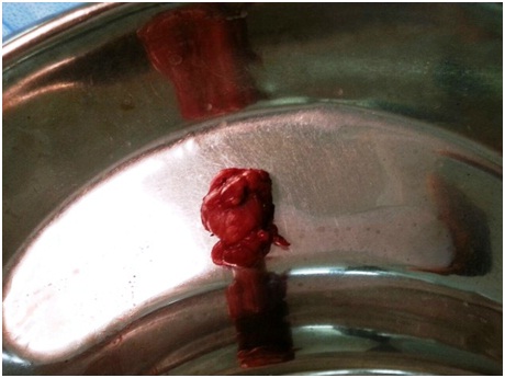
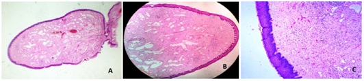
A 12-year-old male child presented to the outpatient services with 6 months history of difficulty in swallowing. There was no history of breathing difficulty, change in voice or history of trauma. The examination of his oropharynx revealed a 2x1 cm small pedunculated polypoid mass arising from the superior pole of right tonsil. The rest of the medical examination and history was non contributory. Excision of the tonsillar mass was done under general anaesthesia and the specimen was sent to the Department of Pathology for histopathological examination [Table/Fig-1]. Macroscopically the pedunculated polypoid mass measured 1.5x0.3x0.2 cm with smooth external surface. Cut section was homogenous tan white and firm in consistency. Microscopic examination showed a polyp lined by non-keratinising stratified squamous epithelium. The subepithelial stroma revealed many variable sized thin walled vascular channels and perivascular infiltration of lymphocytes and plasma cells. These findings suggested a diagnosis of fibroepithelial polyp [Table/Fig-2a-c]. There was no evidence of dysplasia or malignancy. The patient was discharged and postoperative period was uneventful.
Surgical specimen of excised polyp
Photomicrograph showing a polyp lined by stratified squamous epithelium with subepithelial fibrosis, thin walled vascular channels and lymphoplasmacytic infiltration {H&E: (a) whole mount specimen 20x; (b) 40x; (c) 100x}
Discussion
Fibroepithelial polyp is a benign lesion of mesodermal origin and is one of the most common cutaneous lesions. They are also known as acrochordons, soft fibromas or pedunculated lipofibromas. They are usually solitary and reports of multiple, bilateral polyps are extremely rare [1]. Prevelance of these polyps is approximately 12 per 1000 population, with a male predominance [2]. They are benign lesions with extremely low incidence of malignant potential [3]. In a study by Eads TJ et al., among the 1335 specimens submitted as fibroepithelial polyps, only 5 were malignant tumours [4]. In addition to the skin, these polyps can occur anywhere on mucosa, most common on tongue, lips and cheek along the occlusal line [5]. Polyps arising in oral cavity represent a reactive hyperplasia of fibrous connective tissue in response to local irritation or trauma [6]. A 2005 study revealed 61.9% fibrous lesions and 38.1% soft haemorrhagic lesions in the oral cavity. According to their study, fibroepithelial polyp was the most common of the fibrous lesions and of the soft tissue haemorrhagic lesions pyogenic granuloma was most common in oral cavity [7]. The differential diagnosis for fibroepithelial polyp included fibroma, mucocele, giant cell fibroma, peripheral giant cell granulomas, lymphangiomatous polyps, lymphangioma, juvenile angiofibroma and squamous papilloma in the oral cavity including tonsil [5,8]. Many other rare benign lesions were reported in the tonsil [Table/Fig-3] [9]. Infrequently these can occur in the external auditory canal, bronchus or pharyngeal wall [10–12]. These polyps present with an indolent clinical course. However, problems may arise depending on location, especially polyps in the bronchus and pharyngeal wall that cause occlusion and airway obstruction. Surgical excision is the treatment of choice.
Other benign tonsillar lesions [9]
| • Fibroma |
| • Lymphoid polyp |
| • Lymphangiomatous polyp |
| • Lymphangiectatic fibrolipomatous polyp |
| • Hairy polyps (dermoids) |
| • Haemangiomatous hamartoma |
| • Fibrovascular polyp |
| • Lipoma |
| • Neurofibroma |
| • Schwannoma |
| • Plasma cell granuloma |
| • Proteus syndrome |
Conclusion
Fibroepithelial polyps are rare benign tonsillar lesions. Malignant change is very rare. Surgical excision is the treatment of choice. Fibroepithelial polyps can cause choking and airway compromise and should be managed as a medical emergency, securing the airway of paramount importance.
[1]. Lloyd S, Lloyd J, Dhillon R, Chondroid metaplasia in a fibroepithelial polyp of the tongueJ Laryngol Otol 2001 115(8):681-82. [Google Scholar]
[2]. Bouquot JE, Gundlach KK, Oral exophytic lesions in 23616 white Americans over 35 years of ageOral Surg Oral Med Oral Pathol 1986 62(3):284-91. [Google Scholar]
[3]. Quamruzzaman M, Das KK, Khondoker MS, Fibroepithelial polyp/skin tag – unusual presentation - A Case ReportBangladesh Journal of Plastic Surgery 2010 1(1):33-35. [Google Scholar]
[4]. Eads TJ, Chuang TY, Fabre VC, Farmer ER, Hood AF, The utility of submitting fibroepithelial polyps for histological examinationArch Dermatol 1996 132(12):1459-62. [Google Scholar]
[5]. Koppolu P, Mishra A, Kalakonda B, Amara Swapna L, Bagalkotkar A, Macha D, Fibroepithelial polyp excision with laser and scalpel: A comparative evaluationInt J Curr Microbiol App Sci 2014 3(8):1057-62. [Google Scholar]
[6]. Arya S, Singhal P, Vengal M, Patil N, Bhateja S, Fibro-epithelial Polyp: Report of Two Cases with Literature ReviewIJSS Case Reports & Reviews 2015 1(9):9-12. [Google Scholar]
[7]. Bataineh A, Al-Dwairi ZN, A survey of localized lesions of oral tissues: A clinicopathological studyJ Contemp Dent Pract 2005 6(3):30-39. [Google Scholar]
[8]. Kardon DE, Wenig BM, Heffner DK, Thompson LD, Tonsillar Lymphangiomatous Polyps: A Clinicopathologic Series of 26 CasesMod Pathol 2000 13(10):1128-33. [Google Scholar]
[9]. Farboud A, Trinidade A, Harris M, Pfleiderer A, Fibroepithelial polyp of the tonsil: case report of a rare, benign tonsillar lesionJ Laryngol Otol 2010 124(1):111-12. [Google Scholar]
[10]. Tanaka N, Matsunobu T, Shiotani A, Fibroepithelial polyp of the external auditory canal: a case report and a literature reviewCase Rep Otolaryngol 2013 2013:1-4. [Google Scholar]
[11]. Amin PB, Baciewicz F, Benign fibroepithelial polyp arising in the bronchus: a case report and review of the literatureArch Surg 2009 144(11):1081-83. [Google Scholar]
[12]. Mangar W, Jiang D, Lloyd RV, Acute presentation of a fibroepithelial polypJ Laryngol Otol 2004 118(9):727-29. [Google Scholar]