Multifocal Bullous Fixed Drug Erruption Due To Phenytoin: A Lesson Learned!
Ankur Jain1, Naresh Gupta2
1 Post Graduate Student, Department of Medicine, Maulana Azad Medical College, New Delhi, India.
2 Director Professor and Head, Department of Medicine, Maulana Azad Medical College, New Delhi, India.
NAME, ADDRESS, E-MAIL ID OF THE CORRESPONDING AUTHOR: Dr. Ankur Jain, Z.P-1, Maurya Enclave, Pitampura, New Delhi-110088, India.
E-mail: drankur589@yahoo.in
Antiepileptic drugs (AED) are a common culprit of cutaneous eruptions in clinical practice. Phenytoin, lamotrigine and carbamazepine are the commonest offenders. Maculopapular eruptions are the most frequently reported events. However, multifocal bullous fixed drug eruptions have rarely been described in association with AED use. The risk factors for skin rash including its association with the rate of drug administration are unclear in the literature. We report a case of a young alcoholic man, on long term phenytoin therapy since 3 years, who presented to our emergency department with a breakthrough seizure episode. Patient’s routine investigations including serum biochemistry, imaging and toxicology screen were normal. Patient was found to have sub-therapeutic serum phenytoin levels and was prescribed loading with intravenous phenytoin (15mg/kg body weight), which was mistakenly infused at a rapid rate (60mg/minute). Patient developed multifocal bullous lesions over muco-cutaneous regions after 6 hours of drug administration which healed after its discontinuation leaving behind residual hyperpigmentation. Patient was managed conservatively, switched to oral levetiracetam and discharged in a stable condition after one week of hospital stay. Present case highlights a yet uncommon reaction to a commonly used drug and tries to establish the relation between rate of drug infusion and the risk of skin reaction.
Anti-epileptic drugs, Generalized tonic clonic seizures, Stevens-Johnson syndrome
Case Report
We report a case of a 35-year-old man (weight 60 kg), who presented to the emergency department with generalized tonic clonic seizures (GTCS). Patient’s review of old records revealed that he was a regular alcoholic and had a past history of subdural haematoma (SDH) three years back, which he developed after a fall and was on treatment with oral phenytoin since then for seizure prophylaxis at a dose of 300 mg once daily. Further history from his relative revealed that he was non compliant with his anti-seizure medications and continued to consume alcohol regularly after his last hospital admission. Patient was not following up in between and hence his serum phenytoin levels were not known at the time of current presentation. History was negative for other substance abuse, other over-the-counter medication intake, fever, trauma, headache, vomiting and photophobia. Patient was in post-ictal state at the time of examination. He was afebrile to touch and his vitals were: blood pressure-110/70 mm hg, pulse rate- 98 beats per minute. General examination was remarkable for the mild pallor and poor oral hygiene. There was no icterus, cyanosis, clubbing, lymphadenopathy or pedal oedema. Nervous system examination was unremarkable for neck rigidity or meningeal signs, pupils were normal size and normally reacting to light and bilateral planters were flexor. Rest of the systemic examination was unrevealing. Patient was turned in left lateral position, airway was protected and immediate venous access was achieved. Patient’s venous blood sample was sent for complete blood counts, phenytoin levels, ionized calcium, magnesium, sodium, random blood glucose, liver and kidney function tests and toxicology screen. Patient was also planned for non-contrast CT scan of the head.
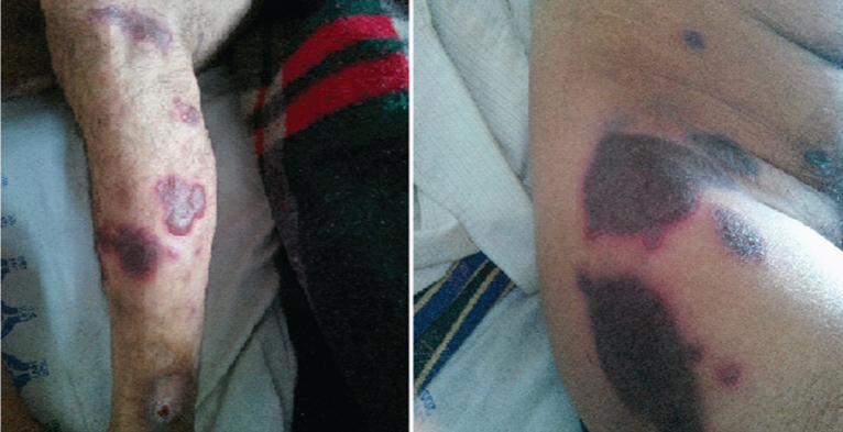
Patient was prescribed intravenous phenytoin 900 mg (15mg/kg body weight) at rate of 30mg/minute awaiting his investigations. However, the prescribed dose was mistakenly administered to the patient within 15 minutes i.e. @60mg/minute by the nursing staff. Patient regained his consciousness after 30 minutes and had no focal neurological deficits. His investigations revealed- Hb-106 gm/L, total leucocyte counts- 5.4 X 10⁹/L, platelet counts-354 X 109/L, serum sodium-140mmol/L, potassium- 3.8mmol/L, calcium-238mmol/L, random blood glucose-92 mg/dL. Liver and kidney function tests, Electrocardiogram, chest X-ray were normal and CT scan of the head revealed old chronic SDH. Patient’s serum phenytoin levels were 32 micromoles/Litre (therapeutic range: 40-79 micromoles/Litre). However, after about 6 hours, patient developed a generalized eruption, consisting of well demarcated, itchy, violaceous macules with a central flaccid bulla and a negative nikolsky’s sign. Lesions were seen on arms, legs, thigh, groin, upper back, genitals and oral mucosa. Dermatology opinion was taken and a possibility of multifocal bullous fixed drug eruption (FDE), likely due to phenytoin was suggested [Table/Fig-1,2]. Phenytoin was discontinued and was replaced by oral levetiracetam (500 mg twice daily). Lesions were managed conservatively by oral levocetrizine (5mg once daily) and topical emollients. Lesions resolved in 7 days with a residual hyperpigmentation at the original site without any sequel. Patient was discharged after one week of hospital stay on oral levetiracetam and was advised to quit alcohol and is following up in our outpatient clinic being currently asymptomatic.
Figure showing well demarcated violaceous plaques with a central flaccid bulla on the back of the patient
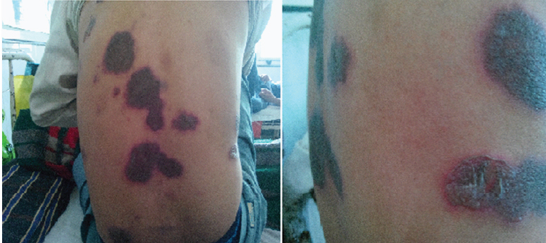
Figure showing well demarcated violaceous plaques with central bulla on the patient’s legs and thigh, groin and genital region
Discussion
Phenytoin, a member of hydantoin group, is one of the most commonly used anti-epileptic and anti-arrhythmic since its first description in 1938 by Merritt and Putnam. Anti-epileptics are notorious for causing cutaneous eruptions [1]. Sharma et al., found an overall prevalence of antiepileptic drug (AED) rash to be 2.5%, the highest rate being seen with phenytoin, lamotrigine and carbamazepine [2,3]. Between 5%-10% of patients taking phenytoin develop cutaneous reactions to it, and according to a review, the risk was 19.4% for the patients with head injury being given phenytoin for seizure prophylaxis, the incidence being higher after the loading dose [4,5]. Apart from hirsuitism and gum hypertrophy, reported reactions to phenytoin includes maculopapular eruptions, generalized exfoliative dermatitis, Stevens-Johnson syndrome (SJS), toxic epidermal necrolysis (TEN), erythema multiforme, vasculitis, follicular or pustular eruptions, fixed drug eruptions (FDE), angioedema, hypersensitivity reactions, purple hand syndrome, cutaneous necrosis, pigmentation changes, porphyria and linear IgA bullous disease [1].
Maculopapular eruption was the most commonly reported event (71.4%), followed by SJS (14.3%), fever (4%), generalized exfoliative dermatitis, TEN, vasculitis and agranulocytosis (2.4% each) [6]. Cutaneous reactions to phenytoin have been classified as Type B i.e. bizarre with unknown pathogenesis [7]. FDE represents about 10% of all adverse drug reactions. FDE presents as sudden onset of well demarcated itchy erythematous macules, evolving rapidly into violaceous plaques over muco-cutaneous regions and characteristically recurs at the same site, 30 minutes to eight hours after the drug re-challenge and heals with residual hyperpigmentation [8]. The exact incidence of FDE due to phenytoin is unknown; however, in a study of patients with FDE, phenytoin was the culprit in 4% of cases and commonly caused generalized reactions [9]. Multifocal bullous FDE is a rare variant of FDE, which is often confused with SJS/TEN complex. Differences between multifocal bullous FDE and SJS/TEN are tabulated in [Table/Fig-3] [8]. The risk factors for phenytoin induced skin reactions are not clearly defined.
Comparison between clinical features and skin biopsy findings of multifocal bullous FDE and SJS/TEN complex
| Parameter | Multifocal FDE | SJS/TEN |
|---|
| Age of the patient | Older | Younger |
| Constitutional symptoms | Less | More |
| Mucosal involvement | Less | More |
| Flag sign | Absent | Present |
| Eosinophils, neutrophilsand melanophages insuperficial and deepinfiltrates in skin biopsy | Present | Absent |
| Recurrence after rechallenge | Characteristic finding | Incompatible with life |
In one study, higher initial serum levels of phenytoin were associated with a greater risk of developing a skin rash. However, since, the adverse cutaneous reactions are seen early in the course of initiating phenytoin (usually within 3 weeks) and chronic side effects of phenytoin are not associated with rash, it is possible that an intermediate metabolite of phenytoin produced during initial treatment is responsible for skin rash, levels of which are higher after loading dose and are reduced after chronic treatment due to auto-induction of metabolism [10].
In this regard, a potential role of an arene oxide intermediates have been proposed for phenytoin induced eruptions [11]. Prior history of another AED rash has been identified as a risk factor for AED rash [3]. The relation between rate of drug administration and development of skin rash has not been widely reported. The maximum rate of infusion of intravenous phenytoin is recommended to be 50mg/minute to avoid cardiovascular side effects.
We could find only one case report whereby a 52-year-old lady, already on long term phenytoin treatment developed a rash after rapid oral loading with phenytoin [12]. Our patient was on phenytoin treatment for past 3 years. However, he was an alcoholic and had poor compliance to antiepileptic medications. Induction of phenytoin’s metabolism by alcohol and poor compliance were responsible for his sub therapeutic serum drug levels resulting in a breakthrough seizure. Rapid intravenous loading of phenytoin done mistakenly in the emergency department was followed by the development of multifocal bullous FDE which resolved after drug discontinuation.
Conclusion
Current case not only highlights a rare adverse event to a commonly used drug, but also alerts the practicing internists that rapid loading of a seizing patient, even if already on chronic antiepileptic, can lead to a widespread cutaneous reaction and thereby needs caution. Cautious use of phenytoin in the emergency department is emphasized in this report.
Financial or Other Competing Interests
The authors disclose no potential conflicts of interests.
[1]. Scheinfeld N, Phenytoin in cutaneous medicine: its uses, mechanisms and side effectsDOJ 2003 9:6 [Google Scholar]
[2]. Sharma VK, Vatve M, Sawhney IM, Kumar B, Clinical spectrum of drug rashes due to antiepilepticsJ Assoc Physicians India 1998 46:595-97. [Google Scholar]
[3]. Arif H, Buchsbaum R, Weintraub D, Koyfman S, Salas-Humara C, Bazil CW, Comparison and predictors of drug rash associated with 15 antiepileptic drugsNeurology 2007 68:1701-09. [Google Scholar]
[4]. Hebert AA, Ralston JP, Cutaneous reactions to anticonvulsant medicationsJ Clin Psychiatry 2001 62(Suppl 14):22-26. [Google Scholar]
[5]. Rapp RP, Norton JA, Young B, Tibbs PA, Cutaneous reactions in head-injured patients receiving phenytoin for seizure prophylaxisNeurosurgery 1983 13:272-75. [Google Scholar]
[6]. Leong KP, Chng HH, Allergic reactions to phenytoin in a general hospital in SingaporeAsian Pac J Allergy Immunol 1996 14:65-68. [Google Scholar]
[7]. Spielberg SP, Jordan GB, Blake DA, Goldstein DA, Herlong HF, Predisposition to phenytoin hepatotoxicity assessed in vitroN Engl J Med 1981 305:722-27. [Google Scholar]
[8]. Patro N, Panda M, Jena M, Mishra S, Multifocal Fixed Drug Eruptions: A case seriesInt J Pharm Sci Rev Res 2013 23:63-66. [Google Scholar]
[9]. Sharma VK, Dhar S, Gill AN, Drug related involvement of specific sites in fixed eruptions: a statistical evaluationJ Dermatol 1996 23:530-34. [Google Scholar]
[10]. Chadwick D, Shaw MD, Foy P, Rawlins MD, Turnbull DM, Serum anticonvulsant concentrations and the risk of drug induced skin eruptionsJ Neurol Neurosurg Psychiatry 1984 47:642-44. [Google Scholar]
[11]. Dwivedi R, Gogtay N, Kharkar V, Amladi S, Kshirsagar N, In-vitro lymphocyte toxicity to a phenytoin metabolite in phenytoin induced cutaneous adverse drug eruptionsIndian J Dermatol Venereol Leprol 2004 70:217-20. [Google Scholar]
[12]. Smetana KS, Suda KJ, Hamilton LA, Fixed Drug Eruption in an Epileptic Patient Previously Receiving Treatment with Phenytoin for Seven YearsJ Inves Med high impact case Rep 2013 :1-3. [Google Scholar]