The Combined use of Diode LASER & Conscious Sedation in the Excision of Pyogenic Granuloma in A Nine-Year-Old Patient
Shankar T Gokhale1, N. Sathyajith Naik2, Deepak Singla3, Akanksha Singh4, Deepankar Bhattacharya5
1 Professor, Department of Periodontics, Institute of Dental Sciences, Bareilly, Uttar Pradesh, India.
2 Professor and Head, Department of Pedodontics, Institute of Dental Sciences, Bareilly, Uttar Pradesh, India.
3 Post Graduate Student, Department of Periodontics, Institute of Dental Sciences, Bareilly, Uttar Pradesh, India.
4 Post Graduate Student, Department of Periodontics, Institute of Dental Sciences, Bareilly, Uttar Pradesh, India.
5 Post Graduate Student, Department of Pedodontics, Institute of Dental Sciences, Bareilly, Uttar Pradesh, India.
NAME, ADDRESS, E-MAIL ID OF THE CORRESPONDING AUTHOR: Dr. Deepak Singla, A – 204, Ext – 2, Shalimar Garden, Sahibabad, Ghaziabad- 201005, Uttar Pradesh, India. E-mail : Deepak.singla2214@gmail.com
This case report is to comprehensively review N2O/ O2 inhalational sedation in the context of conscious sedation for treating a nine-year-old patient with pyogenic granuloma. The excision was carried out by the use of diode laser. The six month postoperative follow up showed complete resolution of the lesion and increased patient acceptance for the future treatment. The use of laser minimizes the pain during the surgery and postoperatively and suturing was not required. Therefore this case report emphasizes the use of combined treatment modalities to increase patient comfort and to obtain a better function and aesthetics of the oral cavity.
Benign, Exophytic, Hamartoma
Case Report
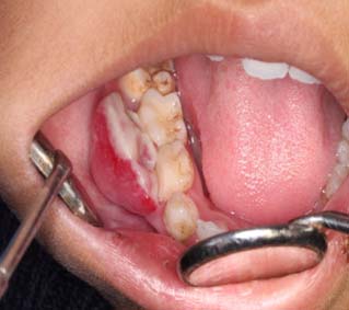
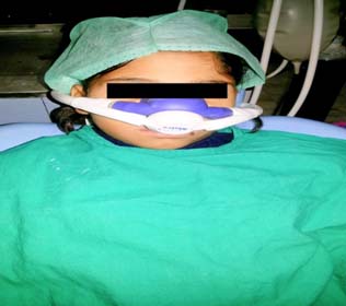
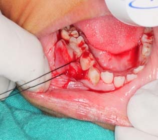
A nine-year-old female patient reported to the Department of Periodontics with a chief complaint of painless swelling in the lower right back teeth region since 1 – 1.5 years [Table/Fig-1]. Patient’s guardian gave the history that the lesion gradually increased in size to a certain extent. On inspection the growth was shiny red in colour present in relation to 46 tooth region measuring about 11mmx 9mm. On palpation it was soft in consistency and was having pedunculated base. After various diagnostic test: radiographic investigations, blood investigations it was provisionally diagnosed as pyogenic granuloma present in relation to 46 tooth region. After treatment planning the patient’s guardian was explained about the treatment and a written consent was obtained. The routine oral prophylaxis was carried out one month prior to the surgery and the patient was uncooperative during the treatment. Therefore it was planned to carry out the surgery under conscious sedation for better patient compliance. The patient was prepared and sedated by using nitrous oxide in 50:50 ratio with oxygen [Table/Fig-2]. After successful sedation was achieved the lesion was held by passing suture through it [Table/Fig-3] and after taking all the safety measures the diode laser of 980 nm wavelength was activated and was applied to the pedunculated base in sweeping motions to avoid over heating or charring of the tissues [Table/Fig-4]. After excision, the lesion was measured using UNC–15 periodontal probe (measuring about 11mm x 9mm) [Table/Fig-5] and the complete debridement of the area was done to prevent re-occurrence of the lesion. After re-evaluation vitamin E capsules were applied to enhance the healing and the patient was given postoperative instructions. The patient was recalled at one week [Table/Fig-6] and six months intervals [Table/Fig-7]. The excised lesion was sent for the histopathological examination for the confirmatory diagnosis. The patient was not kept on antibiotics regimen because the surgery was carried out in a fumigated room with the help of LASER which itself creates a sterilised environment at the surgical site. The patient neither had the painkillers as LASER help in release of the endorphins which help in reducing the pain [1].
Patient under conscious sedation
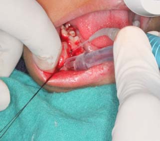
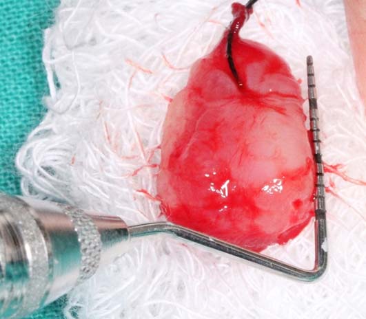
Excised growth measuring 11mm x 9 mm
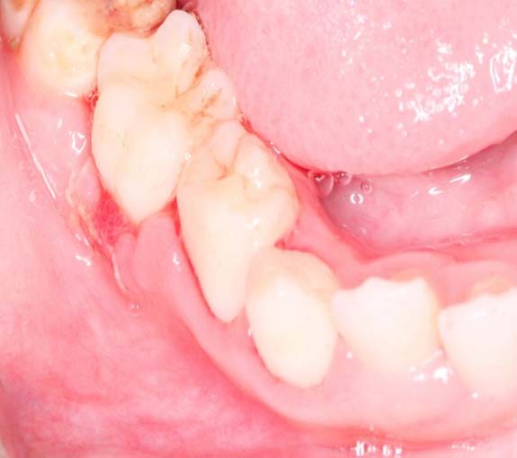
One week postoperative view
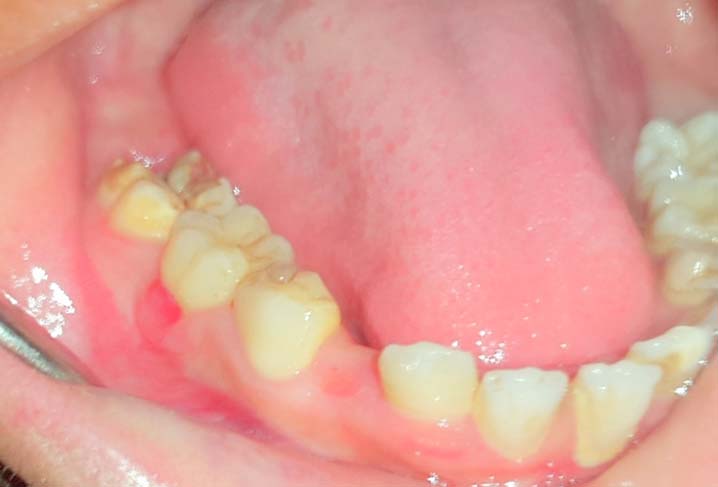
Six Months postoperative view
Histopathological Examination
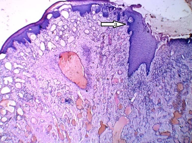
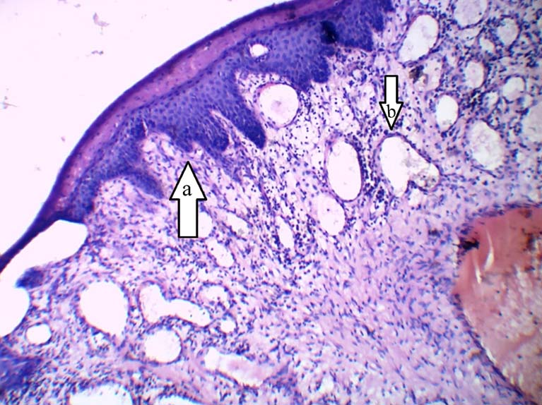
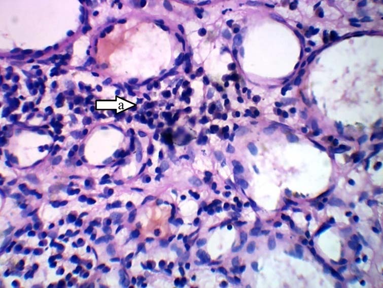
Under scanner view: ([Table/Fig-8]{4x}) Single piece of tissue with discontinuous epithelium overlying inflamed, fibro-cellular and vascular connective tissue stroma. Under low power & higher magnification: {[Table/Fig-9] (10x), [Table/Fig-10] (40x)} Epithelium was stratified squamous parakeratinized type. Connective tissue stroma comprises of loose to dense bundles of collagen fibres with predominantly plump shaped fibroblasts. Numerous small to large endothelial lined vascular spaces (some with RBC’s) and budding capillaries along with endothelial cell proliferation are evident. Diffuse chronic inflammatory infiltrate predominantly comprising of lymphocytes is evident. Based on these histopathological features the final diagnosis was given as pyogenic granuloma.
a) +Discontinuous epithelium over inflamed connective tissue stroma
a) Stratified squamous parakeratinised epithelium. b) Budding capillaries
a) Inflammatory cell Infiltrate with predominant lymphocytes
Discussion
Pyogenic granuloma is a local, benign and vascular lesion which occurs in response to long standing, mild, local irritants. It occurs due to the fibrovascular proliferation of connective tissue [2]. Clinically, it is an exophytic lesion which appears as small, red erythematous papules present on a sessile or pedunculated base. Clinically the lesion is slow in origin, asymptomatic in nature and usually painless. The size varies in diameter from a few millimetres to several centimetres [3]. Frequently, the lesion arises on the gingiva of the maxillary anterior region, but also it has been seen on the lips, tongue, and buccal mucosa [4]. Treatment consists of surgical removal in toto, extending the incision up to the periosteum and periodontal ligament and involving the associated connective tissue as well as any other aetiologic factor present. Providing dental treatment to the young patients is challenging because the child’s may not be co-operative for basic treatment in the mouth [5].
Conscious sedation is a technique which has opened new path to manage uncooperative children in almost all the allied disciplines of healthcare and has rightly become popular with the busy modern day dentist in the developed countries [6]. Nitrous oxide (NO) is being used from decades in the outpatient department as a safe and reliable method of sedation. It is a colourless and sweet smelling gas. NO possesses various properties, it act as sedative, analgesic, and anxiolytic gas when mixed with oxygen in a 50/50 ratio. It can also be combined with opioids or benzodiazepines. It diffuses very rapidly, which provides a sedation which is rapid onset and short in duration [7]. PG is relatively common on the gingiva of the anterior maxillary region, probably due to the prevalence of local irritating factors [4]. In the present case report the PG was present on the posterior aspect of the mandible and the main aetiological factor was the local deposits as a source of irritation at the particular site. Sometimes it often seems to follow a minor injury and grows rapidly over a period of a few weeks [8]. Increased production of angiogenic growth factor also plays a key role in the progression of the lesion [9]. Lesion removal is indicated to alleviate any bleeding, discomfort, cosmetic distress, and incorrect or retarded tooth eruption [10].
Conclusion
In this case the treatment of a nine-year-old patient with PG was done under conscious sedation and laser technique to minimize the pain, discomfort and postoperative complications. The patient showed uneventful healing and satisfactory postoperative results. Adequate amount of sedatives and analgesic medications can make painful situations tolerable. Specifically, conscious sedation is a method which is available to dentists that can improve patient acceptability of unpleasant procedures and the additional use of laser may further reduce the pain and provide a clean field for better treatment results.
[1]. Lim HM, Lew KK, Tay DK, A clinical investigation of the efficacy of low level laser therapy in reducing orthodontic post adjustment painAmerican J Orthod Dentofac Orthoped 1995 108:614-22. [Google Scholar]
[2]. Jensen JL, Barr RJ, Lesions of the facial skinDifferential diagnosis of Oral and Maxillofacial Lesions 1997 5th ednSt Louis, MOMosby Books:549-550. [Google Scholar]
[3]. Waldron CA, Odontogenic cyst and tumors. In: Neville, BW, Damm, DD, Allen, CM, Bouquot, JE, edsOral and Maxillofacial Pathology 1995 1st ednPhiladelphia, PAW.B. Saunders:371-73. [Google Scholar]
[4]. Kneafsey L, Hughes C, Quadhelix appliance therapy resulting in pyogenic granuloma of the tongueDent Update 2002 29:462-63. [Google Scholar]
[5]. Shapira J, Holan G, Botzer E, Kupieztky A, Tal E, Fuks AB, The effectiveness of midazolam and hydroxyzine as sedative agents for young pediatric dental patientsJ Dentist Child 1996 63:421-25. [Google Scholar]
[6]. Milnes AR, Maupome G, Cannon J, Intravenous sedation in Pediatric dentistry using Midazolam, Nalbuphine and DroperidolPediatr Dentist 2000 22:113-19. [Google Scholar]
[7]. Gamis AS, Knapp JF, Glenski JA, Nitrous oxide analgesia in a pediatric emergency departmentAnn Emerg Med 1989 18:177-81. [Google Scholar]
[8]. Espinoza I, Rojas R, Aranda W, Gamonal J, Prevalence of oral mucosal lesions in elderly people in Santiago, ChileJ Oral Pathol Med 2003 32:571-75. [Google Scholar]
[9]. Damm DE, Fantasia JE, Exophytic and ulcerated nodule of the tongue: Pyogenic granulomaGen Dent 2005 53:447-49. [Google Scholar]
[10]. Meissner M, Spieth K, Loffler T, Pyogenic granulomaJ Dtsch Dermatol Ges 2005 3:1007-08. [Google Scholar]