Glandular Odontogenic Cyst of Mandible: A Rare Entity
Ankur Mittal1, Vikram Narang2, Gursheen Kaur3, Neena Sood4
1 Resident, Department of Pathology, Dayanand Medical College and Hospital, Ludhiana, Punjab, India.
2 Assistant Professor, Department of Pathology, Dayanand Medical College and Hospital, Ludhiana, Punjab, India.
3 Resident, Department of Medicine, Dayanand Medical College and Hospital, Ludhiana, Punjab, India.
4 Professor and Head, Department of Pathology, Dayanand Medical College and Hospital, Ludhiana, Punjab, India.
NAME, ADDRESS, E-MAIL ID OF THE CORRESPONDING AUTHOR: Dr. Vikram Narang, Assistant Professor, Department of Pathology, DMCH, Ludhiana, Punjab, India.
E-mail: drvikramnarang@yahoo.com
Glandular odontogenic cyst (GOC) is a rare developmental odontogenic cyst. It is a slow growing and asymptomatic swelling, usually affecting middle aged men and has tendency to reoccur. Here, we report a case of GOC in the anterior portion of mandible diagnosed by histopathology.
Developmental cysts, Jaw cysts, Tumour
Case Report
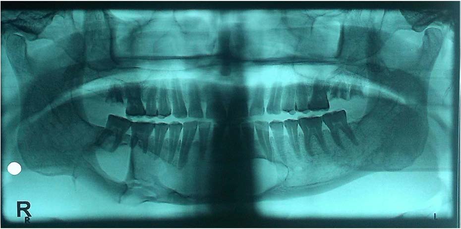
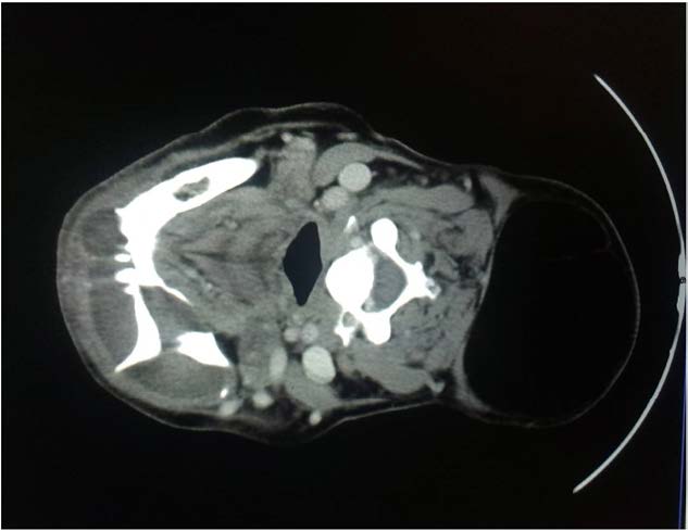
A 17-year-old female presented with a history of pain and swelling in the right mandibular region of 5 year duration. On examination, tender facial swelling measuring 4x6x3 cm was noted. It was associated with bleeding gums and foul smelling discharge. Panoramic view radiograph showed a well defined radiolucency, with irregular margins in the mandible [Table/Fig-1]. A well-defined multilocular radiolucent lesion in the region of anterior portion of the mandible was appreciated on CT scan [Table/Fig-2]. She underwent hemimandibulectomy with clinico-radiological suspicion of ameloblastoma.
Panoramic radiograph showing large multilocular radiolucency
CT section showing well defined multilocular lesion in the anterior portion of mandible
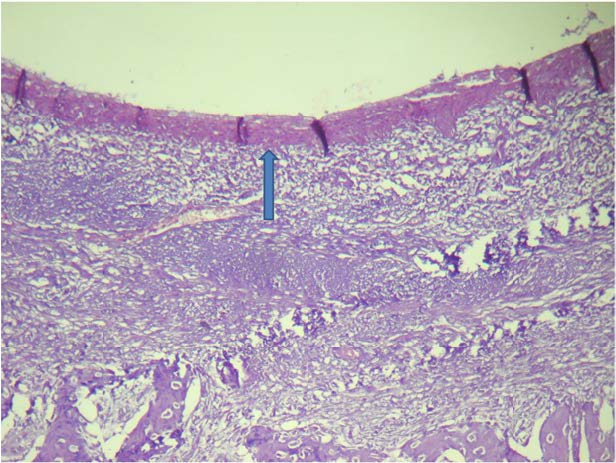
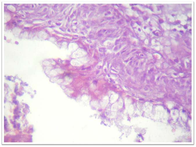
Grossly external surface was unremarkable and on cut section a cystic cavity was identified in the mandible. Histology revealed fragments of cyst wall lined by stratified squamous epithelium of varying thickness along with epithelial whorls in few areas and goblet cells, suggestive of GOC [Table/Fig-3,4 and 5].
Cyst cavity lined by stratified squamous epithelium (arrow) (H&E 100X)
Squamous epithelial lining with numerous goblet cells (H&E 400X)
Goblet cells showing PAS positity (PAS 200X)

Discussion
GOC is a rare lesion with prevalence ranging from 0.012 to 1.3% of all jaw cysts [1]. In 1987, Padayachee and Van Wyk described two cystic lesions with histologic features that did not fit into the known classification of cysts, who called “sialo-odontogenic cyst” [2]. One year later Gardner et al., described eight similar cases and suggested the term “glandular odontogenic cyst” [3]. In 1992, the WHO named the GOC an independent pathologic entity and classified it as a developmental odontogenic cyst [4]. Only 50 cases of GOC have been reported in literature so far [5]. Usually it presents as a painless lesion, but can be painful if neurovascular bundles are compressed or infected secondarily. Most authors agree that there are no radiographic features specific of GOC [1]. Therefore, histopatholgy is essential for diagnosis.
GOC usually occurs in middle age men and only few cases have been reported in females. The differential diagnosis includes lateral periodontal cyst, botryoid cysts, keratocysts, residual cysts, the central mucoepidermoid carcinoma and the ameloblastoma. These can be differentiated histologically [1].
Microscopically, GOC is characterized by a cyst wall lined by non-keratinized epithelium, with papillary projections, nodular thickenings, mucous filled clefts, and ’mucous lakes’. It also includes cuboidal basal cells, which may sometimes be vacuolated. There is histological similarity with central mucoepidermoid carcinoma, however, histological features of GOC like epithelial whorls, ciliated cells and intraepithelial microcytes or duct like structures are not seen in central mucoepidermoid carcinoma and help to make the definitive diagnosis [6–8]. Similarly, presence of ciliated epithelium in GOC helps to differentiate GOC from lateral periodontal cyst and botryoid cysts [9]. Certain authors have suggested that GOC shows positive immuno staining for CK-18, CK-19 and Ki-67 and decreased positivity for p-53, which helps to differentiate these tumours especially from central mucoepidermoid carcinoma [10].
Treatment of GOC includes curettage and enucleation, although some authors believe marginal resection to be a more reliable treatment, due to tendency of the cyst to recur after curettage and enucleation. However, no malignant transformation potential seen even though an increased Ki-67 index reported in certain studies [5,6,11].
Conclusion
Glandular odontogenic cyst is a rare lesion, and its differential diagnosis includes both benign and malignant lesions. The radiopathological correlation is required for definitive diagnosis.
[1]. Manzini M, Deon C, Corte LD, Bertotto JC, Abreu LB, Glandular odontogenic cyst: an uncommon entityBraz J Otorhinolaryngol 2009 75:320 [Google Scholar]
[2]. Shahnaz ST, Freny RK, Archana Y, Kaustubh S, Subodh S, Glandular odontogenic cyst: a case reportImaging Sci Dent 2014 44:75-79. [Google Scholar]
[3]. Noke C, Raubenheimer EJ, The glandular odontogenic cyst: clinical and radiological features; review of the literature and report of nine casesDentomaxillofacial Radiology 2002 31:333-38. [Google Scholar]
[4]. Marco M, Andrea S, Antonio S, Maurizio P, Lorenzo LM, Antonio Z, Glandular odontogenic cyst: review of literature and report of a new case with cytokeratin-19 expressionThe Open Dentistry Journal 2014 8:1-12. [Google Scholar]
[5]. Anuthama K, Sherlin HJ, Karthikeyan R, Anuja N, Premkumar P, Glandular Odontogenic Cyst: Report of Two Cases and Review of LiteratureHead and Neck Pathol 2009 3:153-58. [Google Scholar]
[6]. Gardner GD, Morency R, The glandular odontogenic cyst, a rare lesion that tends to recurJ Can Dent Assoc 1993 59:929 [Google Scholar]
[7]. Regezi JA, Odontogenic Cysts, Odontogenic Tumours, Fibroosseous, and Giant Cell Lesions of the JawsModern Pathology 2002 15:331-41. [Google Scholar]
[8]. Kaplan I, Anavi Y, Manor R, Sulkes J, Calderon S, The use of molecular markers as an aid in the diagnosis of glandular odontogenic cystOral Oncol 2005 41:895-902. [Google Scholar]
[9]. De Soussa SO, Cabexas NT, De Oliveira PT, De Araujo VC, Glandular odontogenic cyst report of a case with cytokeratin expressionOral Surg Oral Med Oral Pathol Oral Radiol Endod 1997 83:478-83. [Google Scholar]
[10]. Pires FR, Chen SY, Perez DEC, Almedia OP, Kowalski LP, Cytokeratin expression in central mucoepidermoid carcinoma and glandular odontogenic cystOral Oncol 2004 40:545-51. [Google Scholar]
[11]. Savage NW, Joseph BK, Monsour PA, Young WG, The glandular odontogenic jaw cyst: Report of a casePathology 1996 28:370-72. [Google Scholar]