Oxidative Stress in Obese Postmenopausal Women: An Additive Burden for Atherosclerosis
Rajesh Bhat Uppoor1, Aparna Rajesh2, Mukund Prathivadibhayankaram Srinivasan3, Bhaskaran Unnikrishnan4, Ramesh Holla5
1 Assistant Professor, Department of Cardiology, Kasturba Medical College (Manipal University), Mangalore, Karnataka, India.
2 Associate Professor, Department of Obstetrics and Gynaecology, K. S. Hegde Medical Academy, Mangalore.
3 PhD Research Scholar, Department of Internal Medicine, Kasturba Medical College (Manipal University), Mangalore, Karnataka, India.
4 Professor, Department of Community Medicine, Kasturba Medical College (Manipal University)Mangalore, Karnataka, India.
5 Assistant Professor, Department of Community Medicine, Kasturba Medical College (Manipal University), Mangalore, Karnataka, India.
NAME, ADDRESS, E-MAIL ID OF THE CORRESPONDING AUTHOR: Dr. Rajesh Bhat Uppoor, Assistant Professor, Department of Cardiology, Kasturba Medical College (Manipal University), Mangalore, Karnataka-575001, India.
E-mail: drrajeshu@gmail.com
Introduction
Coronary Artery Disease are on the rise in the general population and is the leading cause of death in both men and women. The impact of CAD is underappreciated in younger women when compared to men. Women have unique risk factors for CAD and postmenopausal women are at higher risk of developing CAD when compared to normal menstruating women.
Aim
The aim of our study was to find out the difference in oxidative stress levels between obese postmenopausal women and normal menstruating women, also to compare the same in normal weight postmenopausal women.
Materials and Methods
Thirty one normal and 29 obese postmenopausal women with age more than 45 years who visited obstetrics and gynaecology outpatient department for general clinical evaluation at a tertiary care centre were recruited in this cross-sectional study. Thirty normal menstruating women were compared. Anthropometric measurements were recorded and the body mass index was calculated. Serum Malondialdehyde and superoxide dismutase was measured using a spectrophotometer.
Results
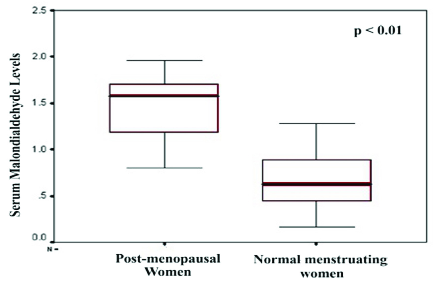
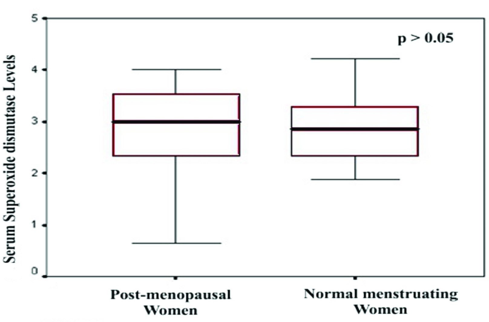
There was a significant difference in mean MDA levels in postmenopausal women (1.477 ± 0.359) when compared to normal menstruating women (0.666 ± 0.302) (p < 0.01). There was no significant difference in mean SOD levels in postmenopausal women (2.836 ± 0.899) when compared to normal menstruating women (2.986 ± 0.686) (p > 0.05). Also, there was a significant increase between mean MDA levels in obese postmenopausal women (2.48 ± 0.52) when compared to normal weight postmenopausal women (1.65 ± 0.36) (p < 0.01). There was a significant difference between mean SOD levels in obese postmenopausal women (1.36 ± 0.96) and normal weight postmenopausal women (2.56 ± 1.03) (p < 0.01).
Conclusion
The oxidative stress was higher in obese postmenopausal women when compared to normal weight postmenopausal women and normal menstruating women.
Coronary artery disease, Cardiac risk factor
Introduction
Coronary Artery Disease are on the rise in the general population and is the leading cause of death in both men and women [1]. The rise of CAD risk in the population is attributed to lifestyle diseases such as diabetes and hypertension. In women the incidence of CAD is low during their reproductive stages; however the presence of diabetes negates this protection [2]. The impact of CAD is underappreciated in younger women when compared to men [2]. Women have unique risk factors for CAD [3,4] and postmenopausal women are at higher risk of developing CAD when compared to normal menstruating women [2,5–7]. In general postmenopausal women are subjected to oxidative stress which is attributed to aging and other related factors [8]. Oxidative stress has known to play crucial role in many human diseases and atherosclerosis is one of them. The reactive oxygen species (ROS) and free radicals produced due to oxidative stress are known to mediate the atherogenesis [9,10].
Studies have shown that age, smoking, diabetes, and body mass index are highly associated with systemic oxidative stress [11]. Studies have also reported that, obesity is known to cause oxidative stress which further contributes for endothelial dysfunction in obese individuals [12]. The presence of oxidative stress in postmenopausal women has been studied earlier [13,14]. However with respect to body weight in postmenopausal women has not been studied so far. If a significant association is established between body weight and oxidative stress in obese postmenopausal women, it will help us to identify high risk individuals who can be further subjected to intense lifestyle modification and diet control, where in future cardiovascular complications can be averted in such individuals.
Thus the present study was designed to find out the difference in oxidative stress levels as measured by serum Malondialdehyde (MDA) between obese postmenopausal women and normal menstruating women, also to compare the same in normal weight postmenopausal women.
Materials and Methods
Sixty postmenopausal women with age more than 45 years who visited obstetrics and gynecology outpatient department for general clinical evaluation at a tertiary care center with no serious gynecological illness were recruited in this cross-sectional study after obtaining the informed consent. Thirty normal menstruating women were compared. Based on body mass index the postmenopausal women were further divided into two groups. The BMI less than or equal to 23 kg/m2 were considered as normal weight and BMI greater than 25 kg/m2 were defined as obese as per Indian standards [15]. Among the 60 postmenopausal women selected for the study 31 had normal weight and 29 were obese. Subjects with hypertension, diabetes, surgically induced menopause, renal diseases, and on exogenous antioxidant supplement were excluded from the study. The study was approved by institutional human ethic committee.
Age and anthropometric measurements were recorded as per WHO norms [16]. The body mass index was calculated. Venous blood samples were collected in a vacutainer. The blood was allowed to clot for 30 min and then centrifuged at 2000 × g for 15 min for separation of serum. The serum then assayed for MDA by thiobarbituric acid method [17] and SOD by nitro-blue tetrazolium method [18] and was measured using a spectrophotometer.
Statistical Analysis
Student t-test was performed to find out whether there is a significant difference between mean MDA and SOD between the groups. p< 0.05 was considered statistically significant. Data were analyzed using SPSS Version 16 (SPSS, Chicago, IL, USA).
Results
The mean SOD level was 2.836 ± 0.899 in postmenopausal women; the mean SOD level was 2.986 ± 0.686 in normal menstruating women [Table/Fig-1]. The mean MDA level was 1.477 ± 0.359 in postmenopausal women; the mean MDA level was 0.666 ± 0.302 in normal menstruating women [Table/Fig-2]. There was a significant difference in mean MDA levels in postmenopausal women when compared to normal menstruating women (p < 0.01) [Table/Fig-3]. There was no significant difference in mean SOD levels in postmenopausal women when compared to normal menstruating women (p > 0.05) [Table/Fig-4].
Mean levels of SOD and MDA in postmenopausal women and normal menstruating women.
| Variable | PostmenopausalWomen (n =60) | Normal menstruatingWomen (n =30) | p-value |
|---|
| SOD |
| Mean ± S.D. | 2.836 ± 0.899 | 2.986 ± 0.686 | p>0.05 |
| MDA |
| Mean ± S.D. | 1.477 ± 0.359 | 0.666 ± 0.302 | p<0.001 |
Mean levels of SOD and MDA in Obese postmenopausal women and normal weight postmenopausal women.
| Variable | ObesePostmenopausalWomen (n =29) | Normal weightPostmenopausalWomen (n =31) | p-value |
|---|
| MDA |
| Mean ± S.D. | 2.48 ± 0.52 | 1.65 ± 0.36 | p<0.01 |
| SOD |
| Mean ± S.D. | 1.36 ± 0.96 | 2.56 ± 1.03 | p<0.001 |
Comparison of serum Malondialdehyde (MDA) level between post menstrual women and normal menstruating women
Comparison of serum Superoxide dismutase (SOD) level between post menstrual women and normal menstruating women
Also, there was a significant increase between mean MDA levels in obese postmenopausal women (2.48 ± 0.52) when compared to normal weight postmenopausal women (1.65 ± 0.36) (p < 0.01). There was a significant difference between mean SOD levels in obese postmenopausal women (1.36 ± 0.96) and normal weight postmenopausal women (2.56 ± 1.03) (p < 0.01).
Discussion
In the present study we found that the obese postmenopausal women are associated with increase oxidative stress when compared to normal menstruating women and normal weight post-menopausal women. This finding is corroborated by the study done at southern part of India; where it has been observed that postmenopausal women had higher proportion of atherogenic markers when compared to premenopausal women [5].
In general postmenopausal women are subjected to oxidative stress which might be attributed to low level estrogen, has estrogen is shown to have antioxidant property [19]. Previous studies have shown that 17-β-estradiol, a nonestrogenic stereoisomer, 17-β-estradiol, and some estradiol derivatives can prevent intracellular peroxide accumulation [20].
Obesity can be the reason for production of atherosclerotic lesions by causing oxidative stress and LDL oxidation. High concentration of ox-LDL can be produced in individuals with greater Waist Circumference which is independent of BMI [21].
As the concentration of MDA, 4-HNE and ox LDL were higher among the postmenopausal women when compared to fertile women; oxidative stress is more among postmenopausal women as depicted in the earlier study (p< 0.001), while GSH-PX concentrations were significantly higher in fertile women than in postmenopausal subjects (p<0.001) [22].
Studies have shown that the decrease in SOD levels and increase in MDA levels is function of age [23]. Similarly in our study we found that there was reduction in mean SOD levels between postmenopausal women and normal menstruating women, which was statistically not significant, but there was a significant decrease in SOD levels between obese postmenopausal women and normal weight postmenopausal women. The mean MDA levels were also comparatively higher in obese postmenopausal women. Thus we hypothesize that significant increase MDA levels in obese postmenopausal women might have caused down regulation of SOD highlighting that obesity playing crucial role in increase oxidative stress in obese postmenopausal women.
It was evident from our study that increase in oxidative stress was observed not only in postmenopausal women but its levels were significantly higher in obese postmenopausal women when compared to normal weight postmenopausal women. This can be explained by the fact that increasing BMI can also play an important role in maximizing the oxidative stress along with increasing age and depleting estrogen level.
With a thorough study on many other oxidative stress markers and antioxidants among postmenopausal women, the oxidative stress can be monitored in the following generation of postmenopausal women which may be helpful in preventing them from developing atherosclerosis complications.
Our study highlighted that obese postmenopausal women are at higher risk of developing atherosclerosis complications when compared to normal weight postmenopausal women. Also, oxidative stress levels can be used as early markers among the premenopausal women and thus and intense life style modifications and diet control should be advised in order to prevent future cardiovascular complications.
Limitations
Limitations of the study were its cross-sectional study design and small sample size. Further estimations of other relevant oxidative stress markers and antioxidant levels might be helpful for monitoring oxidative stress in these high risk individuals.
Conclusion
Our study showed that the oxidative stress was higher in obese postmenopausal women when compared to normal weight postmenopausal women and normal menstruating women. Thus highlighting that obese postmenopausal are higher risk of developing cardiovascular complications and should managed with aggressive lifestyle modification and diet control.
[1]. Epstein FH, Ostrander LD, Johnson BC, Payne MW, Hayner NS, Keller JB, Epidemiological studies of cardiovascular disease in a total community: Tecumseh, MichiganAnn Intern Med 1965 62:1170-87. [Google Scholar]
[2]. Parizad R, Chenaghlou M, Namdar H, Evaluation of Acute Coronary Syndrome and Cardiovascular Risk Factors in Women of Reproductive Age in Northwest IranInternational Journal of Women’s Health and Reproduction Sciences 2015 3(1):56-60. [Google Scholar]
[3]. Sharma K, Gulati M, Coronary Artery Disease in Women: A 2013 UpdateGlobal Heart. World Heart Federation (Geneva) 2013 8(2):105-12. [Google Scholar]
[4]. Blum A, Blum N, Coronary Artery Disease: Are Men and Women Created Equal ? Gender MedicineExcerpta Medica Inc 2009 6(3):410-18. [Google Scholar]
[5]. Bulliyya G, Risk of coronary heart disease in women after menopauseJ Indian Med Assoc 2001 99(9):478-80.:482 [Google Scholar]
[6]. Tandon VR, Mahajan A, Sharma S, Sharma A, Prevalence of cardiovascular risk factors in postmenopausal women: A rural studyJ Midlife Health 2010 1(1):26-29. [Google Scholar]
[7]. Gierach GL, Johnson BD, Bairey Merz CN, Kelsey SF, Bittner V, Olson MB, Hypertension, menopause, and coronary artery disease risk in the Women’s Ischemia Syndrome Evaluation (WISE) StudyJ Am Coll Cardiol 2006 47(3 Suppl):S50-58. [Google Scholar]
[8]. Mittal PC, Kant R, Correlation of increased oxidative stress to body weight in disease-free postmenopausal womenClinical biochemistry 2009 2(10-19):1007-11. [Google Scholar]
[9]. John FK, Martin GL, Ramachandran SV, Peter WFW, Izabella L, Diane C, Obesity and Systemic Oxidative Stress-AtherosclerosisThrombosis and Vascular biology 2003 23:434-39. [Google Scholar]
[10]. Schnabel R, Blankenberg S, Schnabel R, Blankenberg S, Oxidative Stress in cardiovascular Disease Successful Translation from Bench to Bedside ?Circulation 2007 116(12):1338-40. [Google Scholar]
[11]. Roussel AM, Age-related oxidative stress and antioxidant parameters in middle-aged and older European subjectsEuropean Journal of Clinical Nutrition 2005 59(2):S58-62. [Google Scholar]
[12]. Perticone F, Ceravolo R, Candigliota M, Ventura G, Iacopino S, Sinopoli F, Obesity and Body Fat Distribution Induce Endothelial Dysfunction by Oxidative StressDiabetes 2001 50(1):159-65. [Google Scholar]
[13]. Doshi SB, Agarwal A, The role of oxidative stress in menopauseJournal of Mid-Life Health 2013 4(3):140-46. [Google Scholar]
[14]. Leal M, Díaz J, Serrano E, Abellán J, Carbonell LF, Hormone replacement therapy for oxidative stress in postmenopausal women with hot flushesObstet Gynecol 2000 95(6 Pt 1):804-09. [Google Scholar]
[15]. Misra A, Chowbey P, Makkar BM, Vikram NK, Wasir JS, Chadha D, Consensus statement for diagnosis of obesity, abdominal obesity and the metabolic syndrome for Asian Indians and recommendations for physical activity, medical and surgical managementJ Assoc Physicinas India 2009 57:163-70. [Google Scholar]
[16]. Physical status: The use and interpretation of anthropometry Geneva: Report of a WHO Expert Committee WHO technical report series 854, 1995, 324pp [Google Scholar]
[17]. Placer ZA, Cushman LL, Johnson BC, Estimation of product of lipid peroxidation (MDA) in biochemical systemsJournals consult 1966 16(2):359-64. [Google Scholar]
[18]. Vijay K, Das UN, Are free radicals involved in the pathology of human essential HypertensionFree radical research 1993 19:59-66. [Google Scholar]
[19]. Behl C, Skutella T, Lezoualch F, Post A, Widmann M, Newton CJ, Neuroprotection against oxidatve stress by estrogensMolecular Pharmacology 1997 51(4):535-54. [Google Scholar]
[20]. Trevisan M, Browne R, Ram M, Muti P, Freudenheim J, Carosella AM, Correlates of markers of oxidative stress in general populationAmerican Journal of Epidemiology 2001 154(4):348-56. [Google Scholar]
[21]. Attipoe S, Park JY, Fenty N, Phares D, Brown M, Oxidative stress levels reduced postmenopausal women exercise training regardless of hormone replacement therapy statusWomen Aging 2008 20(1-2):3145 [Google Scholar]
[22]. Tanja W, Helmut S, Veronica E, Montserrat F, Roberto E, Joan V, Circulating oxidized LDL is Associated with increased waist circumference independent of BMI in men and WomenAmerican Journal of Clinical Nutrition 2006 83(1):30-35. [Google Scholar]
[23]. Kuldip S, Sandeep K, Kanta K, Gurpreet S, Amrit K, Alterations in lipid peroxidation and certain antioxidant enzymes in different age groups under physiological conditionsJ Hum Ecol 2009 27(2):143-47. [Google Scholar]