Nonpathological Lesser Trochanter Fracture in Adult: Case Report and Brief Review of Literature
Pritish Singh1, Ashok Kumar2, Vishal Shekhawat3, Prateek Singh4
1 Senior Resident, Department of Orthopaedics, Lok nayak Hospital & Maulana Azad medical College, Delhi, India.
2 Professor, Department of Orthopaedics, Maharaja Agresen Medical College, Agroha, Haryana, India.
3 Senior Resident, Department of Orthopaedics, Lok nayak Hospital & Maulana Azad medical College, Delhi, India.
4 Junior Resident, Department of Radiology, Mahatma Gandhi Institute of Medical Science, Sevagram, India.
NAME, ADDRESS, E-MAIL ID OF THE CORRESPONDING AUTHOR: Dr. Ashok Kumar, Professor, Department of Orthopaedics, Maharaja Agresen Medical College, Agroha, Haryana, India.
E-mail: drashokbagotia@gmail.com
Lesser trochanter fractures are rare in adult bones. Few cases have been reported in the literature. When fracture of lesser trochanter is met in patients with closed growth plates, it is likely to be precursor of a silent neoplastic process. A case of lesser trochanter fracture in middle aged female with traumatic aetiology is presented here, which came out to be non-pathological despite high degree of suspicion for contrary. Patient responded positively to conservative line of treatment. Correct evaluation and anticipation of further complication take precedence in case rather than rarity.
Avulsion fracture, Pathological fracture, Silent neoplastic process
Case Report
A 40-year-old female presented to trauma centre after a road traffic accident. Allegedly her torso was pushed forward to recoil again in sitting position after her still standing car being hit by a heavy vehicle from behind simulating dashboard injury. She complained of severe excruciating pain in right groin, which was aggravated on all movements of right lower limb at hip. A swelling appeared on right thigh. She was unable to perform straight leg raising. She had pain on palpation on medial aspect of thigh, which increased in intensity with internal and external rotation of lower limb.
A hip fracture was suspected, pelvis and ipsilateral hip radiograph in orthogonal planes was requested. The radiograph revealed lesser trochanter fracture and contralateral undisplaced inferior pubic rami fracture. Fracture was apparently fresh with sharp margins without any evidence of lysis and periosteal reaction. On plain radiograph, any pathological process in situ was actively sought and ruled out. Inferior pubic rami fracture was considered as incidental association. Obscure intertrochanteric fracture was also ruled out [Table/Fig-1,2]. Skeletal survey also did not reveal any apparent pathology. Initial and follow-up serum calcium, serum phosphate, alkaline phosphatase, parathyroid levels were normal, essentially excluded any lytic lesion involving bone. Patient was admitted and a derotation boot and bar brace was applied to comfort limb. After symptomatic relief, patient was discharged with a boot and bar cast for 6 weeks along with intermittent ambulation using walker and partial weight bearing. On follow up at 6 weeks, patient was symptom free and movements of hip were no longer painful. Boot and bar brace was discontinued and ambulation was started with tripod stick. Serial follow-up radiographs depicted sclerosis and rounding of fracture edges as it was anticipated. It also categorically excluded pathological process despite a high degree of anticipation. Written informed consent was obtained from the patient authorizing treatment, procedure, photographic documentation and use of data for research purposes.
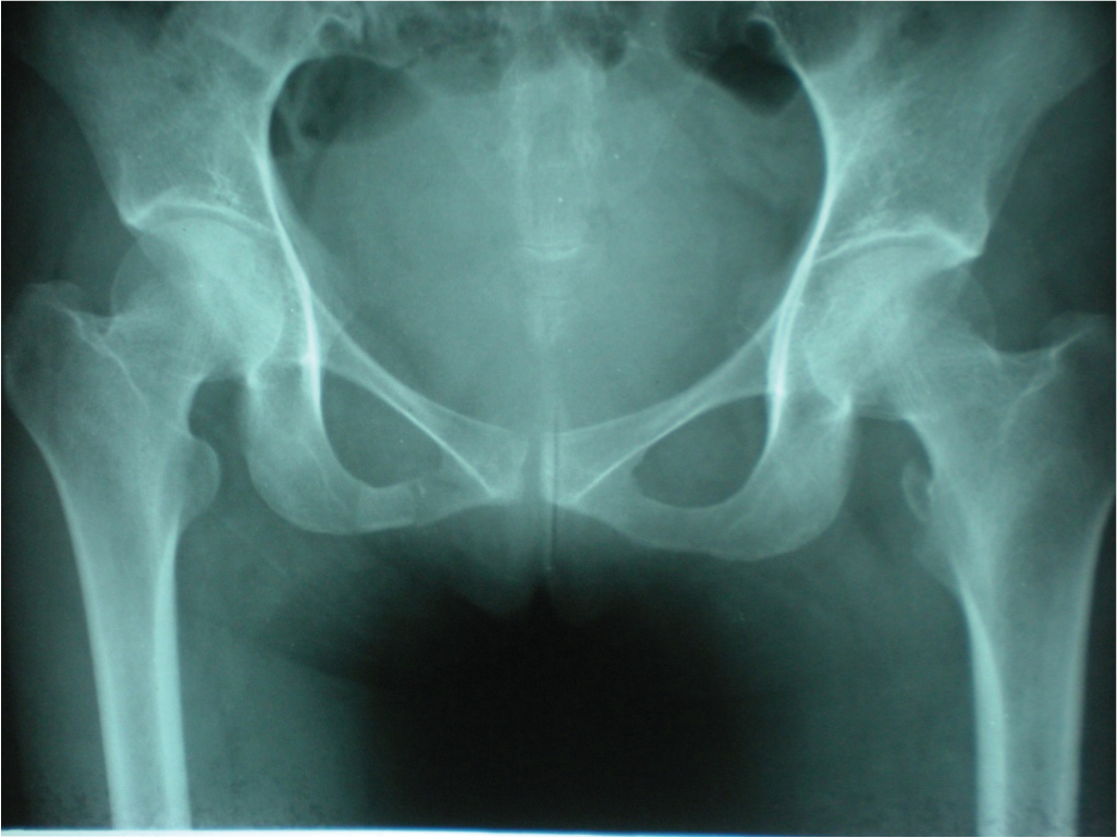
Radiograph of pelvis showing displaced lesser trochanter avulsion without evidence of local pathological bony lesion and contralateral undisplaced inferior pubic rami fracture
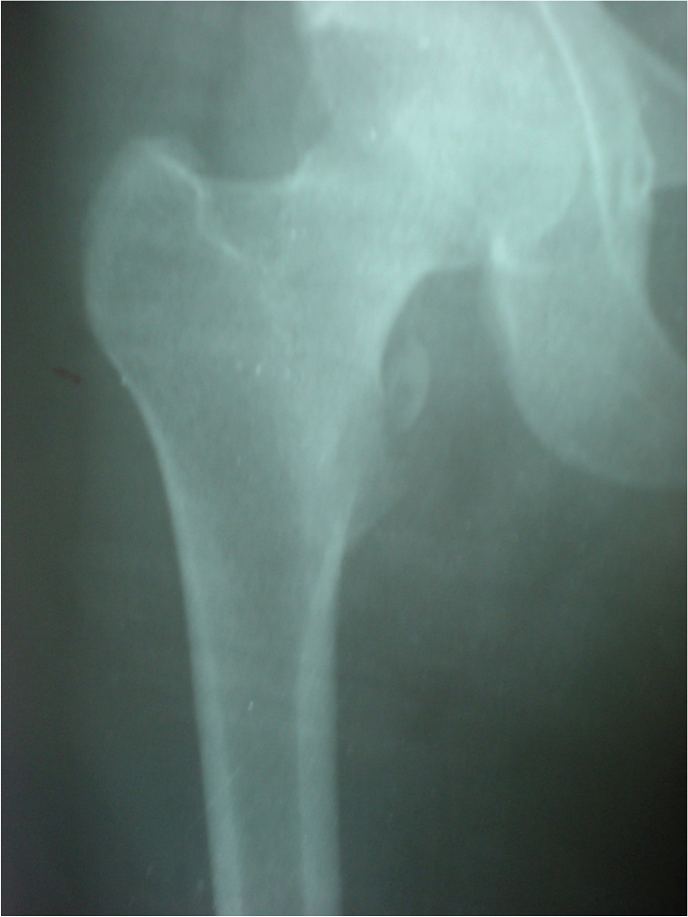
Follow up radiograph of hip at 4 months showing displaced lesser trochantric fracture with some sclerosis at fracture margins but without any evidence of pathological process like lysis, new bone formation
Discussion
Lesser trochanter fracture is not a usual presentation of hip fracture especially in non-paediatric population [1]. Classically, lesser trochanter fractures in adults are associated with antecedent neoplastic change and often related to insignificant trauma [2]. Very rarely, it may be seen as avulsion of lesser trochanteric apophysis in athletic population or very high velocity accidents [3,4]. This avulsion theory of fracture is observed chiefly in young population, when fusion of lesser trochantric apophysis has not occurred [5]. However, number of cases in mature skeleton with lesser trochanteric fracture in absence of underlying oncological process is extremely low, as it was a scenario in our case. This particular case is presented here not due to its rarity, but to stress on importance of correct diagnosis, thorough investigation to uncover clandestine pathological process.
Literature also suggests lesser trochanteric fracture as part of even more complex injury. Fracture line may further propagate to convert or reveal a seemingly simple fracture as intertrochanteric fracture. Bonshashi and colleagues provided a recent report of isolated lesser trochanteric fractures in adults without pathologic process. Two of their 3 cases later developed displaced intertrochanteric fractures [6].
In adult population with a mature skeleton, avulsion of lesser trochantric should be seen as serious event, with a focused vision to rule out any primary or secondary neoplastic process [7]. A high degree of suspicion should be maintained for a neoplastic change on follow up of patient also. A conservative treatment plan for such fractures is often successful and it includes comforting patient in neutral position and protected weight bearing [8]. Fixation with a dynamic hip screw or a intramedullary proximal femoral device can be opted if fracture line propagates to intertrochanteric region in due course of time. Our case is an uncommon presentation of hip fracture. The fracture was not associated with any hidden pathology locally and neither was it part of other injury pattern. A non-operative plan was sufficient to heal fracture without any undesirable sequelae of fracture.
Conclusion
The case and literature presented here offers a message for clinical practice that lesser trochantric fracture in adults is a rare clinical occurrence. When found, high degree of suspicion for clandestine neoplastic process or an occult intertrochantric fracture should be kept.
[1]. Heiney JP, Leeson MC, Isolated lesser trochanter fracture associated with leukaemiaAm J Orthop 2009 38:E56-58. [Google Scholar]
[2]. Fox TP, Lakkol S, Oliver G, Lesser trochanter fracture: the presenting feature of a more sinister pathologyBMJ Case Reports 2014 2014:bcr2013202590 [Google Scholar]
[3]. Bertin KC, Horstman J, Coleman SS, Isolated fracture of the lesser trochanter in adults:an initial manifestation of metastatic malignant diseaseJ Bone Joint Surg Am 1984 66:770-73. [Google Scholar]
[4]. Theologis TN, Epps H, Latz K, Cole WG, Isolated fractures of the lesser trochanter in childrenInjury 1997 28:363-64. [Google Scholar]
[5]. Quarrier NF, Wightman AB, A ballet dancer with chronic hip pain due to a lesser trochanter bony avulsion: the challenge of a differential diagnosisJ Orthop Sports Phys Ther 1998 28:168-73. [Google Scholar]
[6]. Bonshahi AY, Knowles D, Hodgson SP, Isolated lesser trochanter fractures: a case seriesInjury 2004 35:196-98. [Google Scholar]
[7]. Afra R, Boardman DL, Kabo JM, Eckardt JJ, Avulsion Fracture of the Lesser Trochanter as a Result of a Primary Malignant Tumor of BoneJ Bone Joint Surg Am 1999 81:1299-304. [Google Scholar]
[8]. Khemka A, Raz G, Bosley B, Ludger G, Arthroscopically assisted fixation of the lesser trochanter fracture: a case seriesJournal of Hip Preservation Surgery 2014 1:27-32. [Google Scholar]