Multiple Giant Sebaceous Cysts of Scalp
Anand Singla1, Mohinder Singh2, Satpaul Singla3
1 Junior Resident, Department of Surgery, G.M.C., Patiala, Punjab, India.
2 Professor, Department of Surgery, G.M.C., Patiala, Punjab, India.
3 Associate Professor, Department of Surgery, G.M.C., Patiala, Punjab, India.
NAME, ADDRESS, E-MAIL ID OF THE CORRESPONDING AUTHOR: Dr. Anand Singla, Junior Resident, Department of Surgery, G.M.C., Patiala, Punjab–147001, India.
E-mail: anand_singla84@yahoo.co.in
Sebaceous cyst is an epidermal cyst often found on the hairy areas of the body such as scalp, trunk and face. Though commonly encountered in surgical practice, its presentation as multiple giant sebaceous cysts over scalp is rare. However, in long standing cases malignant transformation has also been sparingly reported. We report a case of a 52-year-old male presenting with multiple large sized swellings on the scalp, seven in number. These were present since childhood and gradually progressed to the enormous size of largest measuring 10cm x 8cm. Excision of these cysts was undertaken and specimens were sent for histopathological examination which confirmed the nature of these cysts to be sebaceous cysts. No malignant changes were reported in any of the specimens. The patient was followed up and was doing well.
Epidermal cyst, Malignant, Sebum
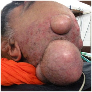
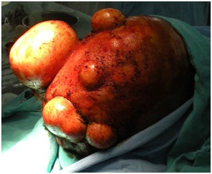
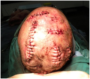
A 52-year-old male presented with large multiple painless swellings on scalp; seven in number since childhood. The swellings increased progressively in size and there was no history of trauma or infection. There was history of associated hair loss on the overlying skin. There was no similar family history. Detailed general and systemic examination was unremarkable. Local examination revealed multiple swellings, cystic in consistency, on the scalp with larger swellings measuring about 10cm x 8cm, 6cm x 4cm and 5cm x 5cm [Table/Fig-1,2]. They were non tender, fixed to the skin and free from the underlying structures, with no local erythema or discharge. Cervical lymphadenopathy was absent. A clinical diagnosis of sebaceous cysts was made. Excision of these giant sebaceous cysts was undertaken [Table/Fig-3]. Histopathalogical examination was consistent with the diagnosis of epidermoid cyst. Thereafter, the patient was followed up in OPD and sutures were removed on the seventh day. There were no fresh complaints and our patient was doing well.
Multiple giant sebaceous cysts scalp
Multiple giant sebaceous cysts
Post excision of the various cysts
In day to day surgical practice several dermatosis are encountered and sebaceous cyst is one of them. Moreover, these giant sebaceous cysts are also more prone for developing malignancy.
Giant sebaceous cysts are a rare entity in clinical practice [1]. These can occur at any age, rarely before puberty, and the most common age of presentation is a young adult male. The most common site of occurrence is the face, trunk, neck, scalp, scrotum, ear lobe, buttock and breast, but location at an unusual site raises concern [2–4]. These are common in females usually on the scalp, more in people working in outdoor conditions with sunlight exposure and unhygienic concerned areas. Normal size varies from a few millimetres to a few centimetres but when the size exceeds 5 cm, it is referred to as a giant sebaceous cyst [5]. The detection of small cysts and evolving into giant cysts takes years and it grows usually at a rate of not more than 0.5 cm per year. In the initial years, growth is more rapid than after attaining large size. These are intradermal in origin and adherent to the epidermis and usually have a central punctum that is often identifiable [1]. These are asymptomatic, painless, dome shaped lesions with overlying smooth skin and contain thick sebum. In our patient too, the cysts were adherent to the epidermis. The lesions were painless with overlying smooth skin. Cysts usually vary in size from 1-4 cm in diameter and arise from a ruptured pilosebaceous follicle. Giant epidermoid cysts have a propensity to develop malignancy [6]. Multiple epidermoid cysts are rarely associated with lipomas or fibromas of the skin and osteomas and should be considered as a part of Gardener’s syndrome with associated premalignant colonic polyps and panchyonchia congenital [7]. In our patient there were no other lesions present elsewhere during the examination and there was no history of bleeding per rectum or altered bowel habits. Various types of malignancy that can arise from a giant sebaceous cyst are squamous cell carcinoma, basal cell carcinoma, mycosis fungicides and melanoma in long standing cases in elderly [8–10]. Our patient had multiple giant sebaceous cysts on scalp for a very long duration which was difficult to excise. He was fortunate enough not to develop any malignant changes but these should not be neglected as done by our patient.
[1]. Venus MR, Eltigani EA, Fagan JM, Just another sebaceous cyst?Ann R Coll Surg Engl 2007 89(6):W19-21. [Google Scholar]
[2]. Handa U, Kumar S, Mohan H, Aspiration cytology of epidermoid cyst of terminal phalanxDiagn Cytopathol 2002 26(4):266-67. [Google Scholar]
[3]. Denison CM, Ward VL, Epidermal inclusion cysts of the breast: Three lesions with calcificationsRadiology 1997 204:493-96. [Google Scholar]
[4]. Momeni MG, Anavim A, Giant epidermal inclusion cyst of buttockSkeletal Radiol 2006 35:864-66. [Google Scholar]
[5]. Basterzi Y, Sari A, Ayhan S, Giant epidermoid cyst on the forefootDermatol Surg 2002 28(7):639-40. [Google Scholar]
[6]. Sumi Y, Yamamoto N, Kiyosawa T, Squamous cell carcinoma arising in a giant epidermal cyst of the perineum: a case report and literature reviewJ Plast Surg Hand Surg 2012 46(3-4):209-11. [Google Scholar]
[7]. Swygert KE, Parrish CA, Cashman RE, Lin R, Cockerell CJ, Melanoma in situ involving an epidermal inclusion (infundibular) cystAm J Dermatopathol 2007 29(6):564-65. [Google Scholar]
[8]. Debaize S, Gebhart M, Fourrez T, Rahier I, Baillon JM, Squamous cell carcinoma arising in a giant epidermal cyst: a case reportActa Chir Belg 2002 102(3):196-98. [Google Scholar]
[9]. Tanaka M, Terui T, Sasai S, Tagami H, Basal cell carcinoma showing connections with epidermal cystsJ Eur Acad Dermatol Venereol 2003 17(5):581-82. [Google Scholar]
[10]. Bauer BS, Lewis VL, Carcinoma arising in sebaceous and epidermoid cystsAnn Plast Surg 1980 5:222-26. [Google Scholar]