Bitewing radiographs, also known as inter-proximal radiographs, are obtained to provide a clear radiographic view of the maxillary and mandibular crowns and the alveolar crest [1]. Due to suitable horizontal angulation of X-ray beam, this radiography can greatly enhance accurate diagnosis of proximal caries and interproximal bone loss [2]. These radiographs are taken using paper loops and film holders [3]. Attempts to standardize dental radiographs lead to inventing a film holder not only to hold the film, but also provide a predictable path for the X-ray beam [4]. Use of film holder as a guiding tool for X-ray beams could result in bitewing radiographs with higher quality. Some researchers have presented new appliances as film holder to facilitate the radiography procedure; however drawbacks of each device lead to not deciding on a specific film holder [5,6]. Choksi et al., compared the efficacy and characteristics of their newly designed all metal film holder with the conventional holder and reported that their designed film holder enabled better positioning of the film but had a poorer performance in terms of frequency of cone cut error [7]. Kositbowornchi et al., compared the efficacy of using paper loop and film holder and showed that use of film holder resulted in less overlap and better positioning of the film but had no effect on cone cut [3]. Dixon et al., conducted a systematic review on the production of film holders and evaluated articles in this regard. They concluded that existing film holders have some advantages and drawbacks. User friendly and patient friendly devices are more acceptable in dental clinics although there is no lack of technical errors in most appropriate film holders [4].
Reducing the exposure dose of the patients is an important issue in diagnostic radiology. Decreasing the exposure dose of the patients lead to reduced severe biological invades of X-ray on organs and the cells. Difficult application of modified film holders like holding the framework by the patient is a major drawback of these innovations which has made them less acceptable by the patients and clinicians. In view of these problems and factors we tried to modify the conventional film holder which is easier to use and reduces the exposure dose of the patients.
This study aimed to design a modified film holder and compare the efficacy of the newly designed film holder with that of conventional film holder (RINN XCP).
Materials and Methods
This study was executed in Department of Oral and Maxillofacial Radiology of Shahaid Beheshti Dental School, Tehran, Iran between April to December of 2014. Seventy volunteer patients with the age between 15 to 54 years referred to the Department of Radiology were included in current study.
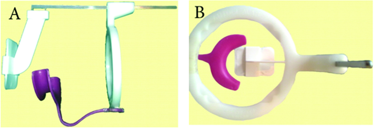
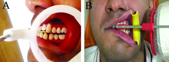
Assembly of the film holder: The new film holder was designed by using a parallel film holder for bitewing radiography and an orthodontic retractor. The conventional orthodontic retractor was cut in halves [Table/Fig-1a] and one half of it was fixed to the loop of the conventional parallel film holder [Table/Fig-1b] in such way to retract the buccal mucosa when taking a radiograph [Table/Fig-2].
a) The newly designed film holder. Orthodontic retractor cut in halves to retract the cheek
b) the parallel loop is used to improve the correct position of the PID

Positioning a bitewing radiograph using the modified film holder Lateral (a) and frontal (b) view

Radiographs for each patient were obtained using the newly designed film holder for one side and a conventional film holder for the other side. All radiographs for each patient were taken by one expert oral and maxillofacial radiologist. Considering the retraction of buccal soft tissue using the newly designed holder, lower exposure settings were applied and the exposure time was decreased by 20%. So the radiographic sites were divided into two groups:
The radiographs obtained by conventional film holders, at 70 Kvp and 8 mA settings in 0.2 seconds.
The radiographs obtained by the customized film holders, at 70 Kvp and 8 mA settings in 0.16 seconds.
Digital bitewing radiographs were taken by photostimulable phosphor plates (PSP No.2, Soredex, Tuusula, Finland) and a Minray intraoral x-ray unit (Soredex). In all cases the patients were made to sit upright with the Frankfort plan parallel to the floor. The cone of radiography unit was placed in line with custom made device in case group. The position indicating device (PID) was angled +10 degrees vertically and perpendicular to the film holder horizontally in control group.
After taking the radiographs, the patients were requested to express their opinion regarding the acceptability of each holder by giving them a score from 0 to 20. The zero score was considered as absolutely not acceptable and annoying device whilst the score of 20 was the most acceptable and comfortable device. A general dentist was requested to assess and compare the two radiographs of each patient and report the degree of overlap (none, slight, moderate, severe) and film positioning (correct or incorrect) for each radiograph. The dentist was blinded to the group allocation of radiographs and the two holders were coded as technique 1 and technique 2. Moreover, the technician was asked to rate his opinion regarding the acceptability of each holder using a 0-20 point scale. Data were collected for each patient by four questions.
The degree of the overlapping was classified as below:
– Slight overlapping: when less than half of the enamel of the adjacent teeth were overlapped.
– Moderate overlapping: when more than half of the enamel of the adjacent teeth were overlapped although the dentin was not overlapped.
– Severe overlapping: the dentins of the adjacent teeth were overlapped.
The position of the radiographic film was defined correctly when there was inadequate coverage for example missing the proximal surface of the examined teeth on the radiographs.
Ethical consideration: The procedures followed were in accordance with the ethical standards of the responsible committee on human experimentation of Shahid Beheshti University of Medical Sciences and with the Helsinki Declaration of 1975 that was revised in 2000.
Statistical Analysis
All the calculations were processed using Statistical Package for Social Science statistical software (version 20; SPSS Inc., Chicago, Illinois). The descriptive statistics, including the tables and graphs, were applied to demonstrate the information. The significance of the categorical findings was compared with that of the normally distributed variables via the Student t-test, and the Mann-Whitney U test was used for the nonparametric data. A p-value of less than 0.05 was considered statistically significant. The data were expressed as mean ± standard deviation.
Results
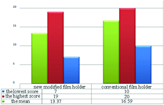
Seventy patients with the mean age of 34.2 years were included in current study. The mean (±SD) satisfaction (acceptance) score of patients was 16.59±3.209 (range of 10 to 20) with the conventional and 13.37±2.503 (range of 7 to 19) with the newly designed film holder [Table/Fig-3]. According to t-test, this difference was statistically significant (p<0.001).
The graph of satisfaction scoring for the patients

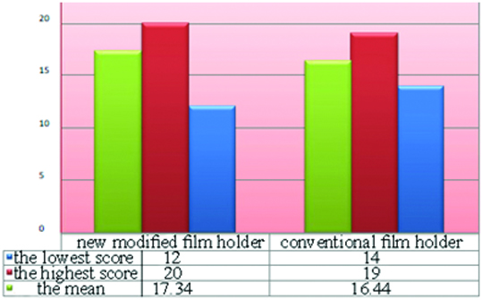
The mean satisfaction (acceptance) score of the technician was 16.44±1.410 (range 14-19) with the conventional and 17.33±1.483 (range of 12 to 20) with the newly designed film holder [Table/Fig-4]. According to t-test, this difference was statistically significant as well (p<0.001).
The graph of satisfaction scoring for the technicians

In radiographs obtained using the conventional film holder, 38.6% (n=27) had no overlap, 37.1% (n=26) showed slight overlap, 18.6% (n=13) showed moderate overlap and 5.7% (n=4) showed severe overlap [Table/Fig-5].
Comparison of the frequency and degree of overlap using the two film holders
| Severe overlap | Moderate overlap | Slight overlap | No overlap |
|---|
| Using conventional holder | 4 | 13 | 26 | 27 |
| Using the newly designed holder | 0 | 0 | 12 | 58 |
In radiographs obtained using the newly designed film holder, 82.9% (n=58) had no overlap, 17.1% (n=12) showed slight overlap, and there was no case of moderate or severe overlap. Non-parametric Wilcoxon test was used to compare the degree of overlap between the two techniques and showed a significant difference (p<0.001, Z=-5.7).
The film was correctly positioned in 58.6% of cases (n=41) and was incorrectly positioned in 41.4% (n=29) when we used the conventional film holder. Using the newly designed film holder, these values were 82.9% (n=58) and 17.1% (n=12), respectively [Table/Fig-6]. Comparison of the two holders in this regard using the kappa agreement revealed no significant difference (p=0.53). However, the McNamara nonparametric test revealed a significant difference in this regard (p=0.005) [Table/Fig-7].
Comparison of correct film positioning using the two film holders
| Groups | Correct position | Incorrect position |
|---|
| Using conventional holder | 41 | 29 |
| Using the newly designed holder | 58 | 12 |
The variables and the statistical tests which were used in current study (SD, Standard Deviation)
| Variable | The new modified film holder (Mean±SD) | Conventional film holder (Mean±SD) | The statistical test | p_value |
|---|
| Satisfaction scoring for the patients | 13.37±2.5 | 16.59±2.2 | Student t test | ≤0.001 |
| Satisfaction scoring for the technicians | 1734±1.48 | 16.44±1.41 | Student t test | ≤0.001 |
| No overlapping | 82.9% | 38.6% | Wilcoxon test | ≤0.001 |
| Correct film positioning | 58 | 41 | McNamara nonparametric test | 0.005 |
Discussion
Improvement of dental radiographic films is an important issue in diagnostic dentistry. The clinician would be able to diagnose the early caries and small cavities in the initiation. Bitewing radiography is a useful method in detection of alveolar bone loss and identification of proximal dental caries [8,9]. In the current study we tried to evaluate the efficacy of a modified film holder for improvement of bitewing radiography technique.
The clinician may be able to diagnose the interproximal problems by increasing the contrast of radiographs [10,11]. Improvement the contrast of the radiographic films by retracting the buccal mucosa is an acceptable method represented in the literature [12]. We tried to design a special radiographic film holder in which the contrast of the films might be increased by retracting the cheek.
Dale et al., stated that techniques and instruments are widely variable in dentistry and a logical comparison among the techniques is very difficult [13]. Considering the decreased frequency of errors using the newly designed holder, it can be routinely used in dental clinical practice resulting in less repeat of radiographs and possibly lower patient radiation dose.
The acceptability of the newly designed film holder was higher than that of conventional film holder from the technicians’ point of view. Using the newly designed film holder, the technician can retract the buccal mucosa and adjust the path of x ray beam reliably as desired. Therefore, although placement of the newly designed film holder in the mouth is more difficult than the conventional one, the technicians prefer using the newly designed holder.
The frequency of overlap was significantly lower in radiographs taken using the newly designed holder and no case of moderate or severe overlap was noted in radiographs taken using the newly designed holder. This further confirms the superiority of the newly designed holder.
Retracting the buccal mucosa provides enhanced vision of the area the clinician can more accurately adjust the path of X-ray beam. As the result, the obtained radiograph will be more accurate with less errors leading to a more accurate diagnosis and treatment planning. This issue is especially important considering the importance of caries detection and prevention in dentistry.
In the current study, film positioning was also compared using the two film holders and the results showed more accurate film positioning using the newly designed holder. The reason is retraction of buccal mucosa enabling better vision of the technician when taking radiographs using the designed holder.
However, patients were less satisfied with the newly designed holder due to the presence of an extra part in this holder. Therefore, the study hypothesis regarding increased patient satisfaction using the newly designed film holder was rejected. The new holder puts pressure and tension on the cheek and retracts the lips and thus, the patients gave a lower score to the newly designed holder.
Considering the obtained results regarding lower technical errors using the newly designed holder and increased satisfaction of clinicians, this holder can be beneficial despite the lower satisfaction rate of patients. Moreover, according to the pilot study 20% reduction in exposure time using this holder helps decrease patient radiation dose, which is in accord with the ALARA regulations [14].
Sakagami et al., proposed a new modified film holder to observe the radiographic changes after root planning [15]. They indicated that their modified system has high accuracy and clinical usefulness. Although it seems that the unique producing technique makes it difficult for the clinician to obtain the radiographs for each patient.
Choksi and Rao evaluated the two film holders for periapical radiography [7]. They showed that the modified film holder was associated with fewer errors in film positioning while it had more errors in cone cutting. In our study we presented a new modified film holder which had fewer errors in film positioning, cone cutting, and horizontal overlapping; however it was not accepted by the patient’s very well.
Conclusion
The newly designed film holder may be able to decrease technical errors in bitewing radiographs and thus, could be successfully used in the clinical practice. It was not generally acceptable by the patients although the clinicians were able to use it easily. Despite the not very high significance of this modified film holder it reduced the horizontal overlapping of the teeth and incorrectly positioned films while in comparison to the conventional one.