Vulvar Haematoma Following Laparoscopic Endometrioma Excision: Report of A Rare Complication
Aytekin Tokmak1, Ilker Gülbasaran2, Serra Akar3, Hüseyin Yesilyurt4
1 Faculty, Department of Obstetrics and Gynecology, Zekai Tahir Burak Women’s Health Research and Education Hospital, Division of Reproductive Endocrinology and Infertility, Ankara, Turkey.
2 Faculty, Department of Obstetrics and Gynecology, Zekai Tahir Burak Women’s Health Research and Education Hospital, Division of Reproductive Endocrinology and Infertility, Ankara, Turkey.
3 Faculty, Department of Obstetrics and Gynecology, Zekai Tahir Burak Women’s Health Research and Education Hospital, Division of Reproductive Endocrinology and Infertility, Ankara, Turkey.
4 Faculty, Department of Obstetrics and Gynecology, Zekai Tahir Burak Women’s Health Research and Education Hospital, Division of Reproductive Endocrinology and Infertility, Ankara, Turkey.
NAME, ADDRESS, E-MAIL ID OF THE CORRESPONDING AUTHOR: Dr. Aytekin Tokmak, Talatpasa Bulvari, Hamamonu, Altindag, 06230 Ankara, Turkey. E-mail : aytekintokmak@gmail.com
Vulvar haematoma as a complication of laparoscopic adnexal surgery has rarely been reported. Indeed, to our knowledge, there are only two case reports describing postlaparoscopic vulvar haematoma in the literature. Although complications associated with laparoscopy are mostly related to bowel or retroperitoneal vessel injury, vulvar haematoma may seldom develop. Vulvar haematoma after laparoscopy may indicate abdominal wall or pelvic vascular injury. We present a case of postoperative vulvar haematoma following laparoscopic endometrioma excision.
Adnexal surgery, Laparoscopic endometrioma, Operative laparoscopy
Case Report
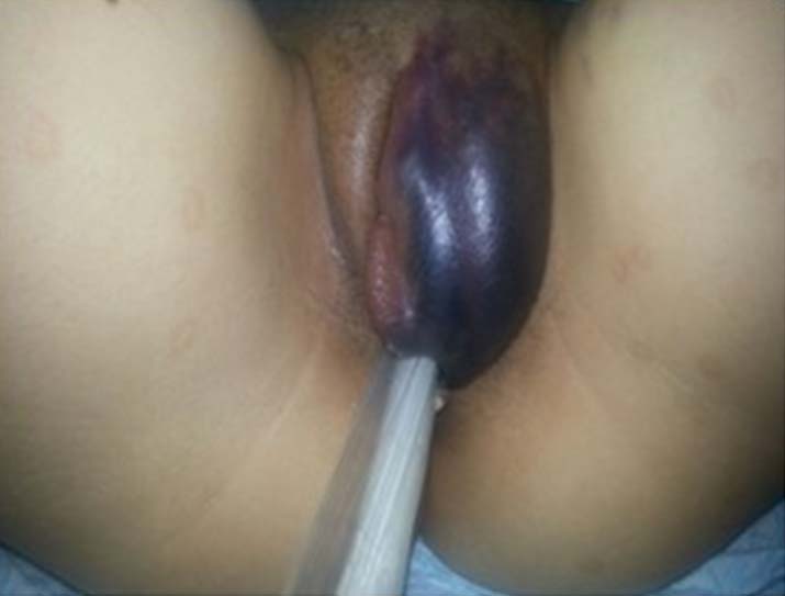
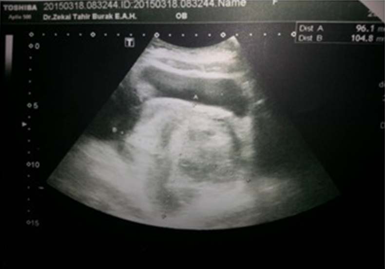
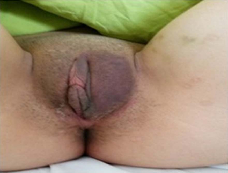
A 26-year–old nulligravida presented with chief compliant of severe dysmenorrhea. Transvaginal ultrasonography revealed an anteverted uterus, an endometrial thickness of 9 mm, homogenous myometrium and bilateral cysts showing acoustic enhancement with internal echoes, consistent with endometrioma, measuring 35x32 mm and 70x50 mm in the right and left ovaries, respectively. On admission, haemoglobin level was 13 g/dl, and CA 125 measured 96 U/ml. The patient underwent an uncomplicated bilateral laparoscopic excision of both endometriomas. Level of 6-hour postoperative haemoglobin was 10.3 g/dl. She was discharged with stable vital signs and normal physical exam findings on postoperative day 1. She presented on postoperative day 2 with a vulvar haematoma. She had a blood pressure of 100/60 mmHg, pulse of 128 beats per minute, and temperature of 36.8°C. Physical exam was unremarkable. Pelvic exam was significant for left vulvar haematoma measuring 10x6 cm in size [Table/Fig-1]. Transvaginal ultrasonography revealed an endometrial thickness of 10 mm, normal appearing right adnexa and a haematoma in the left adnexal area measuring 104x96 mm and minimal fluid in the cul-de-sac [Table/Fig-2]. Haemoglobin level was 6.6 g/dl, white blood cell count 10.700/uL, platelet count 160.000/uL, and coagulation parameters were normal. The patient was managed expectantly. Intravenous ceftriaxone and metronidazole were given. Anti-inflammatory medication and local Eau de Goularde were administered. A control haemoglobin level of 6.4 g/dl was obtained and the patient was transfused with two units of packed red blood cells. After 10 days of treatment, patient made full recovery with regression of the vulvar haematoma [Table/Fig-3]. She was discharged with a haemoglobin level of 10.4 g/dl, white blood cell count of 7.400/uL and sonographic exam showed a decrease in the dimensions (81x50mm) of the haematoma previously noted in the left adnexal area. Written informed consent was obtained from the patient for publication of this case report and accompanying images.
Vulvar haematoma postoperative on day 2
Postoperative ultrasound image of the pelvic haematoma
Spontaneously resolved haematoma postoperative on day 10
Discussion
Although complications associated with laparoscopic surgery are rare, a significant proportion of these occur during the abdominal entry and include bowel and vascular injury [1]. Vulvar haematoma following laparoscopic surgery is extremely infrequent and possibly, secondary to disruption of blood vessels in the abdominal wall or the pelvic viscera. Patients may present with unstable vital signs, vulvar mass, anemia and pelvic haematoma on sonographic exam. Management may be expectant with close patient monitoring if severe vessel injury is not suspected. There have been two reported cases of vulvar haematoma following injury to epigastric vessels after laparoscopic cystectomy [2,3]. We described a case of vulvar haematoma subsequent to a laparoscopic bilateral endometrioma excision. The patient was managed expectantly and fully recovered without repeat surgery.
Interestingly, previously reported cases of vulvar haematoma were nulliparous patients who underwent laparoscopic cystectomy, as the case presented here. Marcovici et al., described a patient who presented with nausea, syncope and vomiting on postoperative day one with orthostasis, tachycardia and a 15% drop in preoperative haematocrit values [2]. The patient, we illustrated presented with tachycardia and about a 40% drop in preoperative haematocrit on readmission. An exploratory laparotomy was undertaken by Marcovici et al., and bleeding was noted from a left superficial epigastric vessel, subcutaneous haematoma and site of cystectomy. Likewise, Valadan et al., reported a vulvar haematoma due to a right epigastric vessel injury following a cystectomy [3]. Although, the exact source of bleeding could not be ascertained in the patient we described, sonography showed a haematoma on the left adnexal area. An injury to the left round ligament and its artery may have resulted in a haematoma in the left labium majus. Alternatively, vulvar haematoma may be explained by the communication between the superficial perineal pouch and superficial fascia of the anterior abdominal wall containing the superficial epigastric vessels that may have been injured during trochar insertion. Knowledge of the course of the specific vessels in the abdominal wall is required to minimize risk of laparoscopy associated vascular injuries. However, a cadaver study showed that half of the trocar sites recommended in the literature for abdominal entry was exposed to the risk of vascular injury [4]. The superficial abdominal wall vessels may also be located by transillumination, particularly in women of normal weight [5]. However, the deep epigastric vessels cannot be effectively located by this method. Other techniques should be used to minimize injury to the deep epigastric vessels [6,7].
The patient described, was managed expectantly as was reported by Valadan et al., Besides compression treatment, antibiotics and packed red blood cells were given. The patient made full recovery without undergoing a repeat operation. Despite a steep drop in the haemoglobin level, increased pulse rate, presence of adnexal haematoma on sonographic exam, a conservative approach to a vulvar haematoma proved successful in this specific case.
Many complications associated with laparoscopic surgery probably are not reported, and sometimes these complications occur after the patient is discharged. Wong et al., reported three cases of intraabdominal bleeding following laparoscopic adnexal surgery and they failed to identify the source of bleeding in two, during postoperative exploratory laparotomy [8]. They concluded that delayed bleeding from vessels tamponaded by the pneumoperitoneum is one of the possible causes of postoperative bleeding. Unnecessary dissections and ovarian injuries that may induce bleeding should be avoided during operative laparoscopy as well as endometriosis surgery and a good haemostasis should be performed meticulously under low-pressure pneumoperitoneum.
Conclusion
In conclusion, laparoscopic complications are commonly related to trochar insertion and port placement, and most of them are associated with bowel and retroperitoneal vessel injury. Vulvar haematoma secondary to superficial epigastric vessel injury is extremely rare. We presented a case of vulvar haematoma following laparoscopic endometrioma excision. Expectant management of the patient resulted in complete recovery. Vulvar haematoma may be a sign of serious vascular injury. Nonetheless, patients may respond to expectant management with close follow-up.
[1]. Chandler JG, Corson SL, Way LW, Three spectra of laparoscopic entry access injuriesJ Am Coll Surg 2001 192(4):478 [Google Scholar]
[2]. Marcovici I, Shadigian E, Operative Laparoscopy and Vulvar Haematoma: An Unusual AssociationJSLS 2001 5(1):87-88. [Google Scholar]
[3]. Valadan M, Ashegh H, Darvish S. Vulvar Haematoma Following Laparoscopic Ovarian Cystectomy. 17th Annual Meeting and Endo Expo. September 17-18, 2008, Chicago, IL, United States [Google Scholar]
[4]. Balzer KM, Witte H, Recknagel S, Kozianka J, Waleczek H, Anatomic guidelines for the prevention of abdominal wall haematoma induced by trocar placementSurg Radiol Anat 1999 21(2):87-89. [Google Scholar]
[5]. Quint EH, Wang FL, Hurd WW, Laparoscopic transillumination for the location of anterior abdominal wall blood vesselsJ Laparoendosc Surg 1996 6(3):167-69. [Google Scholar]
[6]. Whiteley MS, Laws SA, Wise MH, Use of a hand-held Doppler to avoid abdominal wall vessels in laparoscopic surgeryAnn R Coll Surg Engl 1994 76(5):348-50. [Google Scholar]
[7]. Saber AA, Meslemani AM, Davis R, Pimentel R, Safety zones for anterior abdominal wall entry during laparoscopy: a CT scan mapping of epigastric vesselsAnn Surg 2004 239(2):182-85. [Google Scholar]
[8]. Wong SM, Levine AB, JacobsA J, Argov Y, Intraabdominal bleeding following laparoscopic adnexal surgery. A report of three casesJ Reprod Med 1966 41(4):294-96. [Google Scholar]