Fusion Between Maxillary Premolar and A Supernumerary Tooth: A Rare Case Presentation
Vipin Kumar1, Vinisha Pandey2, G Rohini3, Bhuvan Jyoti4
1 Consultant Periodontist and Implantologist, Delhi, India.
2 Senior Lecturer, Department of Conservative Dentistry and Endodontics, Rama Dental College, Kanpur, India.
3 Senior Lecturer, Department of Periodontics, CSI College of Dental Sciences and Research, Madurai, India.
4 Consultant Dental Surgeon, Department of Dental Surgery, RINPAS, Ranchi, Jharkhand, India.
NAME, ADDRESS, E-MAIL ID OF THE CORRESPONDING AUTHOR: Dr. Vinisha Pandey, 117/H-1, 312 Model Town, Pandu Nagar, Kanpur-208005, U.P, India.
E-mail: Vinisha9@gmail.com
Atypical morphology, Developmental disturbances, Double tooth, Schizodontism
Morphological and anatomical aberrations present a great challenge to the endodontic treatment. In such cases, knowledge of anatomical variations and developmental disturbances of teeth is essential for successful root canal treatment. The purpose of this article is to differentiate the diagnostic dilemma between gemination and fusion and to step by step outline the endodontic management of such types of anatomical aberrations. A 21-year-old female was referred to a practicing endodontist for the root canal treatment of maxillary left premolar. The patient’s chief complaint was a throbbing pain with sensitivity to cold and hot associated with the offending tooth since 1 month. Clinical examination revealed three intact crowns with tooth coloured restorations on each of them performed by a general dentist [Table/Fig-1].
Clinical examination revealed three intact crowns with tooth colored restorations on each of them
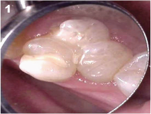
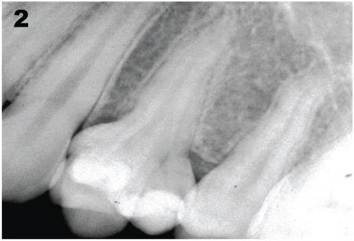
All the crowns were termed as mesial, distal and palatal. Her systemic health was good and medical history was non-contributory. Distal and palatal crowns demonstrated atypical morphology, palatal the most atypical whereas the mesial crown was characteristically & anatomically similar to the maxillary first premolar. Radiographically, it appeared to be a single root at the cemento-enamel junction [Table/Fig-2]. No periapical pathology was evident which led to the diagnosis of irreversible pulpitis with acute apical periodontitis.
Preoperative radiograph shows the presence of separate roots at cement-enamel junction
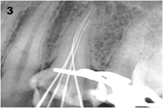
After an informed consent access openings were made into each crown using Endo access bur (Dentsply Tulsa, UK) to allow access into the pulp chamber and preparation of the chamber walls, in one operation. Working length was established [Table/Fig-3] using an apex locator (Root ZX mini, J. Morita, USA) which was confirmed using a radiovisiography (Kodak RVG 5100, Care stream dentals LLC, North America).
Working length was established
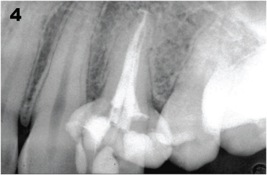
All the canals were prepared with Hyflex CM rotary files (Coltene Whaledent, Switzerland) in a crown down manner. Sodium hypochlorite was used as an irrigant. Obturation was performed with warm vertical condensation using System B and Obtura II (Sybron Endo, CA, USA) for backfilling. A radiograph was taken immediately to verify the obturation [Table/Fig-4]. After a week the patient reported with complete resolution of the symptoms. Gemination is a developmental anomaly of tooth shape that develops when a single tooth bud attempts to form two teeth. It leads to the formation of two or more identical crowns with a single root and root canal radio graphically [1,2]. Fusion is often correlated and confused with gemination. In our case we distinguished between the two on the basis of the following features:
Postobturation radiograph
Morphology- Gemination results in mirror images of coronal portion of the tooth whereas fusion gives a crooked appearance to the oral cavity.
Anatomy- Radio graphically fused teeth has a separate pulp chamber and root canal while gemination has a single pulp chamber.
Count of teeth- Mader in 1979 emphasized on the count of teeth in the arch as a differentiating feature between gemination and fusion. He stated that fusion is counted as one tooth in the arch and in gemination the number is increased. This was called as two-tooth rule. He explained that if the resulting dental anomaly is counted as two teeth with the normal count present in the arch, it is fusion. Whereas the resulting dental anomaly is counted as two with the extra count present in the arch, it is termed as gemination or a fusion between normal and supernumerary teeth [3].
Morphological and anatomical variations are not uncommon finding; however, their simultaneous presentation is. This case presents the occurrence of fusion in a supernumerary tooth, which is a very rare finding.
Yang et al., reported a geminated supernumerary tooth with two crowns and one root in the maxillary premolar region [3,4]. Fusion between supernumerary teeth and permanent teeth occurs less frequently than fusion between other types of teeth. Yuzuwa et al., reported incidence of 0.1% fusion with a supernumerary tooth [5].
Diagnostic tools such as radiographic examinations and an operating microscope played an important role in the diagnosis of this case. Endodontic treatment is usually difficult owing to complex anatomy, tooth position in the arch and difficulty in isolation. Strict adherence to biomechanical principles of root canal preparation provided successful treatment. Multidisciplinary treatment modalities have been suggested for fused teeth. The endodontic treatment depends on the thorough knowledge of the morphological variations of root canal anatomy [5]. Diagnostic tools such as cone-beam computed tomography and better visualization by using an operating microscope are all important aids in detecting communication.
The occurrence of fusion between normal and supernumerary tooth is very rare but the clinician should be conscious of specific treatment needs, canal variations and importance of radiographic diagnosis. Clinicians should also consider the status of pulp of the supernumerary tooth and the tooth to which it is fused.
Proper diagnosis and treatment planning of the fused teeth can ensure predictable and successful results.
[1]. Sharma D, Bansal H, Sandhu SV, Bhullar RK, Bhandari R, Kakkar T, Fusion: A case report and review of literatureJournal of Cranio-Maxillary Diseases 2012 1(2):114-18. [Google Scholar]
[2]. Canger EM, Celenk P, Sezgin OS, Dens Invaginatus on a Geminated Tooth: A Case ReportContemp Dent Pract 2007 5(8):099-105. [Google Scholar]
[3]. Ertas ET, Atici MY, Arslan H, Yasa B, Ertas H, Endodontic Treatment and Esthetic Management of a Geminated Central Incisor Bearing a Talon CuspCase Reports in Dentistry 2014 1(2):1-4. [Google Scholar]
[4]. Yang G, Supernumerary teeth and geminationBr J Oral Maxillofac Surg 2012 50:15-18. [Google Scholar]
[5]. Yuzawa M, Akimoto Y, Omata H, Fusion of a Maxillary Central Incisor with a Supernumerary ToothJ Nihon Univ Sch Dent 1985 27:252-54. [Google Scholar]