Ectopic Supernumerary Tooth at the Anterior Nasal Spine- A Developmental Glitch
Kritika Jangid1, Sheeja Saji Varghese2, Nadathur Doraiswamy Jayakumar3
1 Post Graduate Student, Department of Periodontics, Saveetha Dental College, Chennai, India.
2 Professor and Academic Head, Department of Periodontics, Saveetha Dental College, Chennai, India.
3 Professor and Administrative Head, Department of Periodontics, Dean, Saveetha Dental College, Chennai, India.
NAME, ADDRESS, E-MAIL ID OF THE CORRESPONDING AUTHOR: Dr. Kritika Jangid, Post Graduate Student, Department of Periodontics, Saveetha Dental College, 162- Poonamallee High Road, Chennai-600077, India. E-mail : doctor.kritika@gmail.com
CBCT, Inverted tooth, Mesiodens, Mesiodents, Nasal septum
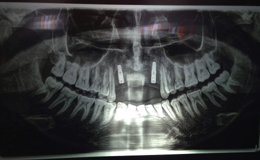
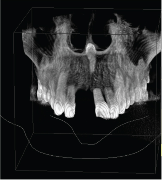
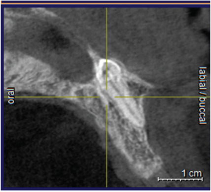
A 25-year-old male reported to the dental clinic with a chief complaint of mobility in the upper front teeth following trauma. On clinical examination, the upper anterior teeth were proclined with grade-III mobility in 11 and grade-II mobility in 12, 21, 22. Orthopantomogram (OPG) revealed bone loss upto the apical third in the upper anterior teeth. Additionally, radio-opacity was seen in the anterior nasal spine which was missed as an artefact [Table/Fig-1]. The anterior teeth were planned for extraction followed by implant placement for which a Cone Beam Computerised Tomogram (CBCT) was taken which showed the panaromic [Table/Fig-2], images of the anterior nasal spine. An inverted tooth like structure was seen in the 3D image. The cross sectional image confirmed the radio-opacity to be a tooth since the enamel, dentin and the pulp were clearly distinguished [Table/Fig-3,4 and 5].
Panaromic View showing a radio-opacity at the lower border of the anterior nasal septum
3D View showing a distal inclination of the root of the supernumerary tooth
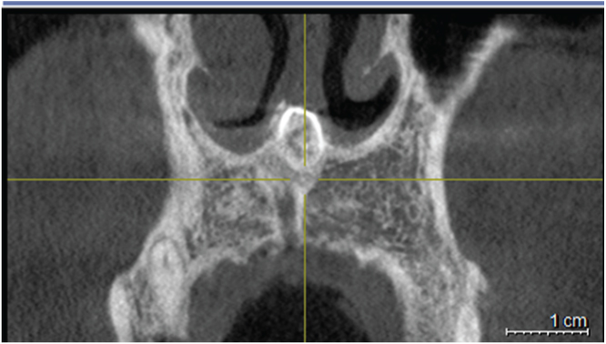
Tangential View confirming the presence of the radio-opacity at the inferior border of the anterior nasal septum
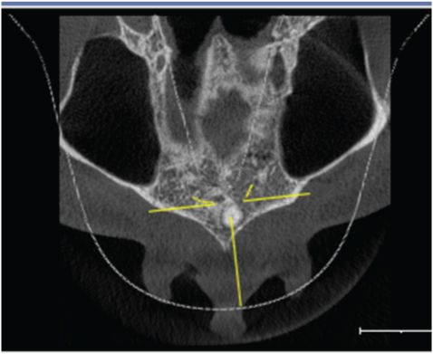
Cross Sectional View which justifies the radio-opacity to be a tooth since the enamel, dentin and pulp structure of the tooth is clearly differentiated
Axial View depicting a slight shift of the ectopic inverted tooth from the midline to the left side
Supernumerary tooth or an addition to the regular number of teeth is a rare developmental anomaly that can occur in any area of the dental arch. When it occurs in between the maxillary central incisors, it is termed as mesiodens. Mesiodens can be presented as labio- palatal impacted, vertical impacted, angular erupted, vertical erupted or inverted. The prevalence of supernumerary teeth in deciduous dentition is 0.3% to 0.8% and 1.5% to 3.5% in the permanent dentition [1]. Supernumerary teeth can be a part of various syndromes like cleft lip/ palate, cleido-cranial dystostosis, Gardner’s syndrome.
The most common complications of mesiodens is crowding, malalignment of teeth, dilacerations of permanent teeth, cyst formation like dentigerous cyst and rarely eruption into the nasal cavity and antrum. Among the supernumerary teeth, the rarest presence is in the anterior nasal spine and anterior septum. These may serve as the nidus for developing rhinoliths. Ectopic teeth in the anterior nasal spine and nasal septum can lead to future complications like rhino-sinusitis, nasal septum abscess, septal perforation, oro-nasal fistula and nasal deformity. Supernumerary teeth may or may not be removed depending on the complications associated with it. A supernumerary tooth warrants its removal if it leads to pathologies like formation of cyst, sinusitis, delay in the eruption of permanent dentition, dental mal-alignment, compromised aesthetics and if it is present in the bone designed for implant placement. [Table/Fig-6] reports the various positions of ectopic supernumerary teeth reported in the literature [2–6].
Ectopic supernumerary tooth reports from the literature
| Author | Position of tooth | Appearance | Complication |
|---|
| Chen [2](Report of3 cases) | Floor of the left nasalcavity between theinferior turbinateand the nasal septum | Straight | Rhinitis |
| Right nasal cavity | Straight | Purulent dischargefrom the nose |
| Left hard palate | Inverted | Aspergillus rhinitis |
| Mohebbi[3] | Intraseptal toothwith nasal obstructionand septal deviation | Inverted | Recurrent sinusitis |
| Clementini[4] | Floor of theright nasal cavity | Inverted withdistal inclinationof the root | Asymptomatic |
| Dhafeeri[5] | Left nasal cavitytouching Little’s area | Straight | Epistaxis,recurrent tonsillitis |
| Prasad[6] | Lateral to the nasalseptum in thenasal cavity | Inverted | Swelling in the nose,nasal obstruction,pain in the upperlabial frenum andphiltrum |
The embryology of the formation of mesiodens is an ongoing debatable topic over centuries. Three theories have been postulated favouring its developmental glitch. The first and the earliest theory of phylogenic reversion postulates that mesiodens could be a phylogenic appearance from our extinct ancestors since they possessed three maxillary incisors, however this theory has now been discarded [7]. The second theory known as the theory of dichotomy suggests that there could be a split in the tooth bud to create two different teeth [8]. The third and the most widely accepted theory is the hyperactivity theory which suggests that mesiodens can occur due to local, independent and conditioned hyperactivity of the dental lamina which results in the proliferation of remnants of dental lamina or the palatal offshoots of the dental lamina which results in an additional tooth bud [9].
The current case presents an inverted mesiodens located at the anterior nasal spine which has been asymptomatic. This was an incidental finding during implant planning of the upper anterior teeth on CBCT and was missed in the routine 2D radiograph OPG. The tooth was however chosen not to be extracted as it was asymptomatic and no associated pathology or developmental defect had been noticed. The patient was informed about the ectopic tooth and was informed for a yearly check-up to monitor the position of the tooth and to manage the complexities if present then.
Conclusion
Ectopic supernumerary teeth have a very rare incidence of being present in the anterior nasal spine which can be missed on a routine radiograph. However in the interest of the patient’s health, we need to be aware and make the patient aware about its presence and the need for its periodic check.
[1]. Mason C, Azam N, Holt RD, Rule DC, A retrospective study of unerupted maxillary incisors associated with supernumerary teethBr J Oral Maxillofac Surg 2000 38(1):62-65. [Google Scholar]
[2]. Chen A, Huang JK, Cheng SJ, Sheu CY, Nasal teeth: report of three casesAm J Neuroradiol 2002 23(4):671-73. [Google Scholar]
[3]. Saleh M, Oveis S, Sedighe E, Ectopic Supernumerary tooth in nasal septum: a case studyIran J Otorhinolaryngol 2013 25(72):183-86. [Google Scholar]
[4]. Clementini M, Morlupi A, Agrestini C, Di Girolamo M, Girolamo S, Ottria L, Endoscopic removal of supernumerary tooth from the nasal cavity of a child: a case reportOral Implantol 2012 5(1):21-25. [Google Scholar]
[5]. Dhafeeri HO, Kavarodi A, Shaikh K, Bukhari A, Hussain O, Baramawy A, Recurrent epistaxis caused by an intranasal supernumerary tooth in a young adultAm J Case Rep 2014 15(5):291-93. [Google Scholar]
[6]. Prasad RG, Nair PP, Gharote H, Hegade K, Agarwal K, Jain A, Intranasal tooth—an ectopic eruption of mesiodens in nasal cavity: a case report and reviewIndian Acad Oral Med & Radiology 2011 23(3):252-55. [Google Scholar]
[7]. Von AT, Anterior maxillary supernumerary teeth: a clinical and radiographic studyAust Dent J 1992 37(3):189-95. [Google Scholar]
[8]. Russell KA, Magdalena AF, Mesiodens- Diagnosis and management of a common supernumerary toothJ Can Dent Assoc 2003 69(6):362-66. [Google Scholar]
[9]. Primosch RE, Anterior supernumerary teeth- assessment and surgical intervention in childrenPediatr Dent 1981 3(2):204-15. [Google Scholar]