Popliteal Artery Thrombosis after Total Knee Replacement: An Unusual Complication
Jayanth Kumar Bangalora Chikkanna1, Deepak Sampath2, Varaprasad Reddy3, Vishnu Motkuru4
1 Senior Consultant and Unit Chief, Department of Orthopaedics, St Martha’s Hospital, No. 5, Nrupathunga Road, Bangalore, Karnataka, India.
2 Consultant, Department of Orthopaedics, St Martha’s Hospital, No. 5, Nrupathunga Road, Bangalore, Karnataka, India.
3 Resident, Department of Orthopaedics, St Martha’s Hospital, No. 5, Nrupathunga Road, Bangalore, Karnataka, India.
4 Consultant, Vascular Surgeon, St Martha’s Hospital, India.
NAME, ADDRESS, E-MAIL ID OF THE CORRESPONDING AUTHOR: Dr. Jayanth Kumar Bangalora Chikkanna, No. 139, 2B Cross, Ombr Layout, Banaswadi, Bangalore, Karnataka-560043, India.
E-mail: drdeepuortho@gmail.com.
Limb ischemia, Popliteal artery occlusion/thrombosis, Total knee arthroplasty
Popliteal artery occlusion after total knee replacement is a rare complication with direct injury being the most common cause. We present a case that developed critical leg ischemia on 2nd post operative day after total knee replacement. The possible cause of the Arterial occlusion was thought to be secondary to atherosclerotic plaque in the popliteal artery. Timely Communication between the orthopedic team and vascular surgeon led to salvage of the limb. This is the first reported case of a post-TKA popliteal artery thrombosis in a patient younger than 60 years without the commonly accepted risk factors.
A 54-year-old female patient with no co-morbidity was admitted in St Martha’s Hospital, orthopedic department for left total knee replacement, she had under gone right total knee replacement 12 months ago without any complications. Physical examination of left knee revealed tenderness on the medial and lateral joint lines, and range of motion (ROM) was 0° to 95° with full extension and a mild varus deformity that was correctable with valgus stress suggesting minimal mediolateral instability. Skin examination was unremarkable, and there were palpable pulses with no sensory deficit. Patient had difficulty in walking and climbing stairs. Radiographs showed tricompartmental osteoarthritis changes with a slight varus deformity and no soft-tissue abnormalities. After obtaining medical fitness patient underwent left posterior stabilized TKA.
The surgery was completed through a standard midline approach and a median Para patellar arthrotomy. The tourniquet was applied and elevated to 330 mm Hg for a total of 70 minutes and was let down after the cement had polymerized. The wound was closed in standard fashion with no significant bleeding. In the recovery room, the patient was found to be stable, distal pulses were well felt in the left leg.
Postoperatively Epidural infusion (Bupivacaine 0.125% and Clonidine 2 μg/ml) was started at 6 ml per hour. Night rounds patient was comfortable with epidural analgesia with bilateral equal pulsations in foot. On next day morning patient complained of severe pain in left calf and numbness in left lower limb, mainly below the knee joint and on examination the left dorsalis pedis and posterior tibial artery pulsation were absent with normal left femoral artery pulsation. Postoperative x-ray in both anterior and lateral view was satisfactory. Toes on the left leg were slightly cooler than on the right and capillary refill time was prolonged. Colour Doppler examination was done which revealed no flow at the level of the knee and below [Table/Fig-1]. Vascular surgeon was immediately consulted and patient was subjected to emergency angiogram followed by thrombectomy and fasciotomy by double incision technique under epidural analgesia on the same day (2nd postoperative day) [Table/Fig-2] and patient was shifted to intensive care unit for next two days with intravenous antibiotics plus heparin infusion, post embolectomy angiogram showed good flow [Table/Fig-3]. On 3rd postoperative day patient had good pulsation on periphery.
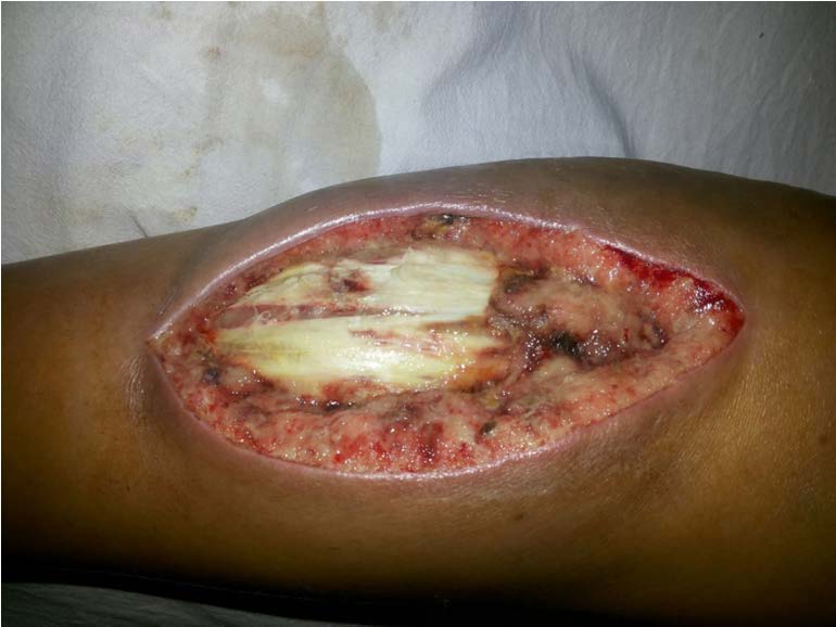
Faciotomy on medial side left lower limb
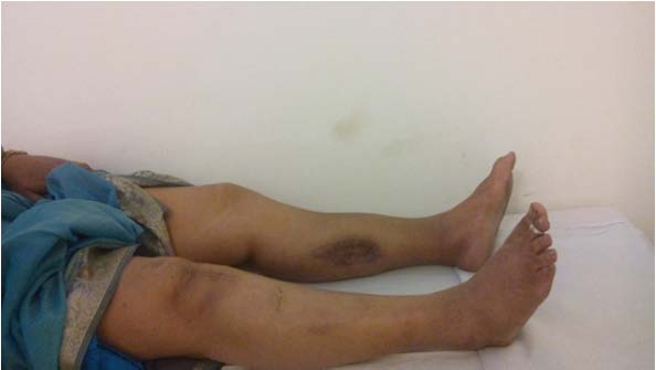
Healed scar of total knee replacement

Healed fasciotomy wound medial side of leg after SSG
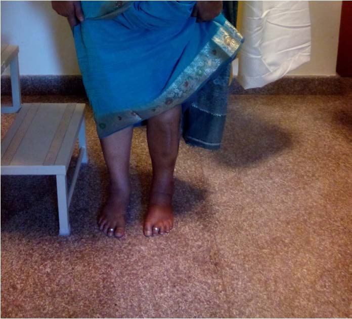
But to our surprise on 4th postoperative day pulses were again feeble with saturation dropping to 85% in left toes, suspecting reblock. Doppler scan showed biphasic flow with no block, vascular surgeons decided to start on prostaglandins infusion to help vasodilatation. After 24 hours of prostaglandin infusion, circulation had established, patient was able to do plantar flexion against resistance, with grade 1 dorsi flexion. After wound was healthier, patient underwent Split Skin Grafting for the fasciotomy wound on lateral aspect, medial aspect wound was sutured [Table/Fig-4,5 and 6]. At the end of one year follow up patient has partially recovered foot drop with wound healed completely [Table/Fig-7].
Healed fasciotomy wound on lateral side of leg
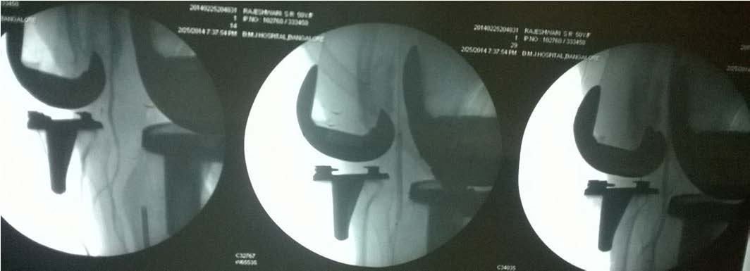
Angiography of left lower limb, demonstrating the decreased flow below the popliteal atherosclerotic plaque
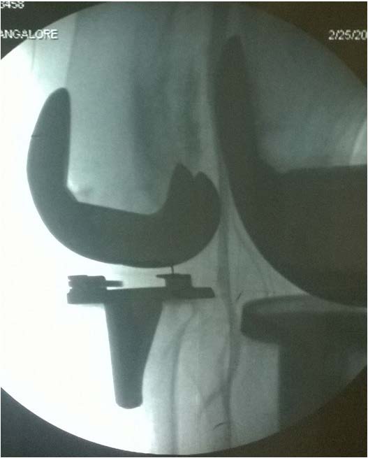
Angiography of left lower limb, post angioplasty flow has increased, with good flow of blood distally
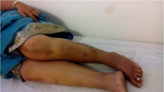
Postoperative function of same lady, walking without foot drop splint, partial recovery of the foot drop noted
Arterial vascular injury is a rare complication of TKA, incidence is about 0.03–0.17% [1,2]. Atherosclerotic occlusion, thrombosis or direct sharp traumas are the most common cause for vascular injury. Preoperative risk factors are history of claudication pain, rest pain, absence of distal pulses, arterial ulcers, popliteal aneurysm, previous arterial reconstruction and calcification of arteries on plain radiographs [3].
Patient had undergone right total knee arthroplasty, one year back in our hospital with same surgeon with similar technique, under epidural analgesia with uneventful postoperative recovery.
We had not done preoperative arterial Doppler for this patient, since patient had good peripheral pulsation, retrospectively analysing after angiogram, vascular surgeon contemplated on stenosis or atherosclerosis plaque of popliteal artery as cause of thrombosis, we were in sighted about our need for arterial Doppler in all subsequent cases, which adds on the cost of TKA, but worth outweighing the risk of vascular complication postoperative.
In our case of popliteal artery occlusion after total knee joint replacement, timely communication between the vascular and orthopaedic team led to diagnosis and management of this serious, debilitating complication. The most important takeaway message from this case is to have high index of suspicion in old atherosclerotic patients, about atherosclerosis of popliteal artery in all TKR [4].
A high index of suspicion and through assessment of risk factors for postoperative arterial complication should be maintained in the form of monitoring limb colour (pallor), temperature peripheral pulses, and if necessary pre-operative Doppler assessment of the limb to prevent this limb-threatening complication.
[1]. Da Silva MS, Sobel M, Surgeons of the Southern Association of Vascular Surgery. Popliteal vascular injury during total knee arthroplastyJ Surg Res 2003 109:170 [Google Scholar]
[2]. Calligaro KD, Dougherty MJ, Ryan S, Booth RE, Acute arterial complications associated with total hip and knee arthroplastyJ Vasc Surg 2003 38:1170-77. [Google Scholar]
[3]. Holmberg A, Milbrink J, Bergqvist D, Arterial complications after knee arthroplasty. 4 cases and a review of the literatureActa Orthop Scand 1996 67:75-78. [Google Scholar]
[4]. Khan Hamid Salam, John Kessels, Popliteal Artery Occlusion After Total Knee Replacement: A Vascular Team Approach for Limb SalvageVascular Disease Management 2014 11(9):300-05. [Google Scholar]