Choriocarcinoma with Uterine Rupture and Shock: A Rare Case Report
Manika Agarwal1, Ritesh Kumar2, Jupirika Pyrbot3, A.S. Singh4
1 Associate Professor, Department of Gynaecology, North East Indira Gandhi Regional Institute of Health and Medical Sciences (NEIGRIHMS), Shillong, Meghalaya, India.
2 Assistant Professor, Department of Oncology, North East Indira Gandhi Regional Institute of Health and Medical Sciences (NEIGRIHMS), Shillong, Meghalaya, India.
3 Senior Resident, Department of Gynaecology, North East Indira Gandhi Regional Institute of Health and Medical Sciences (NEIGRIHMS), Shillong, Meghalaya, India.
4 Professor, Department of Gynaecology, North East Indira Gandhi Regional Institute of Health and Medical Sciences (NEIGRIHMS), Shillong, Meghalaya, India.
NAME, ADDRESS, E-MAIL ID OF THE CORRESPONDING AUTHOR: Dr. Ritesh Kumar, Assistant Professor, Department of Oncology, North East Indira Gandhi Regional Institute of Health and Medical Sciences, Shillong, Meghalaya, India.
E-mail: riteshkr9@gmail.com
Choriocarcinoma is a rare neoplasm and a malignant form of gestational trophoblastic disease. Choriocarcinoma is frequently preceded by a complete mole, ectopic pregnancy, nonmolar intrauterine abortion, and uncommonly by a partial mole. It is treated medically with chemotherapeutic drugs usually. However, we managed to save a life with appropriate and timely surgical intervention in a case of choriocarcinoma who presented with uterine rupture, haemoperitoneum, anaemia and hypovolemic shock. The patient underwent exploratory laparotomy and hysterectomy followed by systemic chemotherapy.
Gestational trophoblastic disease, Hysterectomy, Neoplasm, Partial mole
Case Report
A 28-year-old P2 was admitted with complaints of amenorrhea for 4 months with irregular bleeding per vaginum. She had complaints of difficulty in breathing and abdominal distension since 4 days. Urine for pregnancy test was positive. She had Hb of 3.5gm%. White blood count, platelets, coagulation profile, liver function tests and renal function tests were normal. Ultrasound showed grossly enlarged mass with miscellaneous echogenic shadows in the myometrium with loss of endometrial junction, multi cystic ovaries with massive ascites and a diagnosis of Gestational Trophoblastic Disease (GTD) was made. Her last child birth was 2 years back and she did not have any history of abortion or molar pregnancy in the past. The patient had features of hypovolemic shock with anaemia. She had received three units of blood transfusion and was on ionotropic support. Then the patient was referred to our institute for further management.
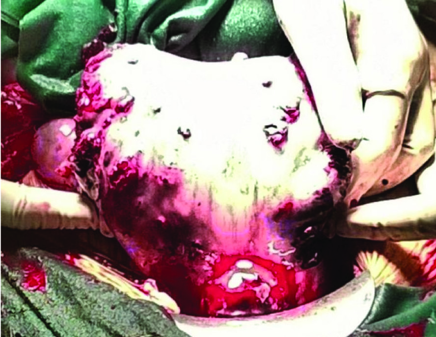
On arrival at the casualty, she was afebrile, had tachycardia, her blood pressure was 80/40 mmHg. On per abdomen examination her abdomen was distended with free fluid present. On per vaginum examination there was no bleeding. Ascitic tapping was carried out and haemoperitoneum was noted. The patient was immediately prepared for an emergency laparotomy. Intraoperatively, there was massive haemoperitoneum and around 4 litres of blood was present, the uterus was enlarged showing multiple haemorrhagic nodules on the surface with rupture at the fundus [Table/Fig-1]. Bilateral ovaries were enlarged with multiple cystic lesions. The omentum was adherent to the fundus of the uterus at the site of uterine perforation. Total abdominal hysterectomy was carried out with partial cystectomy and omental biopsy. Intraoperatively 4 units of packed red blood cells were transfused. On cut section the uterus showed reddish friable haemorrhagic mass 7X6X8 cm involving the myometrium upto the serosa [Table/Fig-2].
Intraoperative photograph showing multiple nodules on surface of uterus
Cut section of uterus showing a reddish friable haemorrhagic mass involving the myometrium upto the serosa (Arrows showing the rupture site)

During her postoperative period the patient recovered well. Histopathological evaluation revealed intimately admixed biphasic pattern of choriocarcinoma, with vascular invasion and bizzare cells with mitotic figures [Table/Fig-3]. No villous structures were identified. Surface nodules on uterus were histologically choriocarcinoma deposits. Omentum and partial cystectomy specimen were infiltrated with choriocarcinoma deposits. Further staging evaluation revealed multiple lung metastases in both lungs and serum βHCG 7,00,000. Thus a final diagnosis of high risk GTD (Stage IV) was made and she was started on chemotherapy with Etoposide, Methotrexate, Actinomycin D, Cyclophosphamide and Vincristine (EMACO regimen) in standard doses. There was a fall in the serum βHCG levels in subsequent follow-up and the patient was responding to the chemotherapy.
Microphotograph showing intimately admixed biphasic pattern of choriocarcinoma (H&E Stain) (100X) with inset-showing vascular invasion and bizzare cells with mitotic figures

Discussion
Uterine choriocarcinoma is a rare neoplasm arising from uterus and is the malignant form of Gestational Trophoblastic Disease (GTD). It is characterized by the invasion of trophoblastic tissue into the myometrium and has high propensity for distant metastasis [1]. It usually presents with amenorrhea and abnormal bleeding. Other less common symptoms are haemoptysis, cerebral bleeding and liver haematoma. It can rarely present with acute abdomen secondary to haemiperitoneum and uterine rupture as in the index case. Choriocarcinoma causing uterine rupture and subsequent haemoperitoneum is a rare presentation [2]. There are case reports of haemoperitonem secondary to hepatic metastasis, pulmonary metastasis and spleen metastasis. Haemoperitoneum secondary to invasive mole is a common presentation but is rarely seen in choriocarcinoma [2]. The exact pathogenesis of rupture of the uterus in choriocarcinoma is not known, however, the various theories have been proposed to explain the mechanism of uterine rupture. Malignant trophoblasts invade the uterine veins and damages the blood vessel. Subsequent to the vascular damage, multiple infarctions do occur due to thrombosis, vascular aneurysms and intratumoral bleeding [3].
The rare presentation in our case is that the patient did not have any preceding history of an abortion, ectopic pregnancy and her last child birth was 2 years back. Significant amount of bleeding occurred due to uterine rupture, and the patient was admitted to the peripheral hospital with an acute abdomen.
Typically a differential diagnosis of ectopic pregnancy with haemoperitoneum, the rupture of a corpus luteum cyst and invasive mole should be considered. Ultrasonography of the pelvis along with serum β-hCG levels helps in the diagnosis of choriocarcinoma. Preoperative β-hCG level and other radiological investigations could not be done in the index case because the patient presented with shock and haemoperitoneum and immediate exploratory laparotomy was planned.
Patients with acute abdomen from uterine perforation and shock should be managed aggressively and bleeding focus has to be managed with surgical intervention. A total abdominal hysterectomy (TAH) is the preferred approach in females especially with family completed. Patients under emergency conditions who are operated with explorative laparotomy due to severe active intra-abdominal bleeding usually requires TAH as life saving procedure. However, conservative surgery is the preferred approach in females with disease limited to the uterus and who wish to preserve their fertility [4]. In the index case, total abdominal hysterectomy was planned as the patient has her family completed. Invasive mole may perforate through the myometrium resulting in uterine perforation and intraperitoneal bleeding. But uterine perforation due to choriocarcinoma is rare [5].
The International Federation of Obstetrics and Gynecology (FIGO) grading and scoring system is recommended for classifying the disease into the low-risk and high-risk groups [6]. It guides the suitable treatment for GTD cases and further chemotherapy regimens to be used. Single-agent chemotherapy with Injectable Methotrexate is used in non-metastatic and low-risk choriocarcinomas (score ≤6). Multi-agent chemotherapy and radiotherapy are used for high-risk, metastatic choriocarcinomas (score >6) [6,7]. In our case multi drug regimen (EMACO) was started in view of high risk disease.
Conclusion
Choriocarcinoma with uterine rupture and shock is a rare clinical presentation. Timely intervention in form of surgery and chemotherapy can save a precious life. A high degree of clinical suspicion is needed in proper management of these cases.
[1]. Froeling FEM, Seckl MJ, Gestational trophoblastic tumours: an update for 2014Current oncology reports 2014 16(11):1-10. [Google Scholar]
[2]. Liberis V, Bouchlariotou S, Ammari A, Psillaki A, Ntatidou M, Sivridis E, Acute abdomen as initial presentation of gestational choriocarcinomaArch Gynecol Obstet 2009 280:859-62. [Google Scholar]
[3]. Ma Y, Xiang Y, Wan X, Chen Y, Feng F, Lei C, The prognostic analysis of 123 postpartum choriocarcinoma casesInt J Gynecol Cancer 2008 18:1097-101. [Google Scholar]
[4]. Xie C, Zheng L, Li Z, Zhao X, Spontaneous uterine perforation of choriocarcinoma with negative beta-human chorionic gonadotropin after chemotherapyMed Princ Pract 2011 20:570-73. [Google Scholar]
[5]. Mackenzie F, Mathers A, Kennedy J, Invasive hydatidiform mole presenting as an acute primary haemoperitoneumBr J Obstet Gynecol 1993 100:953-54. [Google Scholar]
[6]. Kohorn E, The new FIGO 2000 staging and risk factor scoring system for gestational trophoblastic disease: description and critical assessmentInt J Gynecol Cancer 2001 11:73-77. [Google Scholar]
[7]. Bolze PA, Attia J, Massardier J, Seckl MJ, Massuger L, van Trommel N, Formalised consensus of the European Organisation for Treatment of Trophoblastic Diseases on management of gestational trophoblastic diseasesEuropean Journal of Cancer 2015 51(13):1725-31. [Google Scholar]