Temporomandibular joint (TMJ) disorders accounts for about 10% of the population with female predilection [1].
TMJ dysfunction is a therapeutic challenge in the oral and maxillofacial clinic. Although TMJ pain and dysfunction can be caused by many different aetiologic factors, the role played by inflammation as an underlying mechanism of pain and dysfunction of the TMJ has played a major role. TMD patients having pain and tenderness for prolonged period of time will show signs of inflammation biochemically as well as radiographically [2].
Various treatment modalities available for TMD patients are, arthrocentesis, arthroscopic lysis and lavage and arthrotomy [3]. An important change in the therapeutic approach occurred with the introduction of arthroscopic lavage and lysis for the treatment of TMDs. The success of arthroscopy has led to the use of arthrocentesis as a simple therapeutic modality with a satisfactory outcome [4].
The procedure of arthrocentesis involves aspiration of the joint cavity with injection of therapeutic substance in the superior joint space that causes release of the disc and ultimately resulting in increased mouth opening [5,6]. Arthrocentesis has proved to be a minimally invasive treatment modality, relatively safe, repeatable and it can be done on out patients under local anaesthesia which significantly reverts the mouth opening to a normal range. It is an effective method of normal disc-condyle-fossa relationship [6].
Several authors have conducted studies to detect effectiveness of arthrocentesis in various TMJ disc disorders. Hence, in the light of previous studies, the present study evaluated the effectiveness of arthrocentesis in TMJ disc disorder patients.
Materials and Methods
The present study was conducted in the Department of Oral and Maxillofacial Surgery, Al-Ameen Dental College and Hospital, Bijapur, Karnataka. The study population was drawn from the outpatient Department of Oral and Maxillofacial Surgery, Al-Ameen Dental College and Hospital, Bijapur, Karnataka, India.
A minimum of 50 patients with TMJ disc disorder who were clinically diagnosed as per the norms laied down by Kaplan were selected [7]. In all, extreme care was taken to selectively include and exclude the subjects mentioned below.
Inclusion Criteria: We included the patients with recent history of pain, limited mouth opening of less than 30 mm, impeded lateral movement towards unaffected side, deviation towards affected side and patients who are not responding to nonsurgical treatment.
Exclusion Criteria: We excluded the patients having undergone invasive procedures, with psychological problems, with a history of bony or fibrous adhesion, gross mechanical restriction and patients having condylar fractures.
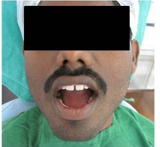
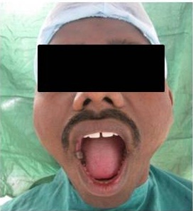
The clinical examination was done including the evaluation of the maximal mouth opening which was measured by the distance between the incisal edges of the upper and lower incisors with the help of a caliper [Table/Fig-1]. Determination of the ‘range of the lateral and protrusive mandibular movement’ measured by the distance between the upper and lower midline on lateral and forward movements by using a caliper.
hotograph showing difficulty in opening mouth
Pain level and location were determined by the patients self assessment using VAS ranging from 0 to 108. After thorough TMJ evaluation and clinical examination, orthopantamograph and transpharyngeal view of the patient were taken. The diagnosis of TMJ disc disorder was made on the basis of history, clinical and radiologic examination as per the norms laied down by Kaplan [7].
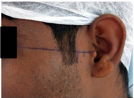
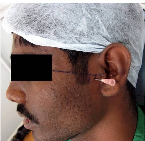
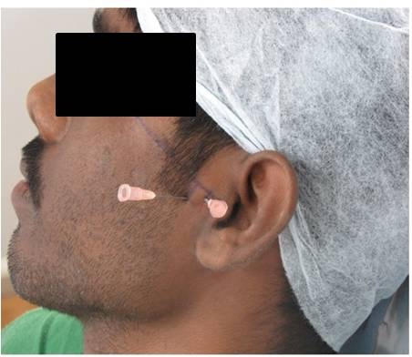
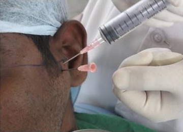
Method of Arthrocentesis: After proper preparation of the target site, external auditory meatus were blocked with cotton soaked in normal saline. Two points of needle insertion were marked over the skin of the affected joint indicating the articular fossa and eminence [Table/Fig-2]. A line was drawn from the middle of the tragus to the outer canthus. The posterior entrance point was located along the canthotragal line, 10mm from the middle of the tragus and 2mm below the line. This posterior point is only for pumping the fluid into the upper compartment to increase the hydraulic pressure within the joint [Table/Fig-3]. The anterior point of entry was placed 10mm farther along the line and 10mm below it [Table/Fig-4]. This was followed by the injection of local anaesthesia to block the auriculotemporal nerve. An 18-gauge needle was then inserted into the superior compartment of the anterior auricular fossa (posterior mark), followed by the injection of 2-3ml of Ringer’s Lactate solution to distend the joint space. We inserted 18 gauge needle for the fluid to come out from the superior compartment. Ringer’s Lactate solution was then connected to one of the needle with sufficient pressure to assure the free flow of 200ml solution during 15-20min period which was achieved by inserting a syringe at an elevation of 1cm above the level of the joint [Table/Fig-5]. During the procedure, exact timing of re-establishment of normal mouth opening determined by having the patient to make the repeated attempts to open the mouth [Table/Fig-6]. On termination of the procedure steroid (dexamethasone 8mg) was injected into the joint space followed by the removal of needle. Postoperatively antibiotic and analgesics with muscle relaxants for 2 weeks were prescribed. Follow up of the patient was done after 1 year.
hotograph of the patient showing markings in the preauricular area
hotograph of the patient showing single needle in position
hotograph of the patient showing two needles in position
hotograph of the patient showing the procedure of arthrocentesis
hotograph of the patient showing mouth opening immediately after the procedure
Results
The present study comprised of 50 subjects with age range of 17 to 66 years. The subjects were followed up for a period of 1 year.
Objective findings Following Treatment
Maximal mouth opening: The maximal mouth opening prior to arthrocentesis ranged from 20-44mm with a mean value of 32.13mm and a SD of 9.86mm. The maximal mouth opening 1 year following arthrocentesis ranged from 41-50mm with a mean value of 46.6mm and a SD of 2.56mm [Table/Fig-7].
Correlation of maximal mouth opening before and after arthrocentesis
| Groups | n | Maximal mouth opening |
|---|
| Range | Mean±SD |
|---|
| Before the procedure | 50 | 20-44 | 32.13±9.86 |
| 1 year after the procedure | 50 | 41-50 | 46.6±2.56 |
| t | 8.05 |
| p* | <0.001 HS |
* Highly significant
Lateral mandibular movements: The right lateral movement prior to arthrocentesis ranged from 5-9mm with a mean value of 7.15mm and a SD of 1.25mm.
The right lateral movement 1 year following arthrocentesis ranged from 8-10.8mm with a mean value of 9.49mm and a SD of 0.61mm.
The left lateral movement prior to arthrocentesis ranged from 5.5-10mm with a mean value of 7.59mm and a SD of 1.26mm. The left lateral movement following arthrocentesis ranged from 8-11mm with a mean value of 9.31mm and a SD of 0.70mm [Table/Fig-8].
Correlation of lateral movements before and after arthrocentesis
| Groups | n | Lateral movements |
|---|
| Right | Left |
|---|
| Range | Mean±SD | Range | Mean±SD |
|---|
| Before the procedure | 50 | 5-9 | 7.15±1.25 | 5.5-10 | 7.59±1.26 |
| 1 year after the procedure | 50 | 8-10.8 | 9.49±0.61 | 8-11 | 9.31±0.70 |
| t | 9.55 | 6.217 |
| p* | <0.001 HS | <0.001 HS |
* Highly significant
Subjective Findings Following Treatment: The degree of pain experienced by patients before arthrocentesis ranged from 6-10 with a mean value of 8.7 and a SD of 1.1. The degree of pain after arthrocentesis ranged from 1-4 with a mean value of 1.13 and a SD of 1.16 [Table/Fig-9].
Correlation of degree of pain before and after arthrocentesis
| Groups | n | Degree of pain (0-10) |
|---|
| Range | Mean±SD |
|---|
| Before the procedure | 50 | 6-10 | 8.7±1.1 |
| 1 year after the procedure | 50 | 1-4 | 1.13±1.16 |
| t | 21.97 |
| p* | <0.001 HS |
* Highly significant
Discussion
Temporomandibular disorders which will present itself as pain, clicking sound and deviation are collectively the disorders of joint and muscles [9]. It has been suggested that classification, diagnosis and treatment of TMJ pain and dysfunction can be based on the position and shape of the articular disc. The question of whether disc displacement is the result, cause, or an accompanying factor in dysfunction remains open to debate [8–10].
The management of refractory pain in the TMJ is both challenging and controversial for Oral and Maxillofacial Surgeons. Controversy continues to surround the role of surgery in the management of pain and dysfunction of the TMJ, although only about 5% of all patients being treated for TMJ disorders are actually operated [11]. The success rate of non-surgical treatments according to many authors is approximately 60%. However, certain group of patients receiving non-surgical treatment may remain unresponsive, thereby prolonging their suffering and treatment dissatisfaction [12]. As the procedures like Arthrocentesis and Arthroscopy are having high success rates with minimal complication, they are used most commonly as a first line of surgical treatment along with nonsurgical treatment [12]. Arthrocentesis is minimally invasive technique with high success rates, very easy to perform and very effective in reliving the pain and ultimately increasing the mouth opening [13].
Thus with the aim to know the efficacy of arthrocentesis in patients suffering from TMJ disorders, the present study was conducted.
The present study showed significant increase in the mouth opening from 32.13mm to 46.6mm. The findings were similar to those of Nitzan DW et al., (24.1mm to 42.7mm), Dimitroulis G et al., (24.6mm to 42.3mm), Fridrich KL et al., (33mm to 41mm), Hosaka H et al., (30.6mm to 44.5mm), Nitzan DW et al., (23.1mm to 44.26mm), Carvajal WA et al., (25.3mm to 43.8mm), Nitzan DW et al., (24.4mm to 43.20mm), and Dhaif G et al., (13.1mm to 43.7mm) [4,6,14–19].
The findings were higher to those of Yeung RWK et al., (38.2mm to 39.8mm), and Onder ME et al., (33.6mm to 38mm) [20,21].
The variations in the values could be due to the differences in the use of intra-articular medications used for the treatment as is the case in the study done by Yeung RWK et al., and Onder ME et al., who used hyaluronic acid for intra-articular injection. But in our study we have used Ringer’s Lactate solution for intra-articular injection [20,21]. There was significant increase in the right and left lateral movement from 7.15mm and 7.59mm to 9.49mm and 9.31mm respectively.
The values were in consistent with that of Nitzan DW et al., (6.30mm to 9.40mm), who used intra articular injection of steroids as was done in our study, however the values were slightly higher to those of Nitzan DW et al., (4.8mm to 8.2mm) and the values were slightly lower compared to the study done by Nitzan DW et al., (3.75mm to 10.5mm) who did not use intra-articular steroid [4,14,19].
The variations in the values could be due to the differences in the use of intra-articular medications used for the treatment as is the case in the study done by Nitzan D W et al., who did not use intra-articular steroids [14]. The degree of pain experienced by patients before arthrocentesis ranged from 6-10 with a mean value of 8.7 and a SD of 1.1. The degree of pain after arthrocentesis ranged from 1-4 with a mean value of 1.13 and a SD of 1.16. The findings were similar to the findings of Hosaka H et al., (6.9 to 1.1), Nitzan DW et al., (9.24 to 1.4), Goudot P et al., (5.6 to 0.9) and Emshoff R et al., (77 to 13.9) [17,19,22,23]. The findings were slightly higher than that of Dhaif G et al., (6.9 to 0.6) [6].
The findings were lower to the findings of Nitzan DW et al., (9.86 to 3.39), Nitzan DW et al., (8.75 to 2.31), Dimitroulis G et al., (8.8 to 2.2), Fridrich KL et al., (66 to 23), Carvajal WA et al., (8.26 to 2.03), Yeung RWK et al., (4.2 to 2.6) and Onder ME et al., (71 to 32) [4,14–16,18,20,21].
The probable explanation that could be given due to the variations in the values is short follow up period and variations in the postoperative medications given. In our study, we have used two needle technique along with injection of dexamethasone as described by Nitzan DW et al., and postoperatively antibiotics and analgesics with muscle relaxants were prescribed for two weeks [14]. In the study done by Dimitroulis G et al., Nitzan DW et al., and Nitzan DW et al., intra articular injection of steroid was not given. In another study done by Hosaka H et al., single needle technique was used [4,15,17,19].
Postoperative medication of Diazepam 10mg was used in the study done by Dhaif G et al., [6]. Intra articular injection of hyaluronic acid was given by Yeung RWK et al., and Onder ME and there was no significant increase in the mouth opening and reduction in the intensity of pain [20,21]. The use of intra articular steroid as done by, Carvajal WA, Emshoff R and in our study yielded a highly significant results [14,18,23].
Limitation
However, these findings need to be carefully interpreted due to small sample size and short follow up period.
Conclusion
The findings of the present study revealed significant increase in maximal mouth opening, lateral movements and significant reduction in TMJ pain after a period of 1 year follow up and suggested potential utility of arthrocentesis in the management of TMJ disc disorders.
Further research involving a larger sample and a longer follow up period is suggested along with extensive work involving specificity of technique and more knowledge on the aetiology of the TMJ disc disorders.
* Highly significant
* Highly significant
* Highly significant