Dermatoglyphics has always been fascinating, not only to anthropologists and medical practitioner, but also to psychologists, writers, printers and palmist [1]. Every individual has unique patterns on the palms and sole which are used for personal identification [2]. Etymologically “Dermatoglyphics” is a harmonious blend of two words Derma which means skin and Glyphe meaning carve. It gives the impression that something has been carved out of the skin [2]. Cummins in the year 1926 coined the term dermatoglyphics to this field of science and is regarded as the “Father of Dermatoglyphics” [3].
The genesis of the dermal ridges occurs with relation to the volar pads. The dermal ridge appear during the 12th week of the intrauterine life and are completed by the 24th week of intrauterine (I.U) life i.e. the same time as that of tooth formation in intrauterine life. This conveys that the genetic meaning contained in the genome, normal or abnormal, is decoded during this stage and could also be replicated by dermatoglyphics [3]. The environmental factors influenced or modified the resulting ridges which are genetically determined [4–7]. The ectoderm, from which the epidermis is derived, plays an important role in the configuration of several structures such as the teeth [8]. If an intrauterine dermal damage take place, a tooth anomaly might be expected [9].
Caries is one of the most common chronic disease present in children worldwide, irrespective of the advancements in oral healthcare, countless adults and children are still affected [10]. Early Childhood Caries (ECC) is a significant public health problem in both developing and industrialized countries which continues to affect babies and preschool children worldwide [10]. It is a chronic, transmissible, infectious disease with a complex and multifactorial aetiology [10]. According to the American Academy of Pediatric Dentistry (AAPD) guidelines the disease of ECC is the presence of 1 or more decayed (noncavitated or cavitated lesions), missing (due to caries), or filled tooth surfaces in any primary tooth in a child under the age of 6 [11]. In children younger than 3 years of age, any sign of smooth-surface caries is indicative of severe early childhood caries (S-ECC) [11]. From ages 3 through 5, 1 or more cavitated, missing (due to caries), or filled smooth surfaces in primary maxillary anterior teeth or a decayed, missing, or filled score of ≥4 (age 3), ≥5 (age 4), or ≥6 (age 5) surfaces also constitutes S-ECC [11]. The cause of caries is multifaceted and includes environmental and genetic factors. The extent of each of these factors causative to dental caries can change significantly on an individual basis [12].
Though early childhood caries is not life threatening its impact on individuals and communities is significant; resulting in pain, mutilation of function, deleterious influence on the child’s growth rate, body weight and ability to thrive, thus dropping the quality of life [13].
Although there are a variety of methods to identify ECC but there is no method to predict it [14]. The foundation of considering dermatoglyphic pattern as genetic marker for dental caries is that the epithelium of finger buds as well as enamel which is the most vulnerable dental tissue to dental caries has an ectodermal origin and both develop at the same point in time of intra Uterine life [14, 15]. Thus with genetic susceptibility and added environmental factors the proneness for caries due to abnormality in the tooth structures may be reflected in the dermatoglyphics namely whorls, loops and arch patterns [16].
The aim of the present study was to assess dermatoglyphic patterns and correlate them with early childhood caries. It was hypothesized that since dental caries has potential genetic contribution an association between dermatoglyphic and dental caries could be compared.
Materials and Methods
The present study was conducted in the Outpatient Department (OPD) of Department of Pedodontics and Preventive Dentistry in Saraswati Dental College, Lucknow. Some of the children were also examined in the schools located in Lucknow. The study protocol was approved by the ethical committee of Saraswati Dental College. Consent was obtained from the parents and respective school authorities.
Study population: Study population included children consulting the Outpatient Department as well as those in different schools examined during dental camps. Children belonging to the age group of 3-6 years were selected. The duration of the study was 6 months and the sample collection was conducted during the school hours and of the Departments OPD timings.
Study design: The study sample consisted of 100 children aged between 3 and 6 years, divided into two groups of 50 children each. The deft score was evaluated to select the experimental group and control group.
Dermatoglyphic pattern recording and interpretation: Cummins and Midlo’s ink method was used to record finger and hand prints [17,18]. Children’s hands were washed with soap and water to eradicate any dirt and oil from the ridged skin and were air dried to improve the quality of finger and palm prints.
Finger prints: Black duplicating ink was used to record finger prints (both right and left hand) of all the subjects which were applied on the fingers with sponge head rolling paint brush. The benefit of using black duplicating ink was that the prints achieved were clear and did not get smudged and the prints could be preserved for an indistinct period of time. The digits were guided and pressed tightly against the white bond paper clipped on to a hard board.
Palm prints: Black duplicating ink was used to record the palm prints (both right and left hand) of all the subjects which was smeared on the palms and were pressed on a bond sheet which was kept firm.
Method of Reading Handprints: The handprints obtained were checked for their clarity with a magnifying glass (×2) and coded [19]. The presence of core and the triradii of the dermatoglyphic pattern were checked thoroughly to include the handprint in the study. A total of 1000 digital prints and 200 palmar prints were obtained.
Qualitative Dermatoglyphic Analysis
Type of dermatoglyphic pattern: The frequency of true patterns of loops, whorls and arches were counted on the fingertips of all the 10 digits of children with ECC and caries-free children. They were assessed for increase or decrease in mean frequencies.
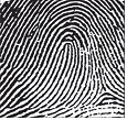
A loop [Table/Fig-1] is documented as a series of ridges that enter the pattern area on one side of digit, recurves abruptly and leaves the pattern area on the same side. A single triradius is present, which is located laterally on the fingertip, where the loop is closed [5].
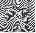
A whorl [Table/Fig-2] varies from the loop in the feature of concentric arrangement of ridges, with two or more triradii in the latter. A whorl may be spiral, symmetrical, double looped, central-pocketed or accidental, depending upon the internal structure of the whorl pattern [5].
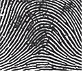
The arches [Table/Fig-3] demonstrate the simplest ridge pattern, which is created by the succession of one or more parallel ridges, which cross the finger from one side to the other without recurving. These patterns generally do not show the presence of triradii, apart from the tented arch is present that will have a triradii point near its midline [5].
Quantitative Dermatoglyphic Analysis
‘atd’ angle
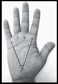
The ‘atd’ angle is a trait of the palm that reveals the position of three triradii-‘a’ and ‘d’, usually located on distal palm just inferior to the 2nd and 5th fingers, respectively and ‘t’ whose location can vary on the proximal palm from just distal to the wrist, up to the center of the palm. The atd angles [Table/Fig-4] were measured for each palm print by drawing two straight lines through the ‘a’ and ‘t’ triradii and the ‘d’ and ‘t’ triradii and measuring the resulting angle. The atd angles were compared and assessed for increase or decrease in mean frequencies between the groups [20].
Results
The evaluation and comparison of patterns in children with ECC and caries-free children in both right and left hands showed the presence of whorls but the presence of whorls in the index finger of the right hand predicts significantly lower risk of caries in children (male and female; combined) [Table/Fig-5]. Presence of whorls in the ring finger of the right hand predicts significantly lower risk of caries in female children [Table/Fig-6]. Statistically no correlation between atd angle and early childhood caries was obtained from the present data [Table/Fig-7].
Digit wise comparison of finger print pattern between cases and controls (Total = Males + Females)
| Right thumb | Case | Control | Total |
|---|
| No. | % | No. | % | No. | % |
|---|
| Loop | 40 | 80.0 | 37 | 74.0 | 77 | 77.0 |
| Whorl | 9 | 18.0 | 13 | 26.0 | 22 | 22.0 |
| Arch | 1 | 2.0 | 0 | 0.0 | 1 | 1.0 |
| Chi sq.=1.844 | p=0.398 | NS |
| Right index | Case | Control | Total |
| No. | % | No. | % | No. | % |
| Loop | 36 | 72.0 | 26 | 52.0 | 62 | 62.0 |
| Whorl | 8 | 16.0 | 20 | 40.0 | 28 | 28.0 |
| Arch | 6 | 12.0 | 4 | 8.0 | 10 | 10.0 |
| Chi sq.=7.156 | p=0.028* | |
| Right middle | Case | Control | Total |
| No. | % | No. | % | No. | % |
| Loop | 41 | 82.0 | 36 | 72.0 | 77 | 77.0 |
| Whorl | 7 | 14.0 | 12 | 24.0 | 19 | 19.0 |
| Arch | 2 | 4.0 | 2 | 4.0 | 4 | 4.0 |
| Chi sq.=1.640 | p=0.440 | NS |
| Right ring | Case | Control | Total |
| No. | % | No. | % | No. | % |
| Loop | 32 | 64.0 | 23 | 46.0 | 55 | 55.0 |
| Whorl | 15 | 30.0 | 26 | 52.0 | 41 | 41.0 |
| Arch | 3 | 6.0 | 1 | 2.0 | 4 | 4.0 |
| Chi sq.=5.424 | p=0.066 | NS |
| Right little | Case | Control | Total |
| No. | % | No. | % | No. | % |
| Loop | 35 | 70.0 | 43 | 86.0 | 78 | 78.0 |
| Whorl | 14 | 28.0 | 7 | 14.0 | 21 | 21.0 |
| Arch | 1 | 2.0 | 0 | 0.0 | 1 | 1.0 |
| Chi sq.=4.154 | p=0.125 | NS |
*significant
Digit wise comparison of finger print pattern between cases and controls
| Right thumb | Case | Control | Total |
|---|
| No. | % | No. | % | No. | % |
|---|
| Loop | 22 | 88.0 | 20 | 80.0 | 42 | 84.0 |
| Whorl | 3 | 12.0 | 5 | 20.0 | 8 | 16.0 |
| Chi sq.=0.595 | p=0.440 | NS |
| Right index | Case | Control | Total |
| No. | % | No. | % | No. | % |
| Loop | 19 | 76.0 | 12 | 48.0 | 31 | 62.0 |
| Whorl | 4 | 16.0 | 12 | 48.0 | 16 | 32.0 |
| Arch | 2 | 8.0 | 1 | 4.0 | 3 | 6.0 |
| Chi sq.=5.914 | p=0.052 | NS |
| Right middle | Case | Control | Total |
| No. | % | No. | % | No. | % |
| Loop | 20 | 80.0 | 18 | 72.0 | 38 | 76.0 |
| Whorl | 4 | 16.0 | 6 | 24.0 | 10 | 20.0 |
| Arch | 1 | 4.0 | 1 | 4.0 | 2 | 4.0 |
| Chi sq.=0.505 | p=0.777 | NS |
| Right ring | Case | Control | Total |
| No. | % | No. | % | No. | % |
| Loop | 20 | 80.0 | 12 | 48.0 | 32 | 64.0 |
| Whorl | 4 | 16.0 | 13 | 52.0 | 17 | 34.0 |
| Arch | 1 | 4.0 | 0 | 0.0 | 1 | 2.0 |
| Chi sq.=7.765 | p=0.021* | |
| Right little | Case | Control | Total |
| No. | % | No. | % | No. | % |
| Loop | 15 | 60.0 | 21 | 84.0 | 36 | 72.0 |
| Whorl | 9 | 36.0 | 4 | 16.0 | 13 | 26.0 |
| Arch | 1 | 4.0 | 0 | 0.0 | 1 | 2.0 |
| Chi sq.=3.923 | p=0.141 | NS |
*significant
Comparison of mean number of different types of patterns between cases and controls (Combined)
| Variable | Cases (n=50) | Controls (n=50) | Statistical significance |
|---|
| Mean | SD | Mean | SD | "t" | "p" |
|---|
| (a) Right side |
| Loops | 3.68 | 1.42 | 3.3 | 1.53 | 1.288 | 0.201 |
| Whorls | 1.06 | 1.38 | 1.56 | 1.53 | -1.720 | 0.089 |
| Arches | 0.26 | 0.75 | 0.14 | 0.40 | 0.995 | 0.322 |
| (b) Left side |
| Loops | 3.72 | 1.21 | 3.5 | 1.31 | 0.870 | 0.386 |
| Whorls | 1.04 | 1.14 | 1.36 | 1.35 | -1.279 | 0.204 |
| Arches | 1.04 | 1.14 | 1.36 | 1.35 | -1.279 | 0.204 |
| (c) Both hands |
| Loops | 7.4 | 2.31 | 6.8 | 2.65 | 1.206 | 0.231 |
| Whorls | 2.1 | 2.24 | 2.92 | 2.72 | -1.643 | 0.104 |
| Arches | 2.36 | 2.21 | 3.06 | 2.68 | -1.424 | 0.158 |
Discussion
Dental caries is one of the most widespread ailments of childhood and is unevenly dispersed in the population with mainly occurring in 20% of children [16]. It is defined as a microbial disease of the calcified tissues of the teeth, characterized by demineralization of the inorganic portion and destruction of the organic substance of the tooth [21]. Dental caries is a chronic, complex, multifactorial illness for which a large number of aetiologies like host and environmental factors have been anticipated [21]. The comparative roles of heredity and ecological (nature versus nurture) in the pathogenesis of dental caries has intrigue clinical and basic researchers for decades. There are abundant of host resistance and risk factors for dental caries that are genetically determined [22].
Since time immemorial the characteristics of the hands has fascinated scholars, sages, theologians, doctors and layman in a similar manner. Through decades of scientific research, the hand has been recognized as a authoritative tool in the diagnosis of psychological, medical and genetic conditions [23]. Dermatoglyphics reflects the study of epidermal ridges and their patterns they make on the fingers, palms and soles [23].
Dermatoglyphic data was collected with Cummin’s and Midlo method of finger print recording using black duplicating printing ink and other methods like stamp pad or liquid dyes were ruled out as it causes smudging and the patterns of the prints are not properly distinguishable [16].
In the present study when the 200 dermatoglyphic patterns of 100 children were analysed, it was found that there was an increased frequency of loops (71%) among this study population followed by whorls (25.1%) and arches (3.9%). Our findings are consistent with the previous studies [18,24] regarding frequency of dermatoglyphic patterns in a population.
Atasu in the year 1998 reported that the caries-free students showed an increased frequency of ulnar loops while the students with dental caries had more whorls on the finger tips [25]. In the present study, frequency of loops was higher in both the ECC group and caries-free group, followed by whorls and arches. The difference in findings of our study with that of Atasu could have been due to variations in the sample size and methodology [25]. Atasu carried out his study on a small sample size of 24 subjects (caries free-12; with dental caries-12) of unspecified age group and the ‘with dental caries’ group involved decay of 10 or more teeth whereas in the present study a sample size of 100 children (caries free children-50; children with early childhood caries-50) aged between 3 to 6 years, deft score of more than or equal to 7 were included [25].
In the present study on individual finger analysis it was seen that the presence of whorls in the index finger of the right hand predicts significantly lower risk of caries in children (male and female; combined) i.e. caries free children showed an increased prevalence of whorls in the index finger of the right hand [Table/Fig-5]. However, children with early childhood caries did not exhibited significant predilection for any specific dermal ridge pattern in our study. The present study don’t corroborate with the findings of Madan et al., who reported that whorls on the 3rd digit (ring finger) of the left hand in females and left hand of males predicts high caries risk [26]. Similarly Abhilash et al., also found that dental caries susceptibility of an individual increases with an increase in the incidence of whorl pattern [16].
In the present study it was also found that the presence of whorls in the ring finger of the right hand predicts significantly lower risk of caries in female children [Table/Fig-6]. The present results do not substantiate with the findings of Atasu, Madan et al., and Rao et al., who state that there is an increased frequency of whorl pattern on 2nd finger (ring finger) of children who have a high caries experience [25–26]. The variation in the findings of our study with the previous studies could be due to difference in caries scoring criteria as the deft indices for the present study was more than or equal to 7 where as in the studies of Atasu and Madan et al., it was more than equal to 10 and Rao et al., had considered a dental caries experience of more than 3 [24–26].
In the present study it was also found that the mean atd angle as well as difference of atd angle of right and left hand showed no significant correlation with ECC. Mean atd angle in caries-free children and children with ECC was almost similar in the present study i.e. no correlation existed between atd angle and ECC (Mean atd angle for children with ECC was 44.60 and caries free children was 44.80). These results do not corroborate with the findings of Atasu who stated that the caries-free children had more ‘t’ triradii (atd angle) than that of the children with extensive caries [25]. Ahmed et al., studied the correlation between dermatoglyphics and dental caries and found that the atd angle was >56° in the control group (caries free) while in the experimental group (with dental caries) it was between 45° and 56° [25].
The disparity of result could be due to the variation in sample size and age of subjects as well as the methodology employed. The sample size for the present study included 100 children of 3-6 years whereas in the study performed by Atasu, a small sample size (24 students) of a different ethnic origin were incorporated [25]. The age of the samples included were not mentioned by Atasu as well as the atd angle was not cited numerically [25]. The literature supports that the atd angle is influenced by the age and with the growth of the hand the atd angle also increases [1]. Also, the size of the atd angle gets affected with the spreading of fingers while recording the pattern [25]. The results of the present study also does not correlate with the findings of Ahmed et al., and the reasons of the variation could be the dissimilarity of the sample size and age of subjects, as in their study [25]. Forty seven patients of unspecified age group were included, furthermore the value of the mean atd angle was not mentioned but a range was stated.
From the present study it was found that there was a significant association between fingerprint patterns and caries free children but no statistically significant correlation of dermatoglyphics was found with early childhood caries. Studies done earlier support that dermatoglyphic pattern is linked with early childhood caries which might be a valuable, non-invasive and lucrative tool for early recognition of caries. However, a complete reliability of dermatoglyphic as a genetic marker for caries detection is questionable because multifactorial aetiology works as a processing unit in the causation of dental caries in mineralized portions of human teeth [7].
Dental Caries is a complex multifactorial disease for which a multitude of aetiologies like host and environmental factors have been proposed. Genetic variations in the host factors may contribute to increased risk for dental caries [16]. Environmental factors, such as diet, oral hygiene habits also play a major role in causing caries. There is no way of finding out whether genetics or environmental factors play a dominating role in the occurrence of dental caries [3].
The result attained from the present study creates a notion on the validity of the results of the previous studies and point them to be chance findings. In the present study, the corroboration of association between dermatoglyphic pattern and caries prevalence provided a weak empirical study. More systematic study on a larger population is defensible. Though dermatoglyphics is considered as an exact science it has moved from obscurity to acceptability as a diagnostic tool for congenital diseases but still further large scale extensive research and studies in this field should be undertaken to evaluate, associate and ascertain the significant role of dermatoglyphic pattern in children with ECC.
Since a lot of studies have been conducted in India and worldwide a comparison of the results of similar studies in literature with present study are tabulated below [Table/Fig-8].
Comparison of the results of similar studies in literature with present study
| S. No | Authors’ Name and Year of Study | Sample Size | Results | Result of the Present Study |
|---|
| 1 | Abhilash PR et al., (2012) [16] | 1250 | The study concludes that the dental caries susceptibility of an individual increases with an increase in the incidence of whorl pattern (83% correlation) and decreases with incidence of loop patterns. | The present study concludes that dental caries resistance increases with an increase in the incidence of whorl pattern in the index finger of right hand in male and female children. |
| 2 | Metin Atasu (1998) [25] | 24 | The results concluded that caries-free students had an increased frequency of ulnar loops on ring finger.Caries free children had more ‘t’ tri radii (atd angle) than that of children with extensive caries | In the present study it was found that the presence of whorls in the ring finger of the right hand predicts significantly lower risk of caries in female children.The mean atd angle as well as difference of atd angle of right and left hand showed no significant correlation with ECC. |
| 3 | VP Thakkar et al., (2014) [27] | 183 | The results showed a statistically significant frequency of whorls in the index finger in children with a dental caries experience of more than 3. | The result of the present study shows the presence of whorls in the index finger of the right hand predicts significantly lower risk of caries in children |
| 4 | C Anitha et al., (2014) [28] | 200 | From the results it was concluded that there was an increased frequency of ulnar loops in caries free children and whorls in children with ECC was observed. Low mean atd angle and low mean. | Presence of whorls in the ring finger of the right hand predicts significantly lower risk of caries in female children.Statistically no correlation between atd angle and early childhood caries was obtained from the present data. |
| 5 | Sharma A et al., (2009) [29] | 90 | Their study concluded that the subject group had a decreased frequency of loops, whereas control group had an increased frequency of loops on all palmar digits. | In the present study caries free children i.e. control group showed the presence of whorls in index finger of right hand wich predicts significantly lower risk of caries in children |
Limitations
The limitations of the present study could be the small sample size of 100 children which were included in the study and more systematic trials on larger sample size should be undertaken in future to assess the role of dermatoglypics in early childhood caries.
Conclusion
The result attained from the present study creates a notion on the validity of the results of the previous studies and point them to be chance findings. Though dermatoglyphics is considered as an inexact science it has moved from obscurity to acceptability as a diagnostic tool for congenital diseases but still further large scale extensive research and studies in this field should be undertaken to evaluate, associate and ascertain the significant role of dermatoglyphic pattern in children with ECC.
*significant
*significant