Urethral Diverticulum Masquerading as Anterior Vaginal Wall Cyst: A Diagnostic Dilemma
Gurpreet Kaur1, Sandhya Jain2, Abha Sharma3, Amita Suneja4, Kiran Guleria5
1 Senior Resident, Department of Obstetrics and Gynaecology, Delhi State Cancer Institute (DSCI), Delhi, India.
2 Assistant Professor, Department of Obstetrics and Gynaecology, UCMS and GTB Hospital, Delhi, India.
3 Specialist, Department of Obstetrics and Gynaecology, UCMS and GTB Hospital, Delhi, India.
4 Professor, Department of Obstetrics and Gynaecology, UCMS and GTB Hospital, Delhi, India.
5 Professor, Department of Obstetrics and Gynaecology, UCMS and GTB Hospital, Delhi, India.
NAME, ADDRESS, E-MAIL ID OF THE CORRESPONDING AUTHOR: Dr. Sandhya Jain, 125, SFS Flats, Phase-4, Ashok Vihar, Delhi-52, India. E-mail : drsandy2010@rediffmail.com
Urethral diverticulum (UD) is a condition in which a variably sized outpouching forms, next to the urethra. Because it connects to the urethra, this outpouching repeatedly gets filled with urine during micturition, thus causing symptoms. In females, it presents as a bulge in anterior vagina, mimicking a vaginal wall cyst. Various aetiologies proposed attributing to urethral diverticulum formation is repeated infection of the periurethral gland, childbirth trauma, iatrogenic and urethral instrumentation. Patients of UD present with non specific irritative lower urinary tract symptoms such as increased frequency, urgency and dysuria; symptoms may not correlate with the size of the diverticulum. Recurrent cystitis or urinary tract infection is seen in one-third of patients. Pain, hematuria, post-void dribbling, dyspareunia, urinary retention or incontinence is other symptoms. In some cases, there may be associated urethral calculi or carcinoma. Magnetic resonance imaging (MRI) is highly sensitive and specific for the diagnosis of UD, although non invasive sonography may be the first line investigation. Treatment is by transvaginal diverticulectomy or marsupialization.
A 60-year-old P9L6 postmenopausal lady, presented with a tender, hard suburethral anterior vaginal wall mass. Cystourethroscopy revealed a small opening in posterior urethra, with stone visible through it. With the final diagnosis of suburethral diverticulum with retained multiple calculi, excision of the diverticulum and repair of urethra was done vaginally. Correct evaluation and treatment of this condition can lead to avoidance of urinary tract injury.
Calculus, Cystourethroscopy, Vaginal cyst
Case Report
A 60-year-old P9L6 postmenopausal lady, presented with complaints of something coming out of vagina for 2 years which was increasing in size. There were no associated urinary or bowel symptoms. She was married for 36 years, had all deliveries in squatting position at home and resumed work immediately after childbirth. She had a smooth menopause 10 years back. There was no postmenopausal bleeding or discharge per vaginum. Patient was a smoker. Her general physical and abdominal examination was normal. Local pelvic examination revealed a bulge in anterior vaginal wall, 1.5 cm proximal to the urethral opening. The external urethral meatus was normal. There was no urinary leak on coughing. On Per-speculum examination, a 4 cm cystic swelling was present in anterior vaginal wall suburethrally. There was 1° uterocervical descent but no cystocele, rectocele or enterocele. On palpation, a 4 cm tender, hard suburethral anterior vaginal wall lump was felt; Uterus was anteverted, normal size and bilateral fornices were free. A provisional diagnosis of anterior vaginal wall cyst/suburethral diverticulum was made. However, the hard consistency of cyst posed diagnostic dilemma.
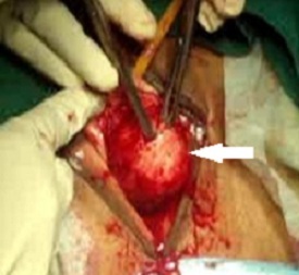
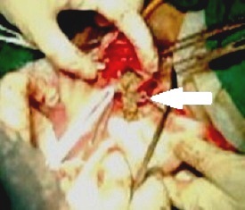
Basic investigations including abdominal ultrasound were normal. Transvaginal scan revealed normal uterus and adnexa; on focusing the anterior vaginal wall, an irregular echogenicity was present. X-ray pelvis showed a dense shadow in region of vagina suggestive of calculi in vaginal cyst, urethral or bladder diverticulum. Examination in OT revealed a 4x3 cm anterior vaginal wall mass suburethrally which was hard in consistency [Table/Fig-1]. On sounding the bladder, calculus was felt. Cystourethroscopy under anaesthesia revealed a small opening in posterior urethra, with stone visible through it. Bladder had evidence of cystitis and ureteric orifices were normal. Final diagnosis of suburethral diverticulum with retained multiple calculi was made. Excision of the diverticulum was planned vaginally. Incision was given over the diverticulum; multiple small calculi along with pus were drained from it [Table/Fig-2]. The diverticulum was excised and repair of urethra was done. Postoperatively, self retaining catheter was kept for 10 days. Patient had uneventful recovery.
Suburethral diverticulum presenting as anterior vaginal cyst

Calculi and purulent secretions coming out of the incised diverticulum

Discussion
Female urethral diverticulum, either congenital or acquired, occurs due to repeated infection of the paraurethral glands leading to obstruction and enlargement into a suburethral cyst or abscess formation. This subsequently ruptures into the urethral lumen, creating a communication in the form of diverticulum [1]. Other proposed aetiologies include childbirth trauma, iatrogenic and urethral instrumentation [2]. The incidence of female urethral diverticulum ranges from 1.5-4% and the most common age of presentation are 30-60 years [1]. The commonest location is mid or distal urethra. Patients present with non specific, irritative lower urinary tract symptoms such as increased frequency, urgency and dysuria; symptoms do not correlate with the size of the diverticulum. Recurrent cystitis or urinary tract infection is seen in one-third of patients. Pain, haematuria, post-void dribbling, dyspareunia, urinary retention or incontinence is other symptoms. Patients may often be misdiagnosed and treated for years for unrelated conditions such as interstitial cystitis, endometriosis, vulvovestibulitis, etc. In some cases, there may be associated urethral calculi or carcinoma.
The differential diagnosis of anterior vaginal mass includes urethral diverticulum, skene gland abscess, ectopic ureterocoele, gartner duct, mullerian remnant or vaginal inclusion cyst [1]. It may even mimic anterior vaginal wall prolapse and cystocele. Billow et al., reported case of a 52-year-old multiparous woman who presented with bleeding ulcerated vaginal mass [3]. It was finally diagnosed as a urethral diverticulum with ulcerated base, which mimicked vaginal cancer. Sometimes patient had a tender anterior vaginal wall mass, which upon gentle compression may reveal retained urine or pus discharge through the urethral opening [3]. Jadhav et al., reported a case of anterior vaginal wall swelling in which UD was misdiagnosed as vaginal wall prolapsed [4]. In one report, the presenting symptom was stress urinary incontinence, when UD was associated with multiple calculi. The symptoms resolved once diverticulectomy was performed and stones removed [5].
Sonography is the first non-invasive investigation to be performed. It can detect the neck of the diverticulum, presence of calculi and adjacent pelvic pathology as well. Magnetic resonance imaging (MRI) is found to be the highly sensitive and specific imaging modality for the diagnosis of urethral diverticulum. It provides detailed information regarding location, number, size, configuration of diverticulum, its communication with the urethra and presence of calculi. It can also differentiate UD from other pathologies such as a mullerian remnant or a vaginal wall cyst [6]. Urethroscopy may help in diagnosing the diverticulum and its location, as in our case. Voiding Computed Tomography, micturating or voiding cystourethrogram (VCUG) are other alternative investigations [7] Balloon or retrograde positive pressure urethrography are no longer recommended as routine investigation for UD [8]. Urodynamic evaluation may be needed in the presence of urinary incontinence or urinary tract obstruction.
Preoperative evaluation for surgical correction includes knowledge of the location, size, number and adjacent anatomy of urethral diverticulum. In patients with urinary incontinence, prior urodynamics are indicated and any infection treated. Except for small asymptomatic fistula, surgical treatment is required. It involves total excision or partial ablation of the diverticulum. Transverse or midline vertical vaginal incision has been used. The basic principles of tension-free meticulous layered closure and good haemostasis are used. Open surgical and endoscopic approaches have been described for the treatment of urethral diverticulum. They include excision of the diverticulum or marsupialization of the diverticular sac into the vagina [9].
A transvaginal technique allows excision of the diverticulum, closure of the urethral communication, reinforced coverage with periurethral fascia, and closure of the anterior vaginal wall. It allows a secure 3-layer closure without overlapping suture lines. Simple marsupialization may be appropriate for diverticula with ostia emptying into the distal one third of the urethra, although it is not popular.
Complications reported following surgery are recurrence of diverticula, urethrovaginal fistula, urethral stricture, urethral pain syndrome, stress incontinence, infection, haemorrhage etc. Urinary catheter is generally kept for a week postoperatively.
Conclusion
Careful preoperative evaluation of any anterior vaginal wall lump or cyst is warranted to avoid surgical mishaps. Once identified, surgical repair is simple by vaginal approach. Any pathology such as stress incontinence may be treated concomitantly and patient satisfaction rates and long term outcomes are favorable.
[1]. Vasavada SP, et al. Urethral diverticula Treatment and Management. E-Medicine World Medical Library. Medscape 2013. http://emedicine.medscape.com/article/443296-overview#a0104 [Google Scholar]
[2]. Blaivas JG, Flisser AJ, Bleustein CB, Panagopoulos G, Periurethral masses: diagnosis in a large series of womenObstet Gynecol 2004 103(5 Pt 1):842-47. [Google Scholar]
[3]. Billow M, James R, Resnick K, Hijaz A, An unusual presentation of a urethral diverticulum as a vaginal mass: a case reportJ Med Case Rep 2013 7:171 [Google Scholar]
[4]. Jadhav J, Koukoura O, Joarder R, Edmonds S, Urethral diverticulum mimicking anterior vaginal wall prolapse: Case reportJ Minim Invasive Gynecol 2010 17(3):390-92. [Google Scholar]
[5]. Lin TC, Siu J-JY, Chou EC-L, Urethral diverticulum with multiple calculi with presentation of urinary incontinence in a female. A case report and literature reviewUrological Science 2014 25(4):129-31. [Google Scholar]
[6]. Dwarkasing RS, Dinkelaar W, Hop WC, Steensma AB, Dohle GR, Krestin GP, MRI evaluation of urethral diverticula and differential diagnosis in symptomatic womenAm J Roentgenol 2011 197(3):676-82. [Google Scholar]
[7]. Macura KJ, Genadry R, Borman TL, Mostwin JL, Lardo AC, Bluemke DA, Evaluation of the female urethra with intraurethral magnetic resonance imagingJ Magn Reson Imaging 2004 20(1):153-59. [Google Scholar]
[8]. Gerrard ER, Lloyd LK, Kubricht WS, Kolettis PN, Transvaginal ultrasound for the diagnosis of urethral diverticulumJ Urol 2003 169(4):1395-97. [Google Scholar]
[9]. Venyo A, Gopall A, Female Urethral Diverticula: A Review of the Literature. Webmed CentralUROLOGY 2012 3(5):WMC003428doi: 10.9754/journal.wmc.2012.003428 [Google Scholar]