Heterotopic Tooth: An Exceptional Entity
Ruchieka Vij1, Meenu Goel2, Puneet Batra3, Hitesh Vij4, Saurabh Sonar5
1 Reader, Department of Oral Pathology and Microbiology, Institute of Dental Studies and Technologies, Modinagar, Uttar Pradesh, India.
2 Senior Lecturer, Department of Orthodontics, ITS Dental College, Muradnagar, Uttar Pradesh, India.
3 Professor and Head, Department of Orthodontics, Institute of Dental Studies and Technologies, Modinagar, Uttar Pradesh, India.
4 Assistant Professor, Department of Diagnostic Sciences and Oral Biology, Division of Oral Pathology, King Khalid University, College of Dentistry, Abha, Kingdom of Saudi Arabia.
5 Professor, Department of Orthodontics, Institute of Dental Studies and Technologies, Modinagar, Uttar Pradesh, India.
NAME, ADDRESS, E-MAIL ID OF THE CORRESPONDING AUTHOR: Dr. Hitesh Vij, Assistant Professor, Department of Diagnostic Sciences and Oral Biology, Division of Oral Pathology, King Khalid University, College of Dentistry, Abha, Kingdom of Saudi Arabia. E-mail – hiteshvij@gmail.com
Coronoid process, Ectopic tooth, Impacted tooth
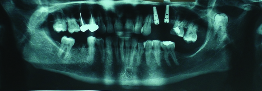
A 30-year-old female patient reported to the dental clinics with a complaint of missing teeth. On clinical evaluation maxillary left pre-molars and mandibular left second molar were absent, the patient was free from any gross pathology and a set of radiographs was advised for analysis of periodontal health and treatment planning. Radiographs revealed the presence of an impacted 3rd molar tooth in the left coronoid process [Table/Fig-1], though clinically no signs or symptoms were observed. Since the location of tooth was abnormal the case was diagnosed as “ectopic 3rd tooth in coronoid process”.
Orthopantomogram showing ectopic 3rd molar in left coronoid process
As the literature suggests such asymptomatic patients can be kept under regular follow-up without any surgical intervention till there is any associated pathology or discomfort. The patient was motivated for placement of implants in edentulous areas and was informed regarding the presence of the ectopic tooth. All the possible complications which could arise in relation to the ectopic tooth were explained to the patient. Further, the patient was advised to have a regular follow-up and visit the dental clinics in case even slight discomfort arose in the concerned region.
Occurrence of impacted 3rd molar tooth is not uncommon but an impacted 3rd molar tooth in an abnormal location (ectopic tooth) is not encountered very frequently [1]. As per the reviews very few cases of ectopic 3rd molar tooth have been reported in English literature [1,2]. Hence, it becomes difficult to comment about the epidemiology of these ectopic teeth. The aetiology of this phenomenon also remains obscure, though many theories have been put forth to explain this exceptional event [2]. This paper presents a case of an ectopic tooth in coronoid process. Only 6 cases of this condition have been reported till date, thus the current case is another milestone of this rare occurrence [1].
A tooth may be impacted, by soft tissue alone or it might be a bony impaction, but the location of the tooth is close to its physiologic position [3]. When such impacted teeth have dislocated to distant regions such as condylar process, mandibular ramus, maxillary sinus, nasal cavity etc these are referred to as ectopic teeth [1,2]. Keros and Susic referred to this condition as “Heterotopic position of tooth” in his report [4]. Occurrence of ectopic tooth as such is an uncommon phenomenon and ectopic tooth in coronoid process is even rarer [1], the latter scenario was observed in the current case.
It is not always possible to determine the exact cause of an ectopic tooth. Various authors have suggested different possible aetiologies which may be either pathologic such as cysts, developmental like an abnormal eruptive pattern or tooth germ development in abnormal location, or sometimes traumatic or iatrogenic in nature [1,2]. In the present case the causative factor appears to be developmental aberration since there was no associated pathology or any history of trauma or surgical interventions in relation to the jaws. Due to the sporadic nature it is not appropriate to definitively comment about the gender predilection, site, clinical signs and symptoms, etc of ectopic 3rd molars. Most of the reported cases in literature are in women and located in mandibular subcondylar or condylar region [1]. In present case too patient was a female but the tooth was located in an atypical position i.e. the coronoid process [Table/Fig-1].
The clinical symptoms may be in the form of pain and swelling, trismus or sometimes fever with acute inflammation and draining sinus [2]. In the case under discussion the patient was asymptomatic and it was an incidental finding. Asymptomatic cases are generally kept under regular follow-up with radiographic evaluation, as was done in the current case. Ectopic tooth associated with cysts are generally surgically treated conservatively until the cyst is very small. In latter case patient is kept under regular follow-up with radiographic evaluation as is done in asymptomatic cases. Intraoral approach is preferred if possible to avoid scarring of facial skin [5]. Based on various reported cases and reviews of ectopic tooth it can be said that coronoid process is not a common location for occurrence of this phenomenon. Further, it is a must that asymptomatic cases are kept under regular follow-up to monitor the development of any new symptoms or pathology or observe any increase or decrease in the associated pathology.
[1]. Iglesias-Martin F, Infante-Cossio P, Torres-Carranza E, Prats-Golczer VE, Garcia-Perla-Garcia A, Ectopic third molar in the mandibular condyle: a review of the literatureMed Oral Patol Oral Cir Bucal 2012 17(6):e1013-17. [Google Scholar]
[2]. Wang CC, Kok SH, Hou LT, Yang PJ, Lee JJ, Cheng SJ, Ectopic mandibular third molar in the ramus region: report of a case and literature reviewOral Surg Oral Med Oral Pathol Oral Radiol Endod 2008 105(2):155-61. [Google Scholar]
[3]. Oikarinen VJ, Altonen M, Impacted third molar in condyloid process: Report of a caseOral Surg 1970 30:7-10. [Google Scholar]
[4]. Keros J, Susic M, Heterotopia of the mandibular third molar: a case reportQuintessence Int 1997 28:753-4. [Google Scholar]
[5]. Bortoluzzi MC, Manfro R, Treatment for ectopic third molar in the subcondylar region planned with cone beam computed tomography: a case reportJ Oral Maxillofac Surg 2010 68(4):870-72. [Google Scholar]