Periodontitis is a destructive inflammatory disease of the supporting tissues of the teeth; this condition is caused by a chronic, mixed infection of gram-negative bacteria and gram-positive bacteria [1]. Periodontal infection triggers an array of events which involves innate & adaptive immunity in the host [2]. The characteristic of this local inflammatory response is infiltration of the periodontal tissue with multiple inflammatory cells which include PMN neutrophils, macrophages, lymphocytes & plasma cells. A large number of inflammatory mediators such as Prostaglandin E-2, Interleukin-1 & Tumour necrosis factor-α are released along with cytokines that are released by activated macrophages. These cytokines bring about the destruction of periodontal connective tissue and alveolar bone [3]. Most severe form of periodontitis is aggressive periodontitis. The aggressive nature of this disease appears to be sufficient to influence the acute phase response despite the localized nature of its occurrence.
Acute phase proteins are defined as proteins whose serum concentrations is altered by at least 25% in response to inflammation and include proteins of the complement, coagulation, fibrinolytic systems, antiprotease, transport proteins, inflammatory mediators and others [4].
The major acute phase proteins includes C-reactive protein, serum amyloid A, fibrinogen and haptoglobin, whose concentration increases with inflammation, where as albumin and transferrin concentration decreases with inflammation [5,6]. C-reactive protein (CRP) was the first protein to be discovered which behaves as an acute phase reactant, and was named for its calcium – dependent interaction with the somatic c-polysaccharide of pneumococci, which recognizes phosphocholine residues. C-reactive protein (CRP) rises in serum or plasma within 24-48 hours following acute tissue damage and decreases with the resolution of inflammation or trauma.
The levels of C-reactive proteins in healthy individuals are about <0.3mg/l, which have been found to increase >100mg/l in the presence of overwhelming systemic infection. This provides a useful matter in tracking the course of infection [7].
Multiple prospective epidemiological studies have shown that CRP is a marker of inflammation in diseases like myocardial infarction, stroke, peripheral arterial disease & sudden cardiac death. The risk of both recurrent ischaemia & death among patients which stable & unstable angina, for patients undergoing percutaneous angioplasty and those patients who present to emergency rooms with acute coronary syndromes can be predicted by using CRP as markers [8]. C-reactive protein (CRP) is an independent predictor of future cardiovascular events that adds prognostic information to lipid screening and to the metabolic syndrome [9]. C-reactive protein (CRP) is currently regarded as a biomarker of systemic inflammation. Most studies to date have evaluated C-reactive protein levels in patients with chronic periodontitis but few have investigated C-reactive protein levels in subjects with aggressive periodontitis [10].
The purpose of this study is to determine the levels of serum C-reactive protein and to compare the C-reactive protein values in patients with health, chronic periodontitis and aggressive periodontitis and to evaluate serum C-reactive proteins levels in patients after periodontal treatment.
Materials and Methods
Source of Data
This comparative quantitative cross-sectional longitudinal study was undertaken by the Department of Periodontology, Sri Siddhartha Dental College, Tumkur (Karnataka) india. Total 150 patients (age ranging between 15 to 50 years including both the genders) have been taken for the study who visited the Outpatient Department of Periodontology. The study spanned over 24 months i.e. from September 2009 to October 2011. A total of 150 patients were selected for the study (58 males and 92 females) were recruited out of which 50 patients constituted Group I Control group (18 males and 32 females) Healthy patients were taken as control group and these were diagnosed as patients not having any clinical evidence of periodontitis (Probing depth ≤ 3mm and the percentage of sites with a Bleeding index of ≥ 3 should be less than 10%), 50 patients constituted Group II Chronic Periodontitis (28 males and 22 females) were diagnosed if patients had at least 4 sites with pocket depth ≥ 5mm and at least 4 sites with attachment loss > 2 mm and 50 patients constituted Group III - Aggressive periodontitis (12 males and 38 females). Diagnostic criteria for aggressive periodontitis were defined according to the classification proposed at the International workshop for the classification of periodontal disease and conditions in 1999. The details are as follows:
The onset of periodontal disease occurred when the patients is under 35 years of age.
Patients having eight teeth with probing depth > 6 mm and radiographic evidence of alveolar bone loss and at least three of these teeth should not be first molars or incisors. We excluded patients with any other bacterial, viral, fungal infections (except periodontitis), diabetic patients, who used tobacco in any form, patients who have taken antibiotics in the past 6 months, patients with systemic diseases such as hypertension, coronary heart disease, rheumatoid arthritis etc. which influences the C- reactive protein level.
Before initiating the study each patient was informed about the purpose and design of the study. An informed written consent and a thorough medical and dental history were taken from all the patients. The study protocol was carried out in accordance with the ethical standards outlined in the 1964 Declaration of Helsinki, as revised in 2008. The Ethics committee, Sri Siddhartha Deemed University, Tumkur (Karnataka), India approved the study protocol. Indicated periodontal management was undertaken for the periodontally diseased after the collection of samples.
All the clinical parameters namely Plaque index (PI) [11], Gingival index (GI) [12], Sulcus bleeding index (Muhlemann HR and Son S 1971), Probing depth (PD), Clinical attachment loss (CA loss) with the help of Williams periodontal probe and blood sample were taken from the patients of all the three groups at the baseline.
Initial therapy consisted of scaling and root planing and oral hygiene instructions for group II and III. The effect of initial therapy was evaluated after 1 month and surgical procedure was carried out for residual periodontal pockets. In surgical procedure, crevicular incisions were given and full thickness flaps were elevated by means of blunt dissection with the help of a periosteal elevator. Lining pocket epithelium was removed and thorough debridement was carried out. Buccal and lingual flaps were approximated using 3-0 non resorbable silk sutures by interrupted suture technique and periodontal dressing was placed. After completion of active therapy (scaling and root planing or periodontal surgery) the patient was clinically examined. All the clinical parameters namely Plaque index, Gingival index, Sulcus bleeding index, Probing depth, Clinical attachment loss were recorded and blood samples were taken for estimation of C- reactive protein levels from group II and group III after 3 months.
Collection of Serum Samples
Skin in the cubital fossa was sterilized with 70% alcohol and allowed to dry. A 2cc disposable syringe and 23 gauge needle was used to draw 2 ml blood from central veins. Blood is centrifuged at 2500 rpm for 10 minutes and the serum thus separated was used to estimate the C-reactive protein levels. Serum C-reactive protein levels were assessed by means of a commercially available highly sensitive immunoturbidimetric assay (Cobas C 111, Tumkur health services lab, Tumkur) at baseline for subjects in all the 3 groups and 3 months after completion of surgical periodontal therapy for subjects in group II and III. Serum levels of C-reactive protein (CRP) were quantified using a high sensitivity C-reactive protein (CRP) enzyme-linked immunosorbent assay (hsCRP ELISA). Lower limits of C-reactive protein (CRP) enzyme-linked immunosorbent assay (CRP ELISA) were 1 mg/l C-reactive protein (CRP), and the upper limits were 150 mg/l C-reactive protein (CRP).
Method of Statistical Analysis
The following methods of statistical analysis have been used in this study.
Kruskal Wallis test was applied to find out significant difference between the study groups.
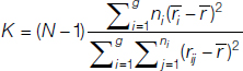
where:
ni is the number of observations in group I;
rij is the rank (among all observations) of observation j from group I;
N is the total number of observations across all groups.
The Wilcoxon signed-rank test is used to compare two related samples or repeated measurements on a single sample to assess whether their population mean ranks differ (i.e. it’s a paired difference test). The steps and formula used as follows
One-way analysis of variance was used to test the difference between groups.
In all above test p-value less than 0.05 was taken to be statistically significant. The data was analysed using SPSS
Results
As per the criteria’s mentioned earlier, the patients were divided in to 3 groups (Group I, II, III) each group consisted of 50 subjects of both sexes, who were in the age ranging from 15 to 50 years.
Age
The mean age of patients in group I, II, III were 23.28 ± 4.946 years, 36.56 ± 10.075 years and 27.72 ± 6.573 years [Table/Fig-1].
Age distribution of the study
| Group | n | Mean Age±SD | Median | F-value | p-value |
|---|
| Group I | 50 | 23.28±4.946 | 24.00 | 20.261 | p<0.001** |
| Group II | 50 | 36.56±10.075 | 40.00 |
| Group III | 50 | 27.72±6.573 | 27.00 |
** Highly significant
Gender
The percentage of males in each group was 36 %, 56% and 24% and that of females were 64%, 44% and 76 % respectively.
C-reactive protein: The mean baseline C-reactive protein (CRP) concentrations in the Groups I, II and III were 1.65±0.57 mg/L, 3.03±2.14 mg/L and 3.09±2.27 mg/L respectively. A significant difference (p<0.05) was found in the C-reactive protein levels between Groups I and II and between Groups I and Group III but there was no significant difference found between Group II and III [Table/Fig-2].
Mean C-reactive protein (CRP) levels in group I, II, III before Treatment (At Baseline)
| Group | N | Mean±SD | Median | ‘Chi Square value* | p-value |
|---|
| Group I | 50 | 1.6520±0.57671 | 1.8000 | 7.563 | 0.023 |
| Group II | 50 | 3.0376±2.14519 | 2.2100 |
| Group III | 50 | 3.0956±2.27050 | 2.3700 |
After treatment, the mean C-reactive protein (CRP) levels in Groups II and III reduced from 3.03±1.67 mg/L to 1.46±1.67 mg/L and from 3.09±1.21 to 1.43±1.21 mg/L respectively. This was statistically (P < 0.001) highly significant [Table/Fig-3].
C-reactive protein levels before and after treatment for group II and III
| | Pre | Post | Z-value* | p-value |
|---|
| Group | N | Mean±SD | Mean±SD |
|---|
| Group II | 50 | 3.0376±1.67994 | 1.4648±1.67994 | -4.373 | p<0.001** |
| GroupIII | 50 | 3.0956±1.21253 | 1.4332±1.21253 | -4.373 | p<0.001** |
** Highly significant
Kruskal-Wallis Test
On comparing the effect of treatment in between group II and group III, the difference was found to be statistically non significant (p>0.05). Both groups showed equal amount of improvement in C-reactive protein levels after treatment [Table/Fig-4].
Effect of treatment on study group II, III.
| Group | | N | Mean | SD | Mean diff | SD | t-value | p-value |
|---|
| Group II | Pre treatment Plaque Index | 50 | 1.4344 | 0.28348 | 0.97600 | 0.29918 | 16.311 | p<0.001** |
| Post treatment Plaque Index | 50 | 0.4584 | 0.23464 |
| Pre treatment Gingival Index | 50 | 1.4644 | 0.34981 | 1.05680 | 0.35400 | 14.927 | p<0.001** |
| Post treatment Gingival Index | 50 | 0.4076 | 016954 |
| Pre treatment Sulcus Bleeding Index | 50 | 2.1284 | 0.33925 | 1.43960 | 0.46859 | 15.361 | p<0.001** |
| Post treatment Sulcus Bleeding Index | 50 | 0.6888 | 0.26858 |
| Pre treatment Probing Depth | 50 | 5.2284 | 0.78431 | 2.05640 | 0.93461 | 11.001 | p<0.001** |
| Post treatment Probing Depth | 50 | 3.1720 | 0.37917 |
| Pre treatment Clinical Attachment Level | 50 | 4.7924 | 1.02548 | 1.60440 | 1.20238 | 6.672 | p<0.001** |
| Post treatment Clinical Attachment Level | 50 | 3.1880 | 0.57974 |
| Group III | Pre treatment Plaque Index | 50 | 1.6124 | 0.63544 | 1.04840 | 0.70321 | 7.454 | p<0.001** |
| Post treatment Plaque Index | 50 | 0.5640 | 0.24980 |
| Pre treatment Gingival Index | 50 | 1.4296 | 0.70162 | 1.04960 | 0.68763 | 7.632 | p<0.001** |
| Post treatment Gingival Index | 50 | 0.3800 | 0.20062 |
| Pre treatment Sulcus Bleeding Index | 50 | 2.1020 | 0.54202 | 1.32120 | 0.47239 | 13.984 | p<0.001** |
| Post treatment Sulcus Bleeding Index | 50 | 0.7808 | 0.37133 |
| Pre treatment Probing Depth | 50 | 6.0940 | 0.61136 | 2.19800 | 0.57013 | 19.276 | p<0.001** |
| Post treatment Probing Depth | 50 | 3.8960 | 0.36683 |
| Pre treatment Clinical Attachment Level | 50 | 4.6164 | 0.45297 | 1.52440 | 0.59105 | 12.896 | p<0.001** |
| Post treatment Clinical Attachment Level | 50 | 3.0920 | 0.37961 |
* Statistically significant
** Statistically highly significant
Periodontal Parameters
Gingival index: At baseline the mean gingival index scores in the Groups I, II and III were 0.61±.21, 1.46±0.34 and, 1.42±0.70 respectively. A significant difference (p<0.001) was found in the gingival index between Groups I and II and between Groups I and III but not between Groups II and III. After treatment the mean gingival index scores in Groups II and III improved from 1.46±0.34 to 0.40±0.16 and 1.42±0.70 to 0.38±0.20 respectively. This was found to be statistically highly significant (p<0.001). A positive effect of the treatment on gingival index was found for Group II and Group III [Table/Fig-5].
Mean gingival Index (GI), plaque index (PI), sulcus Bleeding Index (SBI) among the groups I, II, III before and after treatment.
| Groups | Gingival index | Plaque index | Sulcus bleeding index |
|---|
| Preoperative | Postoperative | Preoperative | Postoperative | Preoperative | Postoperative |
|---|
| Group I | 0.611 | | 0.758 | | 0.698 | |
| Group II | 1.464 | 0.4076 | 1.434 | 0.4584 | 2.128 | 0.6888 |
| Group III | 1.43 | 0.38 | 1.612 | 0.564 | 2.102 | 0.7808 |
Plaque index: The plaque control in all the patients was satisfactory. At baseline the mean plaque index in the Groups I, II and III was 0.75±0.10, 1.43±0.28, 1.61±0.63 respectively. A significant difference (p<0.001) was found in the plaque index between Groups I and II and between Groups I and III but no significant difference was found between Group II and Group III [Table/Fig-5]. After treatment the mean plaque index in the Groups II and III improved from 1.43±0.28 to 0.45±0.23 and from 1.61±0.63 to 0.56±0.24 respectively. This was found to be statistically (p<0.001) highly significant [Table/Fig-5].
Bleeding Index
The mean bleeding index scores in the Groups I, II and III at baseline were 0.69±.14, 2.12±.33 and 2.10±.54 respectively. A significant difference (p<0.001) was found in the bleeding index between Groups I and II and between Groups I and III but no significant difference was found between Group II and III. After treatment the mean bleeding index scores in Groups II and III improved from 2.12±.33 to 0.68±.26 and from 2.10±.54 to 0.78±.37 respectively. This was found to be statistically (p < 0.001) highly significant [Table/Fig-5].
Probing pocket depth: The mean probing depths for the entire mouth at the beginning of the study for II and III were 5.22±0.78 mm and 6.09±0.61 mm respectively. A significant difference (P<0.001) was found in the mean probing depths between Groups II and III [Table/Fig-6]. After treatment, the mean probing depths in Group II and III reduced from 5.22±0.78 mm to 3.17±0.37 mm and from 6.09±0.61 mm to 3.89±0.36 mm respectively. This was found to be statistically (P < 0.001) highly significant. The positive effect of the treatment on mean probing depth was found for Group II and Group III [Table/Fig-6].
Comparison of mean probing depth and clinical attachment level among the groups II, III before treatment
| Groups | Clinical attachment loss | Probing pocket depth |
|---|
| Preoperative | Postoperative | Preoperative | Postoperative |
|---|
| Group II | 4.792 | 3.188 | 5.228 | 3.172 |
| Group III | 4.616 | 3.092 | 6.094 | 3.896 |
Clinical attachment level: There was a significant gain in clinical attachment. The mean attachment loss at the beginning of the study for II and III were 4.79±1.02 mm and 4.61±0.45 mm respectively. This was found to be statistically non significant (p>0.05) between group II and group III. After treatment, the mean attachment loss in Groups II and III reduced from 4.79±1.02 mm to 3.18±0.57 mm and from 4.61±0.45mm to 3.09±0.37 mm respectively. The positive effect of the treatment on mean clinical attachment level was found for Group II and Group III. This was found to be statistically (p<0.001) highly significant [Table/Fig-6].
Discussion
Periodontitis is a chronic, tissue destructive inflammatory state that is predominantly induced by Gram-negative bacteria that have colonized the gingival crevice. The disease degrades the attachment apparatus of the teeth [13]. A number of chemicals can be associated with acute inflammatory reactions. These are haptoglobulin (HPT), α-1-antitrypsin (AT), C-reactive protein (CRP), α-1-acid glycoprotein (AAG), ceruloplasmin (CER). These are also called acute phase proteins (APPs). CRP & cytokines like interleukin 6 & TNF – α have been demonstrated in chronic periodontal infections & other systemic diseases as effective inflammatory markers [14]. It has been demonstrated that CRP, serum amyloid A & α- 2-macroglobulin respond rapidly to inflammatory stimuli & their serum levels increase several 100 folds [15] in the strong acute inflammatory phase. Thus these are said to be strong acute phase proteins. Haptoglobin, α-1-antitrypsin & fibrinogen have been found to increase only about 2-10 fold [16] & thus are said to be moderate acute phase proteins. Complement component C-3 & ceruloplasmin increase only upto 2 fold & thus are called weak acute-phase proteins [17].
The discovery of C-reactive protein (CRP) was reported in 1930 by Tillet and Francis [18]. The swift rise of its serum concentration during the acute phase, the magnitude of the response approaching 1000-fold increase within 24–48 hours, and the equally quick return to the very low normal concentration of a few gm/ml are the most impressive biologic characteristics of C-reactive protein (CRP). Synthesis in the liver is responsible for blood C-reactive protein (CRP) (Gabay and Kushner) but extra hepatic expression has also been documented (Dong and Wright) [19,20]. C-reactive protein (CRP) belongs to the pentraxin family of calcium-dependent ligand-binding plasma proteins. The human C-reactive protein (CRP) molecule is composed of five identical non glycosylated polypeptide subunits. In recent years, a plethora of studies have demonstrated a direct association between slightly elevated C-reactive protein (CRP) serum levels and the risk of developing cardiovascular disease [21–24].
It has been found that CRP activates the complement system. CRP production is activated by interleukin-6 [15] which is a key cytokine in inflammation & immune modulation. Thus elevated levels of CRP & interleukin-6, suggests that there is underlying biological mechanism that associates periodontits & cardiovascular diseases [15].
In case of patients with periodontal disease serum C-reactive protein (CRP) was elevated which was also evident in our study comprising of chronic and aggressive periodontitis patients with increased pocket depth and attachment loss as compared to previous studies conducted [7]. Bacterial load, antibody titer and avidity to the specific pathogens are improved following successful periodontal treatment, there leading to decrease in local inflammation and improvement in clinical parameters [3]. We needed to know whether this improvement in clinical parameters have any role in reducing the elevated C-reactive protein (CRP) which failed to illustrate changes in C-reactive protein (CRP) following treatment in previous studies [25]. The results of our study reinforced the observations of the above studies indicating that periodontal diseases are associated with elevation in serum C-reactive protein (CRP) levels. In the present study, there was a significant reduction in probing pocket depth and gain in clinical attachment level. These results coincide with that of the earlier studies carried, that showed an improvement in these clinical parameters following periodontal therapy [26–28]. In the present study, 7 patients of group II and 8 patients of group III patients showed values of ≥ 3 mg/ l respectively, while only 1 patient had values ≥ 3 mgs/ l in Group I suggesting the possibility that periodontal disease have an effect on C- reactive protein levels. In present study, the C-reactive protein (CRP) levels in chronic and aggressive periodontitis patients reduced significantly after periodontal therapy (p <001). Also, the percentage of subjects with C-reactive protein (CRP) level > 3mg/l decreased significantly. However, some authors have failed to observe any reduction in circulating C-reactive protein (CRP) following periodontal treatment (Scaling and root planing) [29]. In present study surgical treatment was also carried out. The medical significance of these changes is further emphasized by the fact that such changes would substantially decrease the predicted risk for future cardiovascular events based on serum C-reactive protein (CRP) concentrations.
Limitations
Although there are limitations in the present study in which the number of subjects is relatively small and the data for conventional atherosclerotic risk factors are lacking, our data showed that serum C-reactive protein (CRP) levels tended to be higher in untreated periodontitis patients compared with periodontally healthy subjects and demonstrated concomitant reduction of C-reactive protein (CRP) levels with successful periodontal treatment.
Conclusion
C-reactive protein (CRP) is considered a biomarker of systemic inflammation and a marker of subsequent atherosclerosis and cardiovascular disease (CVD). Within the limitations of this study, successful periodontal treatment results in significant decrease in serum C-reactive protein (CRP) levels in otherwise healthy subjects. The potential benefits of periodontal therapy to reduce the serum levels of inflammatory markers needs to be further investigated in greater depth. Also the effect of successful periodontal therapy to reduce the risk of cardiovascular diseases should be further studied. Large multi-centre randomized trials can be suggested. Higher prevalence of periodontal disease & evidence of predictable periodontal treatment leading to high standard of oral health suggests that periodontal treatment should be considered as a preventive practice in cardiovascular disease prevention. This idea should be included in clinical practice as well as in future study designs.