Oral Squamous Cell Carcinoma: Picro Sirius Red Stain
Ketki Kalele1, Noopur Kulkarni2, Rahul Kathariya3
1 Senior Lecturer, Department of Oral and Maxillofacial Pathology, V.Y.W.S Dental College and Hospital, Amravati, Maharashtra, India.
2 Senior Lecturer, Department of Oral Pathology, Microbiology and Forensic Odontology, Pandit Deendayal Upadhyay Dental College and Hospital, Kegaon, Solapur, Maharashtra, India.
3 Assistant Professor, Department of Periodontics and Oral Implantology, Dr. D.Y Patil Dental College and Hospital, Dr. D.Y Patil Vidyapeeth (Deemed University)Pune, India.
NAME, ADDRESS, E-MAIL ID OF THE CORRESPONDING AUTHOR: Dr. Rahul Kathariya, Assistant Professor, Department of Periodontics and Oral Implantology Dr. D.Y Patil Dental College and Hospital, Dr. D.Y Patil Vidyapeeth, Pune-411018, India.
E-mail: rkathariya@gmail.com
Collagen, Malignancy, Metastasis, Special stain
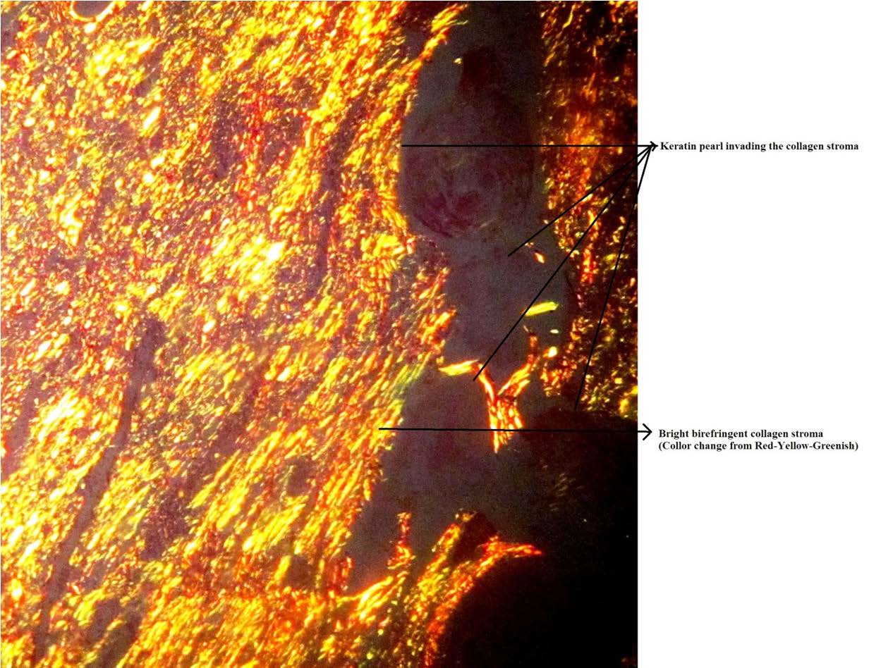
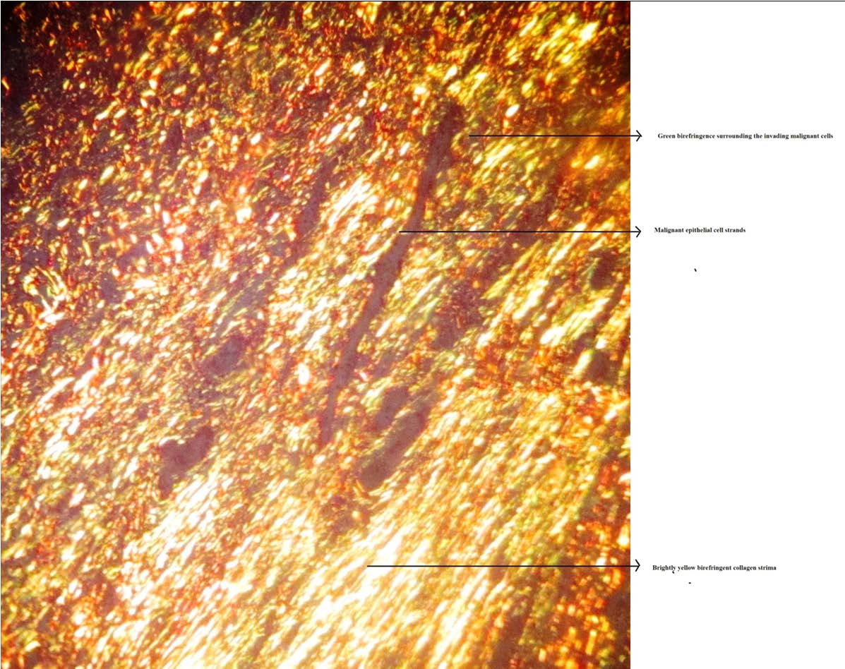
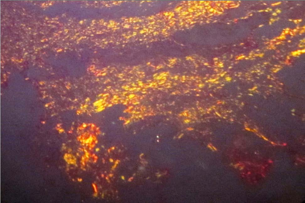
Oral Squamous Cell Carcinoma (OSCC) in Picro Sirius red stain [Table/Fig-1–3]. Recent times have witnessed lot of work on malignancies and related targeted therapies to prevent its invasive and metastatic potential. Collagenous stroma plays a very important role in inhibiting spread of the malignant tumour cells by acting as a barrier. However, in malignancies there occurs stromal remodeling which weakens the stroma to facilitate the spread of the tumour [1,2]. Picro Sirius red is the special stain which can be used to study nature of the collagen fibers by using its birefringence properties [3]. The given images are 40X magnification of a section of oral squamous cell carcinoma, well differentiated variety showing predominantly red birefringence [Table/Fig-1]. Brightly yellow to green birefringent collagen stroma surrounding the malignant epithelial cell strand in moderately differentiated squamous cell carcinoma [Table/Fig-2] and section of poorly differentiated OSCC presenting with green birefringent stroma [Table/Fig-3]. The change in birefringence of collagen fibers (colour-red-golden yellow-green) surrounding the tumour islands [Table/Fig-1–3] with increasing grade of the tumour is highlighted under polarized microscope stained with Picro Sirius red stain; as stroma changes from red-yellow to green when the grade of tumour increases. The changes in the stromal birefringence are attributed to the change from mature collagen fibers to immature collagen [3]. This is because of increased proteolysis of the collagen fibers, by invading tumour islands, which favour metastasis of the tumour [1,2]. Golden yellow coloured collagen fibers are seen along the invading islands and keratin pearls [Table/Fig-1,2] which denotes slight proteolysis in the stroma which is beautifully birefringent in the section. Thus, along with routine Hematoxylin and Eosion staining Picro Sirius red should also be employed to the microscopy of malignancy [1–3].
A 40X magnification of well differentiated Oral Squamous Cell Carcinoma
A 40X magnification of moderately differentiated Oral Squamous Cell Carcinoma
A 40X magnification of poorly differentiated Oral Squamous Cell Carcinoma
[1]. Venigella A, Charu S, Evaluation of collagen in different grades of oral squamous cell carcinoma by using the picrosirius red stain: a histochemical studyJ Clin Diagn Res 2010 4:3444-49. [Google Scholar]
[2]. Conti J, Thomas G, The role of tumour stroma in colourectal cancer invasion and metastasisCancers 2011 3:2160-68. [Google Scholar]
[3]. Rich L, Whittaker PC, Picrosirius RS, A polarized light assessment of fibrillar hue and spatial distributionBraz J Morphol Sci 2005 22:97-104. [Google Scholar]