Bonding is the first choice in practice since the development of numerous painless techniques in restorative dentistry. This technique has the additional advantages of minimal tooth reduction & reversibility [1]. Development of acid etch technique by Buonocore [2] in 1955 and the BIS-GMA based composite resin by Bowen has made possible the direct bonding of composite resin to the facial surface of stained, malposed, fractured and other teeth requiring aesthetic & functional improvement. Resin-based composites have been used for many years but only recently exhibited is the improved wear resistance [2].
The ultimate goal of a bonded restoration is to attain an intimate adaptation of the restorative material with the dental substrate. The interface between restoration and dental substrate is an area of clinical concern that can result in secondary decay, marginal discoloration, and pulpitis [3]. Perfect adaptation is hard to accomplish because of inconsistent physical properties between tooth structure and restorative materials. The hidden leakage is called microleakage. Microleakage may be defined as the clinically undetectable passage of bacteria, fluids, molecules or ions between a cavity wall and the restorative material applied to it [4]. Thus, alterations in the bonding systems and resin-based composites must be achieved to minimize the deleterious effects of polymerization shrinkage of the resin material, gap formation and consequent leakage [3,4].
The bonding of resin-based restorative materials to dentin has always been more challenging. The dentinal tubules are the only pores available for micromechanical retention. These tubules contain fluid, which would be an impediment to bonding. To reduce microleakage, certain procedures such as maintaining a wet dentin [5], applying the adhesive and restoring with resin composite byan incremental technique have been recommended [6]. Hence, this invitro study has been undertaken to investigate the degree of dye penetration as an estimation of microleakage of composites with bonding agents placed under different techniques.
Materials and Methods
The study was designed to evaluate the microleakage of dental composites using bonding agents with different placement techniques in vitro. The study was conducted in the Department of Pedodontics & Preventive dentistry, Guru Nanak Dev Dental College and Research Institute, Sunam in 2012.
Materials
The following materials were selected for the study.
Etchant 37% phosphoric acid gel [Table/Fig-1]Materials used in the study
Etchant, 37 % Phosphoric acid, Meta Etchant – 37
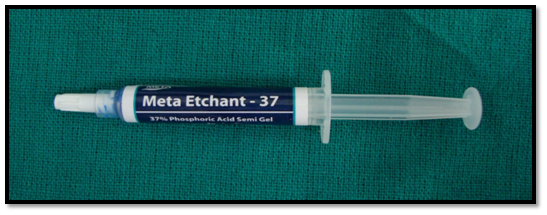
(Meta Etchant _37, Meta Biomed Co. Ltd)
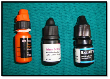
Adhesive system [Table/Fig-2]Bonding agents used in the study
1. Single bond (3M, ESPE)
2. Prime and bond NT (Dentsply)
3. Excite (Ivoclar Vivadent)
Single bond (3M, ESPE)
Prime and bond NT (DENTSPLY)
Excite (IVOCLAR VIVADENT)
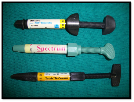
Restorative materials [Table/Fig-3]Composite Materials used in the study
1. Z 100 (3 M, ESPE)
2. Spectrum TPH (Dentsply)
3. Tetric (Ivoclar Vivadent)
Z 100 (3 M, ESPE)
Spectrum TPH (Dentsply)
Tetric (Ivoclar Vivadent)
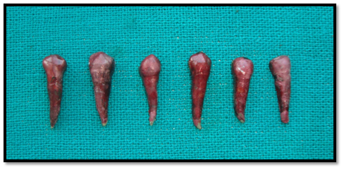
Sample collection and storage: Fifty four noncarious human premolars extracted for orthodontic purpose were collected.
Division and restoration of samples [Table/Fig-4].
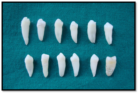
Collected teeth were divided into 9 groups with 6 teeth in each group. These samples were stored in distilled water for further use.
Cavity preparation: Class II cavities were prepared with dimensions 2.5 mm depth occlusally, 2 mm width occlusally, 3.5 mm depth gingivally, and a 3mm wide gingival seat using high speed handpiece with continuous water cooling. The teeth were subsequently stored in distilled water [Table/Fig-5,6 and 7].
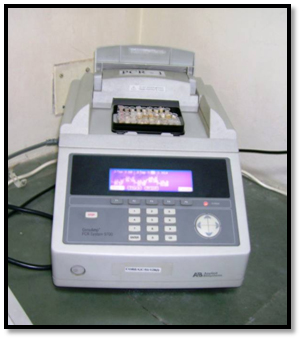
Equipments used in the study
Thermocycling machine
Samples after application of nail varnish
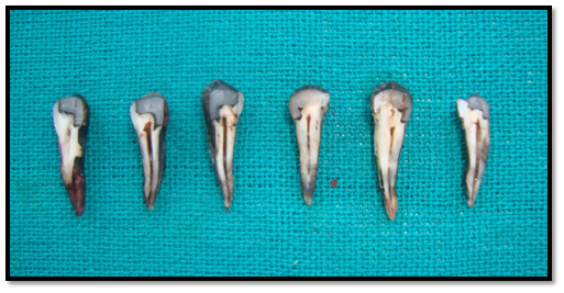
Some of the sectioned samples
The prepared teeth were randomly divided into 9 groups [Table/Fig-8,9 and 10].
| SUBGROUP I | Prepared cavities were acid etched with etching gel (37% phosphoric acid) for 20 seconds, washed in distilled water & gently dried. Z100 composite was placed & light cured for 40 sec. |
| SUBGROUP II | Acid etching for 20 seconds washed & gently dried. Two increments of Spectrum TPH was placed into the cavity and light cured for 40 seconds each. |
| SUBGROUP III | Acid etching with etching gel for 20 seconds washed in distilled water & gently dried. Two increments of Tetric composite were placed & light cured for 40 seconds. |
| SUBGROUP IV | Acid etching, a single coat of single bond bonding agent was applied, gently air dried for 2 to 5 seconds and then light cured for 10 seconds. Z100 composite was placed & then light cured for 40 seconds. |
| SUBGROUP V | Acid etching, single coat of Prime and bond (NT) bonding agent was applied, light cured for 20 seconds. Spectrum TPH composite was placed & light cured for 40 seconds. |
| SUBGROUP VI | Acid etching, Excite adhesive is generously applied to the tooth structure, light activated for 20 seconds. Tetric composite was placed & light cured for 40 seconds. |
| SUBGROUP VII | Acid etching, two coats of Single bond bonding agent was applied one after other; air dried for 2-5 seconds and then light cured for 10 seconds, restored with Z100 composite and light cured for 40 seconds. |
| SUBGROUP VIII | Acid etching, two coats of Prime and bond NT bonding agent was applied, light cured for 20 seconds. Spectrum TPH composite was placed & light cured for 40 seconds. |
| SUBGROUP IX | Acid etching. One coat of Excite bonding agent was applied, light cured for 20 seconds each, then a second coat of excite bonding agent was applied; and then light cured for 20 seconds restored with Tetric composite & light cured for 40 seconds. |
Group A: Without application of bonding agent (Subgroups I,II,III)
Group B: Application of single layer of bonding agent (Subgroups IV,V,VI)
Group C: Application of double layer of bonding agent (Subgroups VII,VIII,IX)
All the specimens were subjected to thermal cycling at 60C, 370C, 540C and again at 370C completing one cycle with a 30 second dwell time. Nail varnish was applied over all the thermally cycled tooth specimens except 1mm around the restoration. Green stick compound was used to seal the apex completely. Each group of samples was placed in 10 ml each of freshly prepared 50% silver nitrate solution for 2 hour in darkness. The teeth were then retrieved, washed thoroughly in distilled water, stored in developing solution and exposed to sun light for 24 hour. The extent of dye penetration was determined.
Results
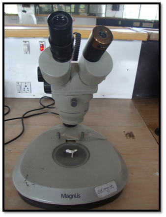
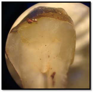
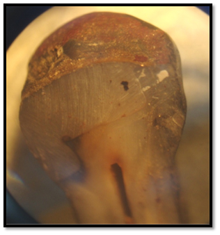
Microleakage for each group was evaluated by stereomicroscope 4X magnification [Table/Fig-11] and recorded using a parametric scale that gives a qualitative measurement of sealing effectiveness of restorative material [Table/Fig-12,13]. The data collected was tabulated accordingly and was statistically analysed using Kruskal Wallis and Chi-Square tests [Table/Fig-14,15].
Equipments used in the study Stereomicroscope (Magnus)
Sample showing microleakage score 0
Sample showing microleakage score 4
Total number of teeth presenting with the microleakage scores along the axial wall
| BONDING AGENT | Score (Axial) | Mean Rank |
|---|
| Grade 0 | Grade 1 | Grade 2 | Grade 3 | Grade 4 | Total |
|---|
| No Bonding | 0 | 0 | 0 | 5 | 13 | 18 | 42.97 |
| Single Coat | 0 | 2 | 8 | 6 | 2 | 18 | 25.11 |
| Double Coat | 3 | 6 | 6 | 3 | 0 | 18 | 14.42 |
Total number of teeth presenting with the microleakage scores along the occlusal wall
| BONDING AGENT | Score (Occlusal) | Mean Rank |
|---|
| Grade 0 | Grade 1 | Grade 2 | Grade 3 | Grade 4 | Total |
|---|
| No Bonding | 0 | 0 | 2 | 9 | 7 | 18 | 43.08 |
| Single Coat | 1 | 3 | 9 | 5 | 0 | 18 | 27.86 |
| Double Coat | 11 | 5 | 2 | 0 | 0 | 18 | 11.56 |
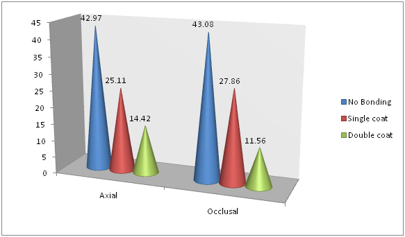
The mean microleakage scores among the individual IX groups were calculated. On comparing the mean microleakage scores among the three groups, maximum microleakage scores have been obtained when no bonding agent was used, while least microleakage scores were obtained with double coat of bonding agent [Table/Fig-16]. Also, the results obtained are highly significant (p < 0.001).
Graph representing the comparison in the mean microleakage scores between the groups with no bonding agent, single coat of bonding agent and double coat of bonding agent
Discussion
Evaluating the importance of adhesive procedures among is an indispensible challenge for most dentists. It is difficult, not to say impossible, to think about modern dentistry without Buonocore’s acid-etching technique, the development of composite resins, or the consequent popularization of various bonding techniques [2]. A restoration should provide a good adhesion to the tooth because of the presence of open margins around restorations which makes teeth to be susceptible to secondary caries. One of the main factors associated with marginal shrinkage and gap formation is the resin composite shrinkage at the tooth-restoration interface (Cenci et al.,) [7]. This polymerization shrinkage causes building up of significant stress in the surrounding tooth structure which may be a major causative factor in bond failure (Amaral et al.,) [8]. Factors affecting the integrity of the tooth-restoration interface are [8]:
Polymerization shrinkage and cavity configuration factor.
Hydroscopic expansion.
Light polymerization concepts and units.
Thermal cycling and occlusal stresses.
Bonding agent & its placement.
To offset the effect of polymerization shrinkage stress and for the prevention of microgap between resin and the tooth, resin to tooth structure bond of 17 MPa is necessary [8].
The stress build up due to polymerisation can cause adhesive or cohesive failure and interfacial gap formation and can eventually lead to deformation of residual tooth structure. Light cured composites develop higher stress than self cured composites, and the use of higher energy curing lights further worsens the situation [9].
The penetration of these newer adhesives into a chemically conditioned dentin creates a mechanical interlocking based on the formation of a hybrid layer and resin tags penetrating into opened dentin tubules (Tay FR et al., Sano H et al.,) [9,10]. Maintaining a moist dentinal surface is of crucial importance in enhancing bond strength in vitro. Moisture prevents collagen fibrils from collapsing into demineralized dentinal surfaces after acid etching, thus allowing the adhesive monomers to penetrate into dentin, forming a hybrid layer. This technique is commonly known as wet bonding technique & has been considered a paramount mechanism in adhesive dentistry. Quantifying the amount of moisture that should be left after acid etching is, however, problematic. Moreover, differing amounts of moisture may be required with specific solvents contained in each adhesive system; over wetting and over drying phenomena may occur and lead to the formation of an altered hybrid layer known as Hybridoid layer (Simone Deliperi) [11]. In this study, particular attention was paid to preserving moist dentin surface prior to the start of bonding procedure. The excess water was removed using an air syringe; this method is particular technique sensitive.
This study used thermocycling to mimic intra-oral temperature variations and subjecting the restorations on the tooth to temperature extremes compatible with oral cavity [12]. Wahab et al., [13] found that thermocycling significantly increased the microleakage of Class V composite restorations. Only when the initial bond is known, thermal stressing of the restoration interface is of value.
Considerable reduction in leakage both at enamel and dentine interfaces with the composite was observed from stereomicroscope, when a recommended bonding agent (Single Bond in the case of Z100, P & B NT in the case of Spectrum TPH, Excite in the case of tetric composite) was applied. The adhesion increased with applying two coats of the bonding agent where the leakage was least in most of the cases observed for all systems studied. Another study compared different bonding agents and concluded that microleakage scores where Clearfill SE bond and Filtek-LS (3M-ESPE) (LS) showed significantly lower microleakage than Prime and Bond NT (Dentsply) (PB), and Scotch Bond Multipurpose (3M-ESPE) had highest mean microleakage score [14].
The adhesion characteristics of the composite resin are improved by the application of a second coat of bonding agent [15]. Moreover, doubling the number of coats could affect the performance of the adhesive system. Conversely, a more uniform hybrid resin layer can result from doubling the number of coats [16]. This procedure can promote the formation of a stress absorbing resin layer which is able to reduce microleakage [17]. Using 5th-generation bonding agent in comparison to 7th-generation bonding agent and not using any bonding decreased microleakage [18].
Conclusion
All the three groups i.e. group A, B, C (No bonding agent, Single layer of bonding agent, Double layer of bonding agent) tested showed microleakage at tooth restoration interface. Highly significant difference was found in the degree of microleakage scores between the group A & C i.e. no application of bonding agent & application of double layer of bonding agent. Significant difference was found in group A & B i.e. with no application of bonding agent & application of single layer of bonding agent. No significant difference was found in group B & C i.e. application of single layer of bonding agent and application of double layer of bonding agent. Though group C showed least microleakage among three groups.