Pneumatization is the formation of air cavities in bone. Apart from the major paranasal sinuses, air cells may arise singly or in clusters in numerous locations in the skull, including the temporal bone [1]. The nomenclature ZACD is used to denote accessory air cells observed in the zygomatic process of maxilla and articular eminence of the temporal bone, which simulate mastoid air cells [2]. It presents as an asymptomatic, non expansile, non destructive radioluceny on radiographs [3]. Recognition of normal anatomy as well as pathologies can be performed effectively with the utilization of widely used imaging modality that is panoramic radiography as it gives more extensive delineation of oro-facial area [2]. On the basis of radiographic appearance on panaromic radiograph there are three types of ZACD: 1. Unilocular type, 2. Multilocular type, 3. Trabecular type. Unilocular type of ZACD presents as radiolucency with well defined borders, whereas multilocular type appears as numerous small cavities within, which resemble mastoid air cells. The third variety is trabecular type which appears as multilocular entity with internal bony striations [1]. At the point when ZACDs have been exhibited preoperatively on a radiograph, these may be contraindications to perform surgical strategies, for example, eminoplasty or eminectomy for the treatment of mandibular dislocations as they get to be potential pathways for intracranial infections [3].
The area of research that is the prevalence of ZACD in North Indian population is under investigated till date and presently has a limited empirical knowledge base. Therefore, the present study was conducted to determine the prevalence of ZACD amongst North Indian population and also to establish the dominant location and type.
Materials and Methods
The present study material comprised of 2500 panoramic radiographs of patients aged between 19 and 78 years, who visited Department of Oral Medicine and Radiology during the period of January 2009 to December 2013. All radiographs were obtained with Gendex Orthoralix 9200 (America) DDE Digital Pan/Ceph System panoramic radiographic machine operating at 65–80 kVp, 10 mA and 16 s. Cases in which the zygomatic arch was not adequately displayed for anatomical or technical reasons were excluded from the sample and did not constitute part of the sample. All persons gave their informed consent prior to their inclusion in a study. All patients had no severe bone disease including tumor, cyst or fracture. Selection criteria for the acquisition of panoramic radiographs included caries, pain, missing or supernumerary teeth, mixed dentition analysis, periodontal disease, third molar extraction, extensive restorative dental procedures, prosthodontic evaluation, orthodontic evaluation and TMJ problems. Subsequently, ZACDs were classified depending on: (1) age and gender, (2) the location: unilateral or bilateral and (3) the appearance: unilocular or multilocular. Radiographs were examined in subdued ambient lighting using transmitted light from a standard viewing box by a single oral and maxillofacial radiologist.
Results
Of the 2500 panaromic radiographs, zygomatic air cell defect was present in 63 radiographs, accounting for prevalence to 2.5%. The overall mean age being 37.4 years. The subjects in the group 19 and 38 years accounted for most number of ZACDs. The youngest subject with ZACD was 19 years and the oldest 75 years. In general, mean age for males with ZACD was 37.5 years and for females it was 37.3 years. Out of 63 ZACDs, 38 were in males and 25 were found in females and of the ZACDs, 44 occurred unilaterally and 19 bilaterally, and 49 presented with unilocular and 14 with multilocular appearance. Amongst the 44 unilateral ZACDs, 25 were noted in males and 19 in females. Out of 19 bilateral ZACDs, 12 were found in males and 07 in females [Table/Fig-1]. Amongst the 49 unilocular, 26 were observed in males and 23 in females [Table/Fig-2]. Of the 14 multilocular, 10 were recognized in males and 04 in females. In the age group of 39-78 years minimum number of ZACDs were found. Among the unilateral variant, 27 were found on left side and 17 on right side [Table/Fig-3]. The variation of ZACDs with gender, locularity and laterality is summarized in [Table/Fig-4] and variation with age is summarized in [Table/Fig-5].
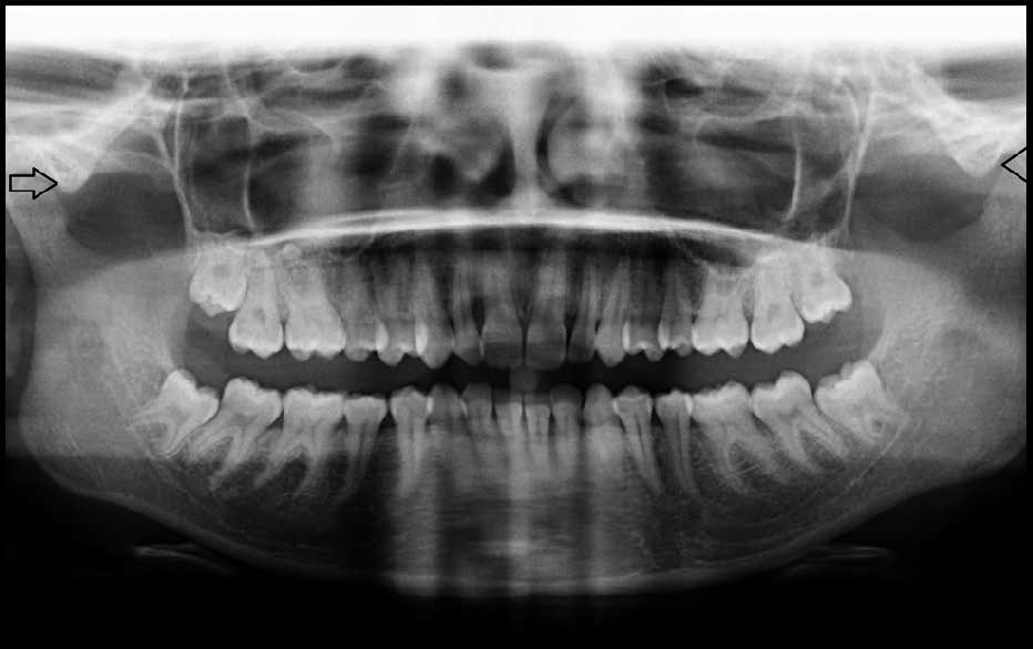
Panoramic radiograph showing bilateral unilocular zacds
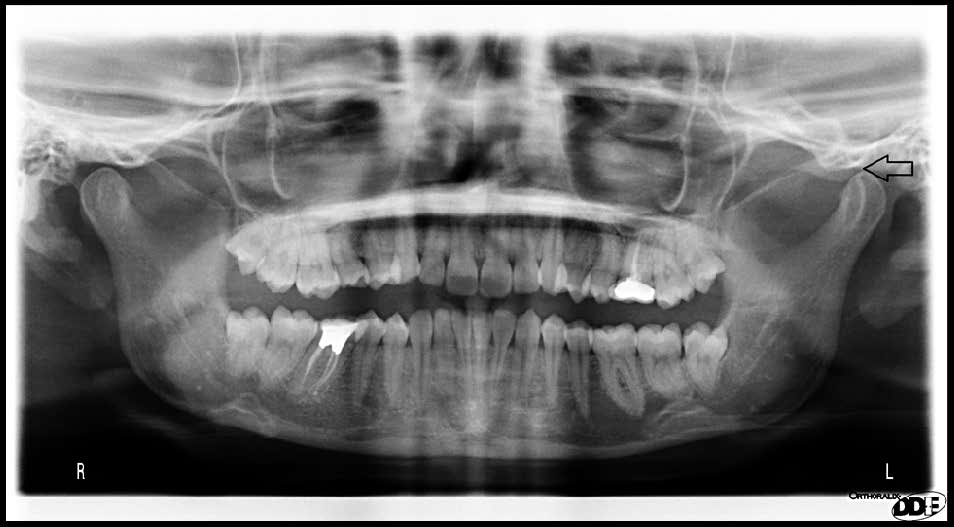
Panoramic radiograph showing right unilocular and left multilocular zacds
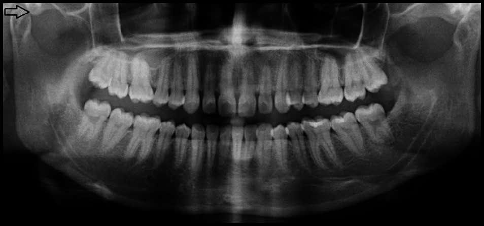
Panoramic radiograph showing right unilocular zacd
ZACD vs Locularity, Laterality and gender
| Gender | Locularity | Laterality |
|---|
| Unilateral (N=44) | Bilateral (N=19) |
|---|
| Unilocular | Multilocular | Right | Left |
|---|
| Males (N=38) | 26 | 10 | 12 | 13 | 12 |
| Females (N=25) | 23 | 04 | 05 | 14 | 07 |
| Total (N=63) | 49 | 14 | 17 | 27 | 19 |
ZACD vs. locularity, laterality and age
| Age Group/S (Years) | Multilocular (N=14) | Unilocular (N=49) | Unilateral (N=44) | Total | Bilateral (N=19) |
|---|
| Right | Left |
|---|
| Group 1 - (19-28) | 07 | 28 | 07 | 15 | 22 | 13 |
| Group 2 - (29-38) | 02 | 09 | 03 | 05 | 08 | 03 |
| Group 3 - (39-48) | 02 | 02 | 01 | 02 | 03 | 01 |
| Group 4 - (49-58) | 01 | 06 | 05 | 02 | 07 | 00 |
| Group 5 - (59-68) | 01 | 02 | 00 | 02 | 02 | 01 |
| Group 6 - (69-78) | 01 | 02 | 01 | 01 | 02 | 01 |
Discussion
The exact etiology of ZACD is not known but it is similar to that of pneumatization of the mastoid process. Normal periosteal activity is responsible for the formation of small osseous cavities which results in pneumatization. Pneumatization of the temporal bone can be divided into three stages: infantile (birth to 2 years), transitional (2 ± 5 years) and adult [1]. Pneumatization of temporal bones occurs in 2 regions- 1- primary region and 2- accessory region. The primary region includes middle ear, mastoid, petrous apex and perilabyrinthine. The accessory region includes the squamous, zygomaticooccipital and styloid. ZACD may also be produced by the extension of tegmental or periantral air cell into the zygomatic arch [4,5].
The overall prevalence of ZACD in the present study was 2.5% which is in accordance with the studies conducted by Srikanth et al., Park et al., and Friedrich et al., [3,6,7]. The results are not consistent with study conducted by Carter et al., Patil et al., Hofmann et al., Asieh Zamaninaser et al., [1,2,8,9]. The variation in the prevalence of ZACD can be due to the difference in the sample size, population studied and methodology.
In our study, ZACDs there was a definite male prediliction which is consistent with the study conducted by Park et al., and in contrast with the studies conducted by Carter et al., and Srikanth et al., where there was no gender predilection [1,3,6]. In a study by Patil et al., ZACD showed the number of males were twice as common as compared to females [2]. In the studies conducted by Asieh Zamaninaser et al., and Friedrich et al., there was female predominance of ZACDs [7,9]. Hence it is difficult to reach a consensus on the gender prevalence of ZACDs.
In our study, the youngest patient reported with ZACD was 19 years of age. Review of the literature demonstrated that the youngest patient reported to date was 16 years [1].
In the present analysis, approximately 2/3rd of the ZACDs were unilateral and 1/3rd were bilateral, which is in accordance with Patil et al., Asieh Zamaninaser et al., Friedrich et al., [2,7,9] 32 were unilateral and 8 were bilateral in a study conducted by Carter et al., [1]. As compared to a study conducted by Parker et al., 24 cases of ZACD (77.4%) were unilateral and seven cases (22.6%) were bilateral [6]. In a study conducted by Srikanth et al., in south Indian population 15 were unilateral and six were bilateral [3]. This investigation exhibited high proportion of ZACDs on left side, opposing to that of other studies where they were present more on right side. In the present study most of the unilateral ZACDs occurred in males. Similar results were found in a study conducted by Patil et al., and Srikanth et al., [2,3].
In the present study 36 subjects were found in the age range of 19-28 years and 10 subjects in the age range of 29-38 years. These results are in accordance with the study conducted by Patil et al., in our study 49 were unilocular and 14 were multilocular as compared to study conducted by Patil et al., in which 45 presented with unilocular and 96 with multilocular appearance [2]. In a study conducted by Srikanth et al., 6 were multilocular and 2 were unilocular.Twenty-six (68.4%) of the defects were of unilocular type while twelve of the defects (31.6%) were of multilocular type in a study conducted by Park et al., [6].
Limitations
The present study has some limitations. The difference in the results as compared to other studies are because of variations in the sample size as well due to inter observer variability in determining the types of ZACDs and also due to superimposition of anatomical structures over the ZACD.
Conclusion
This research shows that prevalence of ZACDs in North Indian population to be 2.5%, with male predominance and dominance of the unilateral and unilocular variants. Most of the dentists are not aware of significance of ZACD, therefore the present study was done to draw attention of oral and maxillofacial radiologists regarding this entity owing to its significance and possible complications. Further studies with increased variables could provide further insights into this little known entity.