Anatomical Variations of Upper Segmental Renal Artery and Clinical Significance
Gyan Prakash Mishra1, Shobha Bhatnagar2, Brijendra Singh3
1 Associate Professor, Department of Anatomy, Career Institute of Medical Sciences and Hospitals, Lucknow, India.
2 Professor, Department of Anatomy, Career Institute of Medical Sciences and Hospitals, Lucknow, India.
3 Additional Professor, Department of Anatomy, Aiims, Jodhpur, India.
NAME, ADDRESS, E-MAIL ID OF THE CORRESPONDING AUTHOR: Dr. Gyan Prakash Mishra, Associate Professor, Department of Anatomy, Career Institute of Medical Sciences and Hospitals, Lucknow, India. E-mail : gyanprakash.mishra88@gmail.com
Introduction
Classically each kidney is supplied by a single renal artery. In its course renal artery divides into anterior and posterior division, both of these division further divide into segmental arteries that are apical, upper, middle, lower and posterior. Segmental arteries are representing an end artery and they form independent renal segments. In their course they are closely related with collecting system. So, a thorough in depth knowledge of the variations in segmental arteries is a basic requirement for surgeons.
Aim
To observe and investigate the anatomical variations in arterial pattern of upper segmental renal artery and its relations with collecting system of ducts in human kidneys.
Materials and Methods
Fifty human kidneys of both sexes were observed and studied by corrosion cast method. Different colour coded cast material (butyl butyrate) like red for artery and black for collecting ducts were used. 20% solution of butyl butyrate in acetone was injected into renal artery and ureter of the kidneys. After polymerization (24 hours) these kidneys were kept immersed in a bath of concentrated KOH solution at 500C for 24-48 hours for corrosion to obtain the endocasts. These endocasts were cleaned and washed under running tap water and photographed.
Results
In present study upper segmental artery was found in 49 (98%) kidneys. It was absent in 1 (2%) kidney. We observed four types of variations in arterial pattern of upper segmental artery namely Upper Segmental Artery Type-1 (USAT1), Upper Segmental Artery Type-2 (USAT2), Upper Segmental Artery Type-3 (USAT3), Upper Segmental Artery Type-4 (USAT4) and they were found in 20 (40%), 14 (28%), 10 (20%), 5 (10%) kidneys respectively. We also observed two different variations in the anatomical relations between upper segmental artery and collecting system namely Upper Segmental Artery Group -1 (USAG1) and Upper Segmental Artery Group-2 (USAG2). USAG1 was found in 40 (80%) kidneys and USAG2 in 9 (18%) kidneys respectively.
Conclusion
Anatomical knowledge of these variations is very important and useful for the uro-surgeons for best outcome and minimal complications in and during nephrectomies, removal of calculi, surgery of renal tumors or other various intrarenal surgeries.
Collecting System, Corrosion cast, Intrarenal surgeries, Kidney
Introduction
Kidney is one of the very vital viscera in human body which is involved in excreting the nitrogenous wastes from the body through urine and so is essential for life. Classically each kidney is supplied by a single renal artery. In its course renal artery divides into anterior and posterior division, both of these division further divide into segmental arteries that are apical, upper, middle, lower and posterior. Renal vascular segmentation was originally recognized by John Hunter [1] and Brodel M [2]. Graves laid the foundation of segmental pattern of intrarenal arterial distribution and its variation [3]. He divided the renal parenchyma into five segments. Kher observed that anterior division gives origin to apical segmental artery first and then to upper, middle and lower segmental arteries [4]. Verma [5] studied the intrarenal branching pattern of renal artery and observed more variations than Graves [3]. Sykes described the relation between the tubules and arterial segments [6]. Saxena has done the radiographic study and described four to five arterial segments according to variations in branching pattern of renal artery [7]. Longia and Ajmani studied the intrarenal arterial pattern of Kidney by corrosion cast method and found variations in arterial pattern of segmental arteries [8,9]. Sampaio studied the anatomical relationship between the upper segmental artery and collecting system [10]. Anatomical knowledge about the upper segmental artery of human kidney is important for performing intrarenal operations, to avoid postoperative complications and injury to the adjacent parenchyma. A serious and troublesome complication of endoscopic intrarenal operation is bleeding from an injured vessel. To diminish the risk of such injury, the surgeon must know and recall the spatial position of the intrarenal vascular structures and their anatomical relations with the collecting system of the kidneys [11]. Objective of our study was to observe variations in the arterial pattern of upper segmental renal artery and its anatomical relations with collecting system in human kidneys.
Materials and Methods
Anatomical variations of upper segmental artery of kidney were observed by corrosion cast method in 50 human Kidneys of both sexes (30 male and 20 female) aged between 18 to 60 years. This study was done in Sarojini Naidu Medical College, Agra during 2009 to 2010. Kidneys were obtained from postmortem bodies within 24 hours after death. Cases with renal trauma, gross renal pathology and signs of decomposition were excluded. We dissected out renal artery, from renal pelvis and ureter at the hilum of the Kidneys. Washed the renal vessel with 0.9% saline solution till the renal vein showed the clear fluid coming out of it. 20% solution of butyl butyrate in acetone was injected into renal artery and ureter. The solution was injected till the increase in resistance was observed. After injection renal artery and ureter were tied by thread and kept in 10% formal saline for polymerization for 24 hour. After polymerization injected kidneys were kept in a bath of concentrated KOH at 500C for 24-48 hours for corrosion to obtain endocasts. These endocasts were cleaned and washed in running tap water, and photographed.
Results
Fifty fresh human kidneys were studied by corrosion cast method and upper segmental artery was seen in 49 (98%) kidneys. It was absent in 1 (2%) kidney. We observed four types of variations in arterial pattern of upper segmental artery namely Upper Segmental Artery Type-1 (USAT1), Upper Segmental Artery Type-2 (USAT2), Upper Segmental Artery Type-3 (USAT3), Upper Segmental Artery Type-4 (USAT4) and they were found in 20 (40%), 14 (28%), 10 (20%), 5 (10%) kidneys respectively, shown in [Table/Fig-1]. USAT1 arose from anterior division of renal artery and found in 20 (40%) kidneys [Table/Fig-2]. Out of these in 4 (8%) kidneys the origin was extra renal, in 15 (30%) kidneys intrarenal and 1 (2%) kidney at the hilum. USAT2 arose with middle segmental artery and found in 14 (28%) kidneys [Table/Fig-3]. Out of these in 10 (20%) Kidneys the origin was intrarenal and in 4 (8%) kidneys at hilum. USAT3 arose with apical segmental artery and found in 10 (20%) kidneys [Table/Fig-4]. Out of these in 1(2%) kidney the origin was extra renal, in 7 (14%) kidneys intrarenal and in 2 (4%) at hilum. USAT4 arose from posterior division of renal artery and found in 5 (10%) kidneys [Table/Fig-5]. Out of these in 1 (2%) kidney the origin was extra renal, in 4 (8%) kidneys intrarenal. We also observed variations in relation between collecting system and upper segmental artery. These variations have been divided into two groups namely Upper Segmental Artery Group-1 (USAG1) and Upper Segmental Artery Group-2 (USAG2). USAG1 ran in front of upper major calyx and seen in 40 (80%) kidneys [Table/Fig-2,3]. USAG2 ran upward medial to the pelvis and upper major calyx and found in 9 (18%) kidneys [Table/Fig-5].
Showing variations in branching pattern of upper segmental artery. In 1 case upper segmental artery was absent
| Sex | Kidneys | USAT1 arose from anterior division of renal artery | USAT2 arose with middle segmental artery | USAT3 arose with apical segmental artery | USAT4 arose from posterior division of renal artery |
|---|
| Male | 30(60%) | 12(24%) | 7(14%) | 6(12%) | 4(8%) |
| Female | 20(40%) | 8(16%) | 7(14%) | 4(8%) | 1(2%) |
| Total | 50(100%) | 20(40%) | 14(28%) | 10(20%) | 5(10%) |
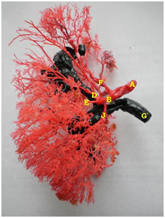
Anterior view showing USAT1 and USAG1. A-Renal Artery, B-Anterior Division, D-Upper Segmental Artery (USAT1 and USAG1), E-Middle Segmental Artery, F-Apical Segmental Artery, G-Ureter, J-Lower Segmental Artery
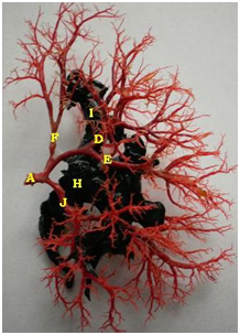
Anterior view showing USAT2 and USAG1. A-Renal Artery, D-Upper Segmental Artery (USAT2 and USAG1), E-Middle Segmental Artery, F-Apical Segmental Artery, H- Pelvis, J-Lower Segmental Artery
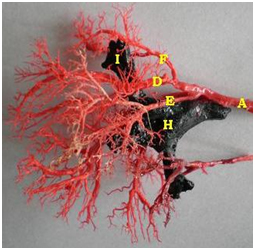
Anterior view showing USAT3 and USAG1. A-Renal Artery, D-Upper Segmental Artery (USAT3 and USAG1), E-Middle Segmental Artery, F-Apical Segmental Artery, H- Pelvis, I- Upper Major Calyx.
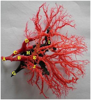
Anterior view showing USAT4 and USAG2. A-Renal Artery, B-Anterior Division, C-Posterior Division, D-Upper Segmental Artery (USAT4 and USAG2), E-Middle Segmental Artery, G-Ureter, H-Pelvis, I-Upper Major Calyx, J-Lower Segmental Artery
Discussion
In present study we found four types of variations in arterial pattern of upper segmental artery namely USAT1, USAT2, USAT3, USAT4 and these variations are seen in 40%, 28%, 20%, 10% kidneys respectively. Variations in arterial pattern of upper segmental artery have been noticed by other authors too, shown in [Table/Fig-6]. H Fine studied 107 Kidneys by corrosion cast method and reported USAT1, USAT2 and USAT3 in 46.72%, 19.62% and 31.77% kidneys respectively [12]. They were not found USAT4 in their study. A study conducted by Ecaterina Daescu on 60 Kidneys by dissection method and observed variations in artery of superior (apical), anterior superior (upper), and anterior inferior (middle) segment [13]. They were seen USAT1 in 40% cases. This was similar to that observed by us. They were also found USAT3 and USAT4 in 51.67% and 1.67% cases respectively. In their study USAT2 was not reported but in one case they were found a different branching pattern as upper segmental artery arose with apical and middle segmental arteries at one point. In our study we found percentage of USAT3 was lesser and USAT4 was greater than that reported by them [13]. A study of 40 Kidneys by angiography and corrosion cast methods and reported USAT3 in 60% cases [14]. This percentage was greater than that observed by us. In our study we found USAT3 only in 20% cases. In their study they were not reported USAT1, USAT2 and USAT3. Chandragirish S studied 100 Kidneys by corrosion cast technique and found variations in anterior division of renal artery, superior (apical), anterior superior (upper) and anterior inferior (middle) segmental arteries [15–17]. They reported USAT1, USAT2 and USAT3 in 19%, 16% and 12% kidneys respectively. In their study USAT1 was reported only in 19% cases while in our study we found it in 40% cases. On the basis of discussion with study of other authors it was observed, USAT3 was most common variation of upper segmental artery which arose with apical segmental artery. Prior apical segmental resection surgeon should exclude the presence of USAT3 to avoid injury of it, because this end artery can lead ischemia in upper renal segment of human kidney. We also observed the two different variations in relation between upper segmental artery and collecting system namely USAG1 and USAG2. USAG1 found in 80% kidneys and USAG2 in 18% kidneys. One corrosion cast study of 107 Kidneys observed USAG1 and USAG2 in 16% and 73% kidneys respectively [12]. Another corrosion cast study of 82 kidneys observed USAG2 in 86.6% kidneys [10]. These variations are of utmost importance during puncture in endourological procedures and intrarenal operations to avoid risk of bleeding. Knowledge of the variations of renal vascular anatomy has importance in exploration and treatment of renal trauma, renal transplantation, renal artery embolization, surgery for abdominal aortic aneurysm and conservative or radical renal surgery [18]. Anatomical knowledge of these variations is helpful for uro surgeon in preoperative planning and prevents injury of arteries and collecting system during intrarenal operations.
Comparative study on arterial pattern of upper segmental artery
| Types | H Fine et al., [12] | Ecaterina Daescu et al., [13] | Chandragirish et al., [15–17] | Rani Neerja et al., [14] | Present study |
|---|
| USAT1, arose from anterior division of renal artery | 46.72% | 40% | 19% | ------- | 40% |
| USAT2, arose with middle segmental artery | 19.62% | ------ | 16% | -------- | 28% |
| USAT3, arose with apical segmental artery | 31.77% | 51.67% | 12% | 60% | 20% |
| USAT4, arose from posterior division of renal artery | ------ | 1.67% | ------ | ------ | 10% |
Conclusion and Clinical Significance
In present study, we found variations in branching pattern of upper segmental artery. It arose with or from apical, middle and posterior segmental artery in 20%, 28% and 10% cases. Prior upper segmental resection surgeon should exclude the origin of apical or middle or posterior segmental artery with or from upper segmental artery to prevent the injury of that, during ligation of upper segmental artery or other surgical procedures. In our study, we also found variations in relation between upper segmental artery and collecting system. These variations are of utmost importance during puncture in endourological procedures and intrarenal operations to avoid risk of bleeding.
The precise knowledge of renal vasculature is of valuable contribution for surgeons in performing more and more conservative renal surgeries like partial and segmental resection of renal tissue instead of going for radical nephrectomy which cause extensive damage and can also lead to intraoperative and postoperative complications. These conservative methods can thus lead to preservation of healthy and functional renal parenchyma which is the main aim of modern surgical procedures and this aim can be achieved only after knowing precisely about the arterial pattern of upper segmental artery and its relation with collecting system. Moreover, it is of valuable contribution of development of new and different techniques for removal of renal calculi like percutaneous nephrolithotomy, further open surgical nephrectomy can be replaced by laproscopic nephrectomy and easy removal of renal tumours.
[1]. Hunter J, Vasculature of the bodyBr J Surg 1794 38:1-8. [Google Scholar]
[2]. Brodel M, The intrinsic blood vessels of the kidney and their significance in nephrotomyBull John Hopkin’s Hosp 1901 12:10 [Google Scholar]
[3]. Graves FT, The anatomy of intrarenal arteries and its application to segmental resection of the kidneysBr J Surg 1952 42:132-40. [Google Scholar]
[4]. Kher GA, Bhargava I, Makhni FS, Intrarenal branching of the renal arteryInd J of Surg 1960 22:563-79. [Google Scholar]
[5]. Mahesh V, Chaturvedi RP, Pathak RK, Anatomy of renal vasculature segmentsJ of Anat Soc of Ind 1961 10:34-37. [Google Scholar]
[6]. Sykes D, The arterial supply of the human Kidney with a special reference to the accessory renal arteriesBr J Surg 1963 50:368-70. [Google Scholar]
[7]. Saxena SK, Dikshit CS, Saxena HN, A radiographic study of intrarenal arteriesMediscope 1973 16(1):19 [Google Scholar]
[8]. Longia GS, Kumar V, Gupta CD, Intrarenal arterial pattern of human kidneys-corrosion cast studyAnat Anz 1984 155:183-94. [Google Scholar]
[9]. Ajmani ML, Ajmani K, To study the intrarenal vascular segments of human kidney by corrosion cast techniqueAnat Anz 1983 154(4):293-303. [Google Scholar]
[10]. Sampaio FJB, Aragao AHM, Anatomical relationship between the intrarenal arteries and the kidney collecting systemThe Journal of Urology 1990 143:679-81. [Google Scholar]
[11]. Sampaio FJB, Anatomic background for intrarenal endourologic surgeryJ Endourol 1992 6(5):301-04. [Google Scholar]
[12]. Fine H, Keen EN, The arteries of the human kidneyJ Anat 1966 100(pt-4):881-94. [Google Scholar]
[13]. Daescu E, Zahoi DE, Motoc A, Alexa A, Baderca F, Enache A, Morphological variability of the renal artery branching pattern: a brief review and an anatomical studyRom J Morphol Embryol 2012 53(2):287-91. [Google Scholar]
[14]. Neerja R, Seema S, Pushpa D, Rani K, Surgical importance of arterial segments of human kidneys: an Angiography and corrosion cast studyJournal of Clinical and Diagnostic Research 2014 8(3):1-3. [Google Scholar]
[15]. Chandragirish S, Nanjaiah CM, Shirur SY, Sahed SH, Study on variations of anterior division of renal arteryInt J Anat Res 2014 2(4):697-700. [Google Scholar]
[16]. Chandragirish S, Nanjaiah CM, Shirur SY, Sahed SH, Study of variations in superior segmental branch of renal arteryInt J Anat Res 2014 2(4):701-04. [Google Scholar]
[17]. Chandragirish S, Nanjaiah CM, Shirur SY, Sahed SH, Study on variations of anterior inferior segmental branch of renal arteryInt J Anat Res 2014 2(4):705-08. [Google Scholar]
[18]. Gupta V, Kotgirwar S, Trivedi S, Deopujari R, Singh V, Bilateral variations in renal vasculatureInternational Journal of Anatomical Variations 2010 3:53-55. [Google Scholar]