Introduction
The design of tooth preparations can have an effect upon the success of individual restorations [1]. Restoration of the lost tooth structure with adequate marginal fit has been the goal of a conscientious prosthodontist. The term angle of convergence can be applied to denote the respective relationship between the two opposing walls of a preparation [2]. The convergence angle is valuable to visualize preparation walls, prevent undercuts, compensate for inaccuracies in the fabrication process, and permit more nearly complete seating of restorations during cementation [3].
In fixed prosthodontics, the interface between the restoration and the tooth is evident as the weakest link [4]. Marginal adaptation is one of the most significant factors influencing the clinical acceptability of the cast restoration [5]. It is a critical parameter as the dissolution of the luting agent and the inherent roughness may result in secondary caries. This loss of the underlying tooth structure will lead to the failure of the restoration [6]. Also, lack of adequate fit is potentially detrimental to the supporting periodontal tissues [7]. Attempts are made by the clinician to compromise the fit of the restorations by occlusal and internal adjustments leading to a threat to the cement-margin failure [8].
Tylman said that “it is apparent that every effort should be made to approach parallelism not exceeding “2° to 5° gingivocclusally” [9], whereas Johnston recommended “5° to 7° parallelism” [10]. Numerous studies have been done to evaluate the marginal fit of single crowns with different tapers but not many studies have been done to evaluate the taper which provides a better marginal adaptation for multiple unit castings. Hence, this study was carried out to verify the marginal fit of single crown, three-unit FPD and multiple-unit FPD with pier abutment with different degrees convergence angles.
Materials and Methods
The present study was a prospective study, conducted over a period of 3 years at A.B. Shetty Memorial Institute of Dental Sciences, Mangalore, Karnataka, India.
Description of Master Die
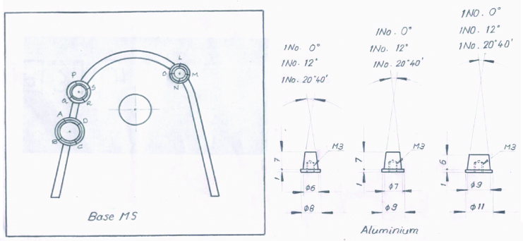
Three metal dies having the same convergence angles were made of aluminum. These dies were placed on an iron square block with the help of threaded retainer shaft in the position of first molar and first premolar on one side of the arch and canine in cross-arch relation. In the same way, four arch forms were made, each arch form having three dies (in the position of first molar and first premolar on one side of arch and canine in cross-arch relation), with the dies on each arch form having similar convergence angle. [Table/Fig-1] depicts schematic representation of the dies.
The dies for the molars had the diameter of 9 mm at the finish line and the height of 6 mm. The dies for the premolar had the diameter of 7 mm at the finish line and the height of 7 mm. The dies for the canine had the diameter of 6 mm at the finish line and the height of 7 mm. Each die had a 1 mm 900 shoulder finish line. The dies with 0°, 6°, 12°, and 20° convergence angles were mounted on the four arch forms.
Custom made moulds were fabricated of aluminum for each die in such a way that there was a uniform spacing of 1mm between the die and the mould. This was done to obtain standardized wax patterns. It is important that the copings be standardized so that no observed changes could be attributed to difference in contour, collar size or thickness of the coping. The dies with 0°, 6°, 12°, and 20° convergence angles and the corresponding moulds are shown in [Table/Fig-2,3,4,5] respectively.
Wax Pattern Fabrication
To fabricate the coping wax patterns, type II inlay wax (Unident, India) was used. Die lube (Dentecon. Inc, Los Angeles, CA) was used as a wax separating agent on the inner aspect of the mould. Molten inlay wax was poured into the mould, and the die was seated in the mould. After cooling, the wax pattern was carefully separated from the mould. Excess wax was removed and the margins were burnished to ensure that they would be closely adapted. A 2.0 mm diameter (12-gauge) wax sprue (Schuler-Dental GmbH And Co. KG., Johannesstraße 6-8, D-89081 Ulm, Germany) was attached at a 45° angle to the occlusal surface of each wax pattern.
The sprue formers length was adjusted so that the pattern was 6.0 mm from the end of the ring, when kept in place. The point of attachment was flared and not restricted to decrease porosity and increase mould filling. Five wax patterns were prepared for single full veneer copings on molars; five wax patterns were made by joining the copings at the position of the molar and the premolar with the help of a connector of 2 mm diameter to form a three-unit FPD; and five wax patterns were made by joining the wax patterns at the position of the molar, the premolar and the canine with the help of connectors of 2.6 mm in diameter to form a multiple-unit FPD with a pier abutment, for each convergence angle.
Investing the Wax Pattern
Each wax pattern was immediately invested (Deguvest GF, Degussa, Germany) after marginal refinement to minimize distortion. Casting rings (Degussa, Germany) of the size no. 3 and no. 5 were lined with one non-overlapping layer of cellulose ring liner (Degussa, Germany), which was maintained 3 mm below the top of the ring, all ring liners were wet when used. A 6mm distance was provided between the margin of the crown and the top of the casting ring. Surfactant (Lubrofilm, Dentaurum, GmbH & Co. KG Turnstraße 31 D-75228 Ispringen) was sprayed on the wax pattern and allowed to dry for 3 minutes. Investing was then carried out, in which the investment was hand mixed for 20 seconds, followed by 90-second mechanical mixing under vacuum (Multivac 4, Degussa). The wax patterns were invested with a camel-hair brush and allowed to bench set for 30 minutes. All base formers were then removed.
Wax Pattern Elimination and Casting
Conventional lost wax technique (Kavo EWL Type 5630) was carried out for complete elimination of wax from the moulds. The moulds were placed in a cold furnace and heated up to 250°C at a rate of 5°C/min and the temperature was maintained for 30 minutes. The mould was then heated to a final temperature of 950°C at the rate of 7°C/minutes and maintained at that temperature for 30 minutes. All casting procedures were carried out in induction casting machine (Degutron, Degussa, Germany) to make nickel chromium copings (Wirolloy, Bego). All castings were cooled to room temperature before removal from casting ring. After the castings were removed, they were sandblasted (Superstrahl, Degussa) with 50 μm Al2O3 at 30 psi pressure at a distance of approximately 5 cm. All sprues were removed with an abrasive disk, cleaned in ultrasonic cleaning solution (Sonorex Super RK102P, Bandelin) for 15 minutes, rinsed and dried. [Table/Fig-6] depicts the castings of single crown, three-unit FDP and multiple-unit FDP.
Method of Measurement
While the castings were seated on their respective dies, indentations were made, with the help of a rotary instrument, at four points on each die, i.e. mesiobuccal, distobuccal, distolingual and mesiolingual. These indentations were used as the reference points to check for marginal fit. An optical microscope (Labomed) with a filar eyepiece was used at 100x magnification to record the measurements of the vertical marginal discrepancy. [Table/Fig-7] represents a photomicrograph showing marginal discrepancy.
A transfer block was constructed to hold the castings for measuring the marginal discrepancy in the optical microscope. The castings in place were embedded in a layer of softened impression compound which was allowed to harden. When the dies were removed from 58the base, the castings remained with the transfer block maintaining the same relationship on the apparatus.
Stastical Analysis
The data was subjected to appropriate statistical analysis. The mean values obtained for discrepancy were compared using t-test and ANOVA.
Results
The results of the present study are presented in [Table/Fig-8-9]. [Table/Fig-8] depicts the mean values obtained for discrepancy in marginal fit observed for single crown, three-unit FDP and multiple-unit FDP with pier abutment. These mean values were obtained by analyzing the mean values obtained at four points on each die, i.e. mesiobuccal, distobuccal, distolingual and mesiolingual.
The results of the marginal discrepancy obtained for the single crown denote that the marginal discrepancy was minimum for 200 convergence angle, i.e. 29.482 μm and was maximum for 00 convergence angle, i.e. 36.395 μm. Similar results were observed for three-unit FDP. The discrepancy for molar was 26.236 μm and that for premolar was 24.061 μm for 20° convergence angle which was much less than the discrepancy seen for 0° convergence angle where the discrepancy for molar was 35.720 μm and that for premolar was 35.453 μm. Multiple-unit FDP with a pier abutment also revealed minimum discrepancy values for 20° convergence angle and maximum discrepancy values for 0° convergence angle. The discrepancy for molar was 29.467 μm, for premolar was 29.519 μm and that for canine was 27.633 for 20° convergence angle which was much less than the discrepancy seen for 0° convergence angle where the discrepancy for molar was 42.900 μm, for premolar was 42.230 μm and that for canine was 42.47.
[Table/Fig-9] depicts the statistical analysis of variance applied on the means of the vertical marginal discrepancies for single crowns, three-unit FDP and multiple-unit fixed partial denture with pier abutment. These included the comparison of the vertical discrepancy between the units of 20° convergence angle with the units of 0°, 6°, and 12° convergence angles. A very high statistically significant difference (p<0.001) was observed for the vertical marginal discrepancy of the single crown, three-unit FDP and multiple-unit fixed partial denture with pier abutment.
Discussion
Convergence angle is mainly associated with the retention, resistance and the marginal fit of the FDP restoration [11]. The marginal fit of any restoration is vital for its long term success [12]. Geometry of the tooth preparation, including the type of finish line and the degree of taper is an important factor in obtaining close marginal adaptation [13]. Dodge et al., concluded that the resistance form is more sensitive to changes in convergence angle [14]. Also, they reported that there is no significant difference in retention values between preparations with 100 total convergence angle as compared with 160 convergence angle. Wilson et al., concluded that the crowns cemented on preparations with a convergence angle between 60 and 120 had the highest retentive values [15]. Many researchers have stated that parallelism of axial walls is important in retention of the crowns; however, parallel walls are impossible to create in the mouth without producing preparation undercuts [16,17].
Poor marginal adaptation is one of the reasons for the failure of crowns and FDP [18]. The reason for failure may be microleakage which may further lead to secondary caries [19]. Also, with poor marginal fit, the plaque accumulation potential of the fixed partial restoration increases leading to periodontal breakdown[20]. Goodacre et al., concluded that the reason for failure of single crown was periodontal disease (0.6%) and caries (0.4%) [21]. The reason for failure of fixed partial denture was caries (18%) and periodontal disease (4%). Walton et al., concluded that there was no apparent relationship between the span of prosthesis and its length of service [22]. Thus, the present study was undertaken to determine the marginal fit of single crown, three-unit FDP and multiple-unit FDP with pier abutment with different convergence angles.
In this study, four convergence angles were used, i.e. 00, 60, 120, and 200 in order to determine as to which convergence angle gives a better marginal fit. These convergence angles were selected for the study as these have been advocated and used by most of the authors in the previous literature. The dies with the convergence angle of 00 had all the axial walls parallel; 60 had 30 taper on each axial wall; 120 had 60 taper on each axial wall; and 200 had 100 taper on each axial wall. The machined metal dies enabled to accurately control the variables of preparation design, degree of convergence angle and the finish line dimensions, which was a 1 mm 900 shoulder finish line. The metal dies were placed at the position of molar and premolar on one side of the arch and canine in cross arch relation, in order to simulate the arrangement of natural teeth in the oral cavity. In this study, the marginal fit was evaluated for the single crowns, three units fixed partial denture as well as multiple unit fixed partial denture with a pier abutment.
The results of the marginal discrepancy obtained for all the units assessed in our study i.e. for single crown, 3 unit FDP and multiple unit fixed partial denture with a pier abutment, denote that the marginal discrepancy was minimum for 200 convergence angle, and was maximum for 00 convergence angle. Thus, as the convergence angle increases from 00 to 200, the accuracy of marginal fit also increases which indicates that there is more complete seating of the single crown with 200 convergence angle. Further, the statistical comparison of the each of the three units assessed in our study, a very highly significant difference (p<0.001) was observed when 200 convergence angle was compared with other convergence angles i.e. 00, 60 and 120.
For the multiple unit FDP with a pier abutment, it has been theorized that the pier abutment acts as a fulcrum, causing forces that are transmitted to the terminal abutments and leads to the failure of the weaker abutment [23]. Construction of a one piece multiple unit FDP has distinct advantages like maximum strength of the rigid connector and elimination of the soldering procedure which may be the cause of failure in due course of time. Garlapo et al., reported that four unit castings were possible without appreciable warpage [24].
From the results, it can be seen that 200 convergence angle gives better marginal fit than any other convergence angle. However, Tylman advocated the convergence angle of 2-50 considering the prime factor retention [9]. Further, according to Woosley and Matich [25] proximal grooves are effective partially to increase the resistance of single crown; and also, Reisbick and Shillinburg state that proximal grooves did not improve retention of single crowns [26]. According to Mack, unless special intraoral jigs are used, it is not possible to prepare teeth with a taper less than 120 [27].
Thus, the above discussion implies that it is not known as to what is the minimum required retentive figure, clinically. But at the same time it is a well known fact that most teeth are prepared with tapers in excess of 120 and they still function adequately [28]. Moreover, there is a minimum difference between the marginal discrepancy of 120 and 200. Hence, without compromising the retention and resistance, it is recommended to provide a maximum axial wall taper of 120 for crowns and FDP prosthesis.
Schematic representation of the dies
Dies with 00 convergence angles and the corresponding moulds

Dies with 60 convergence angles and the corresponding moulds

Dies with 120 convergence angles and the corresponding moulds

Dies with 200 convergence angles and the corresponding moulds

Castings of single crown, three-unit fixed partial denture and multiple-unit fixed partial denture

Photomicrograph showing marginal discrepancy

The mean values and standard deviation of discrepancy in marginal fit noted for single crowns, three-unit fixed partial denture and multiple-unit fixed partial denture with pier abutment (discrepancy values in μm)
| Convergence angle | Single crown | Three-unit fixed partial denture | Multiple-unit fixed partial denture with pier abutment |
|---|
| For molar | For premolar | For molar | For premolar | For canine |
|---|
| 00 | Mean | 36.395 | 35.720 | 35.453 | 42.900 | 42.230 | 42.477 |
| SD# | 0.9891 | 0.6975 | 0.8530 | 0.8250 | 0.7102 | 0.6665 |
| 60 | Mean | 33.990 | 34.758 | 34.976 | 42.564 | 42.084 | 42.805 |
| SD# | 0.5924 | 0.8543 | 0.6178 | 0.5761 | 0.6917 | 0.4014 |
| 120 | Mean | 32.406 | 32.121 | 30.630 | 38.659 | 36.924 | 38.129 |
| SD# | 0.8341 | 0.6175 | 0.9915 | 0.5992 | 0.6672 | 0.4957 |
| 200 | Mean | 29.482 | 26.236 | 24.061 | 29.467 | 29.519 | 27.633 |
| SD# | 0.8325 | 0.9218 | 0.9214 | 1.4144 | 1.6175 | 0.6154 |
#SD: Standard Deviation
Statistical comparison between mean values of discrepancy in marginal fit of single crown, three-unit fixed partial denture and multiple-unit fixed partial denture with pier abutment, with four different convergence angles
| Convergence angle | Single crown | Three-unit fixed partial denture | Multiple-unit fixed partial denture with pier abutment |
|---|
| For molar | For premolar | For molar | For premolar | For canine |
|---|
| t-value | p-value | t-value | p-value | t-value | p-value | t-value | p-value | t-value | p-value | t-value | p-value |
|---|
| 0° and 20° | 23.914 | 0.001* | 23.218 | 0.001* | 31.156 | 0.001* | 36.688 | 0.001* | 31.207 | 0.001* | 35.725 | 0.001* |
| 6° and 20° | 10.739 | 0.001* | 13.542 | 0.001* | 29.156 | 0.001* | 29.127 | 0.001* | 35.781 | 0.001* | 92.348 | 0.001* |
| 12° and 20° | 11.096 | 0.001* | 11.347 | 0.001* | 21.704 | 0.001* | 26.761 | 0.001* | 18.927 | 0.001* | 59.543 | 0.001* |
Limitations
The present study has certain limitations, some of which are stated as follows: this is an invitro study and hence the exact resemblance of the oral cavity environment could not be incorporated in the study, though effort was made to do so by arranging the dies in an arch form and selecting the dimensions of the teeth as that of the ideal natural teeth; also the convergence angles were studied only with respect to the marginal fit of the crowns and fixed partial dentures, the resistance and retention was not taken into consideration. Hence, a further invivo research is recommended to compare the convergence angle with the retention, resistance and the marginal fit of the crowns and fixed partial dentures.
Conclusion
From our study, the following conclusions could be drawn: the 200 convergence angle showed the minimum marginal discrepancy whereas 00 convergence angle exhibited maximum marginal discrepancy. Statistical analysis showed that the difference between the four convergence angles for single crowns, three unit FDP, and multiple unit FDP with pier abutment was very highly significant. From the discussion, it can be concluded that though 200 convergence angle gives better marginal fit, it may compromise the retention and resistance of the crowns and FDP restorations. As there is minimum difference between the marginal discrepancy of 120 and 200, hence, without compromising the retention and resistance, it is recommended to have a maximum axial wall taper of 120 for crowns and fixed partial denture prosthesis. Further research should be conducted to determine the effect of convergence angle on the marginal fit as well as the retention and resistance of fixed partial restoration. Also instead of metal dies, natural teeth should be considered for the future study.
#SD: Standard Deviation
[1]. HT Shillingburg, S Hobo, DW Fisher, Preparation design and margin distortion in porcelain-fused-to-metal restorationsJ Prosthet Dent 2003 89:527-32. [Google Scholar]
[2]. JW Soukup, CJ Snyder, TL Karls, J Riehl, Achievable convergence angle and the effect of preparation design on the clinical outcome of full veneer crowns in dogsJ Vet Dent 2011 28:72-82. [Google Scholar]
[3]. MH Parker, JR Ivanhoe, JS Blalock, KB Frazier, KD Plummer, A technique to determine a desired preparation axial inclinationJ Prosthet Dent 2003 90:401-05. [Google Scholar]
[4]. DA Felton, BEd Kanoy, JT White, SC Bayne, Porcelain-fused-to-metal surface oxidation effects on cemented casting retentionJ Prosthet Dent 1987 58:677-86. [Google Scholar]
[5]. M Vojdani, K Torabi, E Farjood, AAR Khaledi, Comparison the marginal and internal fit of metal copings cast from wax patterns fabricated by CAD/CAM and conventional wax up techniquesJ Dent 2013 14:118-29. [Google Scholar]
[6]. L Sandu, F Topal, S Porojan, FEA for teeth preparations marginal geometryWorld Acad Sci Eng Technol 2011 5:6-23. [Google Scholar]
[7]. BB Bugurman, SB Turker, Clinical gap changes after porcelain firing cycles of zirconia fixed denturesJ Adv Prosthodont 2014 6:177-84. [Google Scholar]
[8]. L Howe, V Barrett, P Palmer, Basic restorative techniquesBr Dent J 1999 187:473-79. [Google Scholar]
[9]. SD Tylman, In: Tylman SD, editor. Theory and Practice of Crown and Fixed Partial Prosthodontics 1970 6th EditionSt. Louis, MissouriC.V. Mosby Company [Google Scholar]
[10]. JF Johnston, RW Phillips, RW Dykema, In: Johnston JF, Phillips RW, Dykema RW, editors. Modern practice in crown and bridge prosthodonticsTetracyclineTeratology 1971 3rd EditionPhiladelphiaW.B. Saunders Co:68 [Google Scholar]
[11]. FM Blair, RW Wassell, JG Steele, Crowns and other extra-coronal restorations: Preparations for full veneer crownsBr Dent J 2002 192:561-71. [Google Scholar]
[12]. AA Sakrana, In vitro evaluation of the marginal and internal discrepancies of different esthetic restorationsJ Appl Oral Sci 2013 21:575-80. [Google Scholar]
[13]. IS Schwartz, A review of methods and techniques to improve the fit of cast restorationsJ Prosthet Dent 1986 56:279-83. [Google Scholar]
[14]. WW Dodge, RM Weed, RJ Baez, RN Buchanan, The effect of convergence angle on retention and resistance formQuintessence Int 1985 16:191-94. [Google Scholar]
[15]. AH Wilson, DCN Chan, The relationship between preparation convergence and retention of extracoronal retainersJ Prosthodont. 1994 3:74-8. [Google Scholar]
[16]. R Makker, V Choukse, M Upadhyay, V Srivastava, S Kumar, V Upadhyay, Assessment and comparison of convergence angle of tooth preparations for fullveneer crowns among practitioners with different levels of experienceInt J Prev Clin Dent Res 2014 1:7-10. [Google Scholar]
[17]. A Sharma, GR Rahul, ST Poduval, K Shetty, Short clinical crowns (SCC)- treatment considerations and techniquesJ Clin Exp Dent 2012 4:e230-36. [Google Scholar]
[18]. NA Wilson, SA Whitehead, IA Mjör, NHF Wilson, Reasons for the Placement and Replacement of Crowns in General Dental PracticePrim Dent Care. 2003 10:53-9. [Google Scholar]
[19]. R Arora, R Kapur, N Sibal, S Juneja, Evaluation of Microleakage in Class II Cavities using packable composite restorations with and without use of linersInt J Clin Pediatr Dent 2012 5:178-84. [Google Scholar]
[20]. S Gudapati, HG Jagadish, RK Alla, S Sajjan, K Ramya, D Naveen, . Evaluation and comparison of marginal fit of provisional restoration fabricated using light cureacrylic resin with other commercially available temporary crown resin materialsTrends Biomater Artif Organs 2014 28:47-51. [Google Scholar]
[21]. CJ Goodacre, G Bernal, K Rungcharassaeng, JYK Kan, Clinical complications in fixed prosthodonticsJ Prosthet Dent 2003 90:31-41. [Google Scholar]
[22]. JN Walton, FM Gardner, JR Agar, A survey of crown and fixed partial denture failures: Length of service and reasons for replacement J Prosthet Dent. 1986 56:416-21. [Google Scholar]
[23]. HT Shillinburg, S Hobo, DW Fisher, Preparation design and margin distortion in porcelain-fused-to-metal restorationsJ Prosthet Dent 1973 29:276-84. [Google Scholar]
[24]. D Garlapo, SH Lee, CK Choung, SE Sorensen, Spatial changes occurring in fixed dentures made as one piece castingsJ Prosthet Dent 1983 49:781-85. [Google Scholar]
[25]. GD Woosley, JA Matich, The effect of axial grooves on the resistance form of cast restorationsJ Am Dent Assoc 1978 97:978-80. [Google Scholar]
[26]. MH Reisbick, HT Shillinburg, Effect of preparation geometry on retention and resistance of cast gold restorationsJ Calif Dent Assoc 1975 3:51-59. [Google Scholar]
[27]. PJ Macky, A theoretical and clinical investigation into the taper achieved on crown and inlay preparationJ Oral Rehabil 1980 7:255-65. [Google Scholar]
[28]. CP Owen, Retention and resistance in preparations for extracoronal restorations. Part II: Practical and clinical studiesJ Prosthet Dent 1986 56:148-53. [Google Scholar]