Prosthodontic practice demands the placement of a temporary crown after tooth preparations which are usually fabricated with acrylic or composite resin. They provide a temporary seal for the prepared dentinal tissues by preventing them from the harmful effects that can occur if they are exposed to the oral environment. The requirements of a provisional restoration are to provide pulpal protection, positional stability, maintenance of occlusal function, cleansability, strength, retention, and aesthetics [1]. Adequate amount of tooth reduction including enamel and superficial layers of dentin has to be done for the specified thickness of the restorative material which will expose numerous dentinal tubules. These exposed tubules have to be sealed with luting cement to block the passage of substances from the dental pulp and outside environment [2]. However, irrespective of the luting cement used for both permanent and provisional crowns microleakage with different extends has been found to occur.
Dentin bonding agents have been introduced as a new way of sealing dentinal tubules [3]. They seal off the dentinal tubules by forming a hybrid layer between the tooth and the restoration. Dental varnish on other hand provides a better marginal seal for dentinal tubules thus by decreasing the postoperative sensitivity for the patient [4]. The purpose of the study was to determine the effect of dentin-bonding, dentin sealing agents on the microleakage of temporary crowns made by tooth colored auto polymerizing resin fabricated with direct and indirect technique.
Materials and Methods
Material Standardization
Four types of materials that were evaluated in the study include-
Dentin bonding agent – VOCO Solobond M [Table/Fig-1]
Cavity varnish – Dental varnish [Table/Fig-1]
Autopolymerising Poly Methyl Methacrylate – DPI Self- cure tooth moulding powder
Tooth coloured cold polymerizing paste – VOCO structur 2 SC/QM.
Commercial name and composition of Dentin Bonding Agent and Cavity Varnish
| S.No | Material | Commercial Name | Manufacture | Contents | Application |
|---|
| 1 | Dentin bonding agent | VOCO Solobond M | VOCO Germany | BIS-GMA HEMA BHT Acetone Organic-acid | Applicator brush |
| 2 | Cavity varnish | Dental varnish | SAMIT products Newdelhi | Fluorides Silicone- varnish Polystyrene-lining agents | Applicator brush |
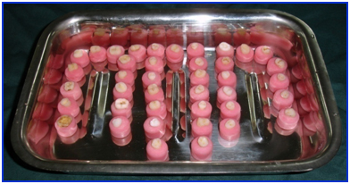
Recently extracted 30 premolar and molar human teeth without any morphological damage were collected for the study. Any remaining soft tissue present on the surface was cleaned by running water. Deposits like plaque and calculus were removed by hand scalers (Hu-Friedy, Germany). It was stored in saline (NS 500ml, Fresenius Kabi, India) and 1% thymol (Fresenius Kabi, India crystals) solution at a temperature between 40- 100. They were individually mounted with self-cure acrylic resin exposing the Cemento Enamel Junction [Table/Fig-2].
Teeth samples after mounting in self cure acrylic resin
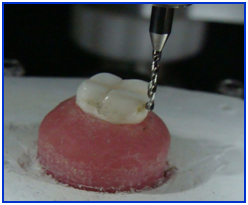
The teeth were marked and divided into 3 groups each containing 10 nos. The specimens were mounted onto the surveyor table of milling machine (AMANNGIRRBACH, af 350, Austria) so that the long axis of the test specimen was parallel to the long axis of a round end Tungsten Carbide bur and crown preparations were carried out [Table/Fig-3]. Tooth preparations with shoulder finish line were made above the cemento-enamel junction by manually rotating the bur against the stationary tooth specimen. A flat occlusal reduction was carried out by means of a diamond wheel so that the vertical height of the axial wall measured 4mm. After crown preparation, the test specimens were arranged in a base former and base is poured with model plaster (Type-II); each containing 6-7 specimens. Impressions were made with soft putty/regular set (Aquasil, Dentsply, Germany) and (Aquasil/LV Dentsply USA) impression material. A single–stage putty-wash technique was advocated to make the impressions with perforated stock trays and was poured with Die Stone (Type-IV) which is commercially available and individual dies were fabricated.
Crown preparations done in a milling machine
Temporary crowns were fabricated for each tooth preparation by direct and indirect method with two types of tooth coloured auto polymerizing resin. In the direct method, Tooth coloured cold polymerizing paste (VOCO structur 2 SC/QM) was packed into the occlusal index of corresponding tooth specimen made by soft putty/regular set (Aquasil, Dentsply, Germany) which was made prior to tooth preparation and pressed against the corresponding die by applying normal thumb pressure. It was allowed to set according to manufacturer’s specifications. Once set it is trimmed, polished and made fit in the corresponding prepared tooth. In the indirect method single layer of heated modelling wax-No. 2(HDP India) was manually adapted over the dies. After waxing-up, the dies were invested in dental flasks using standard investment procedures. After a lost-wax technique, the moulds were packed with Autopolymerising PMMA (DPI Self- cure tooth moulding powder) using standard laboratory procedures according to manufacturer’s instructions and were allowed to polymerize by keeping the flask under pressure in a hydraulic clamp (Sirio dental Snc, P 300, Italy) for 10 minutes. It was then deflasked and divested. A reline procedure was carried out on the temporary crowns with Autopolymerising PMMA (DPI Self- cure tooth moulding powder). It was then trimmed with acrylic burs (pink colour, medium coarseness) and polished with pumice powder (Dento-kem, India).
In the first group (Group A, Control group), the temporary crowns fabricated with both direct and indirect method were cemented directly with temporary luting cement (provical – eugenol free temporary cement voco) according to manufacturers specifications. In the second group ( group B, Test group 1) dentine-bonding agent (VOCO – Solobond M) was applied once evenly on the prepared tooth surface of each tooth specimen after etching (Etching gel 3ml,Detrey, Dentsply, Germany. Composition- 36% phosphoric acid) for 10 seconds and light cured (3M ESPE, Germany) for 10 seconds. Half of the samples were cemented with crowns fabricated by direct method and other half with crowns fabricated by indirect method with the eugenol free temporary cement (provical – eugenol free temporary cement voco). In the third group (Group C test group 2), a single layer of cavity varnish (Dental varnish, Samit India) is applied on the prepared tooth surface. After application of the cavity varnish, a light air pressure was used for at least two seconds to spread the varnish. Half of the samples were cemented with crowns fabricated by direct method and other half with crowns fabricated by indirect method with the eugenol free temporary cement (provical – eugenol free temporary cement voco).
Crowns were cemented at room temperature and were then cleaned of excess cement. Thermal treatment of the specimens was carried out with Cryostat (Inserf, India) in 1% basic methylene blue, pH 7.2 from 50C to 550C with a dwell time of 30 seconds. Specimens were rinsed with water and lightly pumiced to remove any superficial dye. After thermal treatment each test specimen was embedded into a clear acrylic resin PMMA matrix (DPI – RR cold cure) block [Table/Fig-4] prior to sectioning with a hard tissue microtome (LEICA 1600, Copenhagen, Denmark). Three sections of 300 micrometers thickness were made bucco-lingually with a blade thickness of 300 micrometers. All the sections from individual tooth were observed under a microscope (Wild Heerbrugg WildM2OEB) at 40X magnification by one observer after mounting it in a glass slide. All the surfaces of the tooth (buccal, palatal or lingual) were examined for the amount of dye penetration on the tooth and restoration interface. It was measured by comparing it with the microleakage scale used [Table/Fig-5].
Embedding specimens prior to sectioning
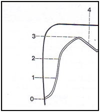
Microleakage scale used (adapted from Tjan et al.,) [5]
0 - No microleakage.
1- Microleakage to one third of axial wall.
2- Microleakage to two thirds of axial wall.
3- Microleakage along full length of axial wall.
4 - Microleakage over occlusal surface.
The extend of dye penetration into dentine tubules of each section was evaluated with the help of a visual scale and it was recorded according to the following values.
0- No tubule infiltration.
1- 1-33% tubule infiltration.
2- 33-67% infiltration.
3 - More than 67% infiltration.
Individual score was recorded on proforma’s (addendum 1) and was analysed statistically. The scores were entered in an excel spreadsheet as proportions and frequencies due to the ordinal nature of the data. SPSS Version 13 software was used for non-parametric data analysis by a qualified statistician. P-values less than 0.05 (p-value<0.05) were considered to be statistically significant.
Results
The data obtained through the study were formulated in different tables. The results were statistically analyzed using one way analysis of variance (ANOVA) and then Tukey honestly significant different (HSD) tests for comparisons among groups at 5% level of significance and were plotted in a graph [Table/Fig-6].
Overall graph showing values of microleakage
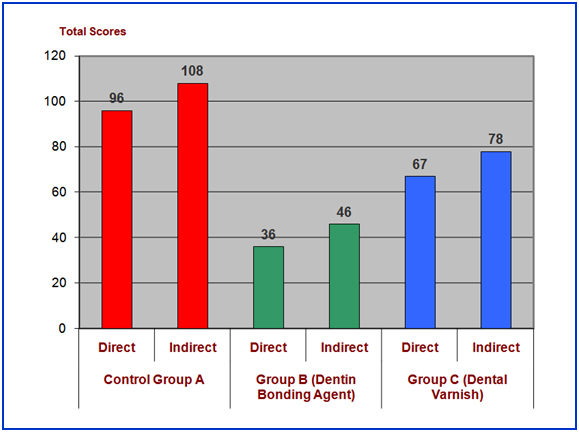
Group A (control group) specimens showed the maximum amount of microleakage in which both the dentin bonding and dentin sealing agent were not applied. When compared between direct and indirect technique, provisional crowns fabricated with direct technique showed less amount of microleakage than with that of indirect technique.
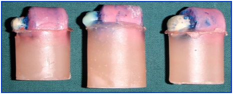
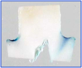
Group B (Dentin Bonding Agent) specimens showed the least amount of microleakage when compared with Group A and Group C [Table/Fig-6,7]. When compared between direct and indirect technique, provisional crowns fabricated with direct technique showed least amount of microleakage than with that of indirect technique.
Group B specimens showing the least amount of microlekage
Group C (Dental Varnish) specimen showed comparatively more amount of microleakage than that of Group B. When compared between direct and indirect technique, provisional crowns fabricated with direct technique showed less amount of microleakage than with that of indirect technique.
Discussion
Patients undergoing complex fixed prosthodontic treatment over extended periods of time should be provided with properly adapted provisional restorations and they should be evaluated at regular intervals for microlekage. Out of all the temporary restorative materials available poly methyl methacrylate (PMMA) is used for both direct and indirect technique for the fabrication of provisional restoration in this study [1].
Dentin is the living, vital, sensitive portion of teeth that has been described as a biologic composite made up of a collagen matrix filled with nanofillers of apatite crystallites and called as dentinal tubules and are sealed peripherally by enamel and cementum [6]. As dentin is made thinner by tooth preparation, it becomes more permeable to oral fluids. This gives an indication of the centripetal penetration into the dentin of the various fluids present in the mouth [7], which can be biologically and chemically aggressive, triggering hypersensitivity, demineralization, and this process is called as microleakage [8]. Crown preparation of a molar tooth can expose up to 2 million dentinal tubules. These exposed tubules will allow to and fro movement of substances between the pulp and outside oral environment. This can result in microleakage with variable intensities, with luting cements even for permenant crowns.
The use of dyes for detecting microleakage is oldest and commonest invitro method. According to Poloa Baldisierra if a material responds positively to the dye tests, it is likely to respond even better on a clinical level [9]. This is probably because the dye gets diffused easily than the bacteria. Even, calcification of the marginal area due to accumulation of protein debris can also improve the seal. This method involves placing a restoration in an extracted tooth and immersing it in a dye solution and after thermal treatment [10] is done it is taken out, sectioned and examined visually for dye penetration [11].
The two main technique involved in temporization are direct and indirect method. In direct technique the use of a mould or matrix which is made from a preoperative diagnostic cast is used. After tooth preparation, the matrix is trial fitted over the tooth. The prepared tooth is cleaned, gently dried, and lubricated by applying petroleum jelly. The selected provisional restorative material is mixed according to the manufacturer’s specification and placed into the matrix carefully without any air bubble incorporation. The matrix with the provisional restorative material is then placed over the prepared tooth in the exact position and allowed to polymerize. It is removed, trimmed, polished and cemented to the respective tooth finally with provisional cement. In the indirect technique, an impression is made after the tooth preparation and the cast is poured. Wax up is done with modeling wax on the prepared tooth in the cast. It is then flasked, dewaxed, packed and cured by keeping it under pressure. Finishing is done by trimming and polishing the provisional crowns after de flasking it. This technique consists of multiple steps which can make the provisional crowns weaker in its marginal adaption. However, provisional crowns fabricated with direct technique always have better marginal adaption and fit thus making it superior.
Introduction of dentin bonding agents for sealing the exposed dentinal tubules proved to be efficient. This is due to the formation of mechanically retentive hybrid layer which can withstand the moisture than the smear layer [12]. The bonding mechanism via hybridization to dental hard tissues relies on the diffusion of the applied monomers into partly demineralised substrates, which subsequently form the hybrid layer after the polymerization. Acid etching of dentine substrate removes the smear layers and plugs formed during tooth preparation [13]. Later demineralization of the mineral phase results in the porosity of these hard tissues into which adhesive monomers can penetrate to prepare a hybridized layer. Dentin adhesion depends on many factors such as dentine thickness, regional variations in permeability and pulpal pressure where as the factors that affect the bond strength are presence of moisture, blood, saliva and eugenol present in some provisional luting cements. All these factors make dentin bonding agent’s very technique sensitive.
One beneficial effect derived from dental varnish is that a better marginal seal is obtained for many restorative materials [14]. The deleterious products from cements, temporary filling materials, restorative filling materials, and oral fluids have less effect on the pulp because of the sealing effect which the varnish has on dentinal tubules. The end result of restorative dentistry with judicious use of dental varnish is better, longer lasting restorations with fewer traumas to the vital structures of the tooth and a resultant decrease in postoperative sensitivity for the patient.
Limitations of the Study
In this study, dentin-bonding agent and dental varnish are used as interim sealants for the prepared abutments. There were some unexpected problems encountered during the study which would have affected the results. Firstly, maintaining a proper shoulder finish line was difficult by rotating the dental bur around the test specimens manually. Secondly, the occlusal reductions for some of the specimens were within enamel and dentin due to the natural curvature of the cemento-enamel junction. Thirdly, standardization of the thickness of provisional crowns fabricated was very much compromised since the base plate wax was manually adapted to the dies. Fourthly, the marginal discrepancy was not measured after cementation of the provisional crowns.
Summary and Conclusion
Application of Dentin bonding, Dentin sealing agents failed to prevent the microleakage completely in temporary crowns. However Dentin Bonding agent (Solobond M) reduced microleakage to the maximum. In light of the above findings, the application of a single layer of Dentin sealing agent (Dental varnish) appears to be of no significant benefit when compared to crowns cemented with the application of Dentin bonding agent (Solobond M) on the tooth surface. However, based on the limitations of this study the application of a single layer of Dentin bonding agent (Solobond M) and provisional crowns fabricated with direct technique may be of some benefit for crown preparations as an interim measure prior to the luting of final crown.
Further study is necessary for the evaluation of the effectiveness of dentin-bonding agents as a sealant for exposed dentinal tubules. The different techniques by which the exact composition and thickness of the dentin- bonding agent has to be researched for a better prognosis of the provisional restorations.