Retrieval of Iatrogenically Pushed Pulp Stone From Middle Third of Root Canal in Permanent Maxillary Central Incisor: A Case Report
Kanika Gupta Verma1, Suruchi Juneja2, Sohajpreet Randhawa3, Tejal Malay Dhebar4, Anupama Raheja5
1Reader, Department of Pediatric and Preventive Dentistry, Surendra Dental College and Hospital, Sri Ganganagar, Rajasthan, India.
2Reader, Department of Pediatric and Preventive Dentistry, Surendra Dental College and Hospital, Sri Ganganagar, Rajasthan, India.
3Post Graduate Student, Department of Pediatric and Preventive Dentistry, Surendera Dental College and Research Institute, Sri Ganganagar, Rajasthan, India.
4Professor, Department of Oral Pathology, Sardar Patel Dental College and Hospital, Lucknow, Uttar Pardesh, India.
5Reader, Department of Prosthodontics, K.D Dental College and Hospital, Mathura, Uttar Pardesh, India.
NAME, ADDRESS, E-MAIL ID OF THE CORRESPONDING AUTHOR: Dr. Kanika Gupta Verma,Reader, Department of Pedodontics &Preventive Dentistry, Surendera Dental College & Research Institute, H.H Gardens, Power House Road, Sri Ganganagar-335001, Rajasthan, India.Email: drguptakanika@yahoo.co.in
The pulp stones are the discrete nodular calcified masses commonly existing in coronal and occasionally in radicular pulp, placed freely, attached or embedded into the dentine. The present case report revealed the iatrogenic pushing of pulp stone and blockage of root canal that caused endodontic failure. The case enlightens the proper use of ultrasonic instruments with irrigating solutions to manage the calcifications in root canal for successful endodontic therapy.
Calcified mass, Radicular pulp, Ultrasonic
Case Report
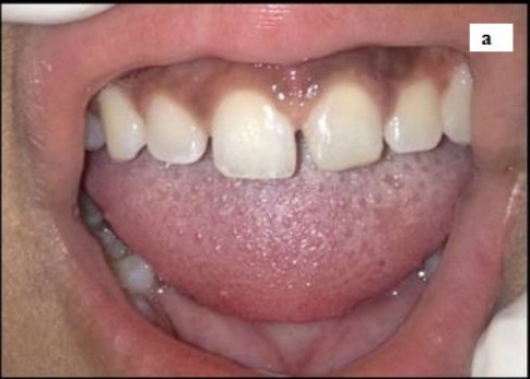
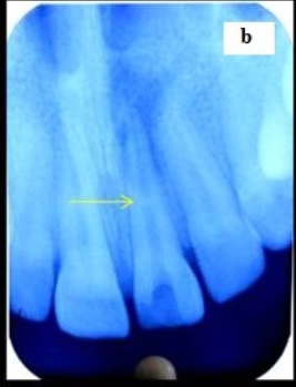
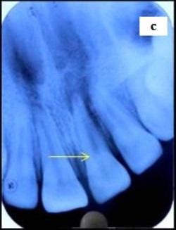
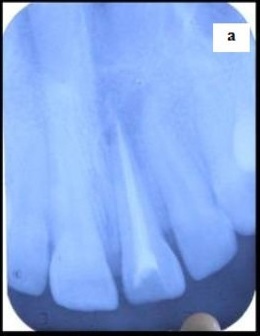
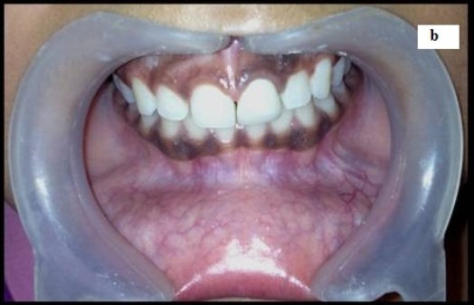
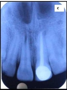
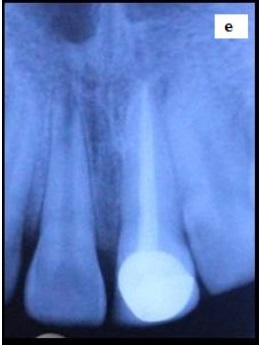
A 13-year-old patient reported to the Department of Paediatrics and Preventive Dentistry, with the chief complaint of pain in upper front tooth since two months. History of present illness revealed that pain was sharp and continuous in nature. Ten days back, a local dental practitioner was consulted by the patient with same chief complaint and he started with endodontic intervention of maxillary left central incisor. As during the course of treatment, his pain got aggravated, so he was referred to the institute for the necessary intervention. Intraoral examination revealed normal gingival status with no intraoral sinus [Table/Fig-1a], patent access cavity but pain on percussion in relation to maxillary left central incisor. Radiographic examination revealed that access cavity was patent with a pulp stone in middle third of the root canal of related tooth [Table/Fig-1b]. Patient got a preoperative IOPA taken before the start of treatment by local dental practitioner that revealed a pulp stone at cervical third of root canal [Table/Fig-1c]. Thus it was concluded that during endodontic procedure, the pulp stone was iatrogenically pushed in the middle third of root canal, leading to incomplete removal of pulpal tissue, causing spontaneous pain. General physical examination revealed no metabolic disturbances and syndrome, which suggested that pulp stone was of idiopathic origin.
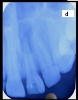
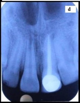
In present case, the conventional approach of dissecting pulp stone was not considered, as dissection would have risked the obliteration of rest of the canal. A no. # 10 K-file was used with EDTA to bypass the pulp stone and a successful retrieval of pulp stone [Table/Fig-1d] was done with the help of ultrasonic endodontic tip. The retrieved pulp stone was in a single mass approximately 3mm in size [Table/Fig-1e] and was then sent for histopathological investigation.
After clearing the canal, endodontic management was done [Table/Fig-2a] and tooth was restored with porcelain fused to metal crown [Table/Fig-2b & Table/Fig-2c]. Follow up was done after 6 months [Table/Fig-2d] and one year [Table/Fig-2e]showing successful endodontic therapy and healthy periodontal status.
Intraoral examination revealing normal gingival status in relation to maxillary left central incisor;
IOPA revealing pulp stone in middle third of the root;
Pulp stone present in cervical third of root;
successful retrieval of pulp stone from canal;
retrieved pulp stone (3mm)

restoration of 21 with porcelain fused to metal crown;
IOPA showing fit of PFM wrt 21;
IOPA revealing no pathology after 6months;
IOPA showing no pathology after one year
Discussion
Pulp stones are the discrete nodular calcified masses of calcium phosphate, calcium carbonate and magnesium phosphate [1], commonly existing in coronal and occasionally in radicular pulp, either placed freely in the pulp tissue or become attached or embedded into the dentine [2]. Structurally, they are classified as true, false and amorphous; the true are made of dentine, false are formed from degenerating cells of pulp and amorphous are irregular in shape occurring close to blood vessels [3]. The aetiological factors implicated in stone formation include pulp degeneration, inductive interactions between epithelium and pulp tissue, age, circulatory disturbances, orthodontic tooth movement, idiopathic factors, genetic predisposition and in certain syndromes such as Van der woude syndrome [4,5].
In spite of higher occurrence of pulp stones in adult population, their presence in young children is rare. For a patient requiring endodontic treatment, negotiation of calcification and retrieval of pulp stone is of paramount importance for successful therapy.
Pulp stones are discovered on routine investigations or described as symptoms of changes in the pulp tissues. An aggravated or reduced response to pulp sensitivity tests can be explained with radiographs showing calcifications in root canals. As in the present case, patient encountered aggravated pain during subsequent visits of endodontic therapy, because of iatrogenically pushing of pulp stone in middle of root canal.
Langeland discussed the clinical relevance of pulp stones in term of their effects upon root canal treatment [6]. The presence of pulp stone may alter the internal anatomy and confuse the operator by obscuring or totally blocking the path of root canal. The tip of exploring instruments get deflected by free denticles, thus preventing their passage down the canal [7,8], as observed in our case.
Many methods have been used for pulp stone removal from pulp chamber and root canals. The large pulp stones are usually dissected using burs and then removed from canals, but special ultrasonic tips makes their retrieval easier [8 - 10]. These tips are sharp with pointed ends, having longitudinal rounded microblades separated by grooves that remove calcifications with ease. The use of ultrasonics should preferably be coupled with sodium hypochlorite irrigating solution, that cause an additive effect by dissolving collagen based tissues [11,12]. In our case, pulp stone was retrieved using START-X TM (Dentsply Maillefer) ultrasonic tips along with the combination of 17% EDTA solution and 5.2% sodium hypochlorite as irrigants.
The retrieved pulp stone was send for histopathological evaluation that showed a calcified mass without dentinal tubules, thus diagnosing it to be a free false pulp stone or partially calcified pulp tissue. During pulp stone removal with burs and endodontic instruments, potential complications may arise like weakening of tooth due to excessive tooth structure removal or perforations [13,14]. Whereas, the ultrasonic instruments producing vibrations can aid in easier, safer and faster pulp stone removal from root canals.
Conclusion
The present case reported the iatrogenic pushing of pulp stone and blockage of root canal that caused endodontic failure. The case enlightens the proper use of ultrasonic instruments with irrigating solutions to manage the calcifications in root canal for successful endodontic therapy.
[1]. WG Shafer, MK Hine, BM Levy, A Textbook of Oral Pathology 1983 4th EditionPhiladelphiaSaunders; [Google Scholar]
[2]. R Goga, NP Chandler, AO Oginni, Pulp stones: a reviewInt Endod J 2008 41(6):457-68. [Google Scholar]
[3]. DH Pashley, RE Walton, HC Slavkin, Histology and physiology of the pulp. In: Endodontics 2002 5th EditionHamilton, OntarioBC Decker Inc:25-61. [Google Scholar]
[4]. S Ranjitkar, JA Taylor, GC Townsend, A radiographic assessment of the prevalence of pulp stones.Aust Dent J 2002 47(1):36-40. [Google Scholar]
[5]. SK Bahetwar, RK Pandey, An unusual case report of generalized pulp stones in young permanent dentition.Contemp Clin Dent 2010 4(1):281-83. [Google Scholar]
[6]. K Langeland, The histopathologic basis in endodontic treatmentDent Clin North Am 1967 11:491-520. [Google Scholar]
[7]. GS Nanjannawar, H Vagarali, LG Nanjannawar, B Prathasarathy, A Patil, N Bhandi, Pulp Stone - An endodontic challenge: Successful retrieval of exceptionally long pulp stone measuring 14 and 9.5mm from the palatal roots maxillary molars.J Contemp Dent Pract 2012 13(5):719-22. [Google Scholar]
[8]. PJ Vyver, F Paleker, Endodontic and restorative management of a lower molar with a calcified pulp chamberSouth Afr Dent Assoc 2013 68(10):450-6. [Google Scholar]
[9]. TR Pitt Ford, JS Rhodes, HE Pitt Ford, Endodontics problem-solving in clinical practic 2002 London. UK:Martin Dunitz Ltd:85 [Google Scholar]
[10]. P Jain, P Patni, H Hiremath, N Jain, Successful removal of a 16 mm long pulp stone using ultrasonic tips from maxillary left first molar and its endodontic.J Conserv Dent. 2014 17(1):92-5. [Google Scholar]
[11]. M Hulsmann, M Heckendorff, A Lennon, Chelating agents in root canal treatment: mode of action and indications for their useInt Endod J. 2003 36(12):810-30. [Google Scholar]
[12]. WC Ngeow, YL Thong, Gaining access through a calcified pulp chamber: A clinical challengeInt Endod J. 1998 31(5):367-71. [Google Scholar]
[13]. MK Iqbal, Nonsurgical ultrasonic endodontic instrumentsDent Clin N Am 2004 48(1):19-34. [Google Scholar]
[14]. PS McCabe, PM Dummer, Pulp canal obliteration: an endodontic diagnosis and treatment challenge.Int Endod J 2012 45(2):177-97. [Google Scholar]