Surgical Management of a Non-healing Intra-alveolar Root Fracture Associated with Pulpal Calcification and Root Resorption: A Case Report
Sonali Kapoor1, Parul Bansal2, Sarath Chandran3, Vineet Agrawal4
1Professor and Head, Department of Conservative Dentistry and Endodontics, Manubhai Patel Dental College, Vadodara, Gujarat, India.
2Reader, Department of Conservative Dentistry and Endodontics, Subharti Dental College, Meerut., Uttar Pradesh, India.
3Professor and Head, Department of Periodontics, Manubhai Patel Dental College, Vadodara, Gujarat, India.
4Reader, Department of Conservative Dentistry and Endodontics, Manubhai Patel Dental College and Hospital, Vadodara, Gujarat, India.
NAME, ADDRESS, E-MAIL ID OF THE CORRESPONDING AUTHOR: Dr. Sonali Kapoor, 30, Nirman Society, Alkapuri, Vadodara, Gujarat - 390005, India.
E-mail: docsonali@yahoo.com
Radicular fractures are very challenging to address due to various complications like periodontal communication, increased mobility, and continued pulpal infection leading to necrosis and its long term sequelae like root resorption and pulp canal obliteration. This paper present a case of a long standing horizontal mid root fracture with root resorption and pulp canal obliteration, which was preserved functionally and aesthetically by surgical approach using MTA (mineral trioxide aggregate) and PRF (platelet rich fibrin).
Canal obliteration, Mineral trioxide aggregate, Platelet rich fibrin
Case Report
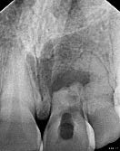
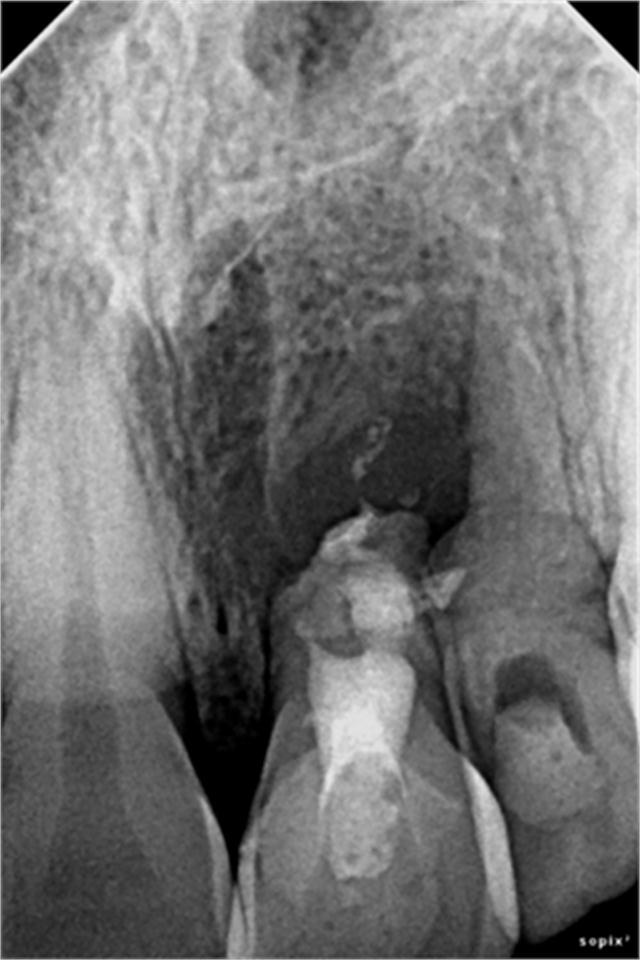
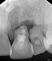
An 18-year-old male patient came to the Department of Conservative Dentistry and Endodontics at Manubhai Patel Dental College, Vadodora, Gujarat, India with the chief complaint of pain in his upper left front teeth since last 3-4 months. Patient gave history of trauma few years back. On vitality tests tooth was non responsive. On palpation vestibular tenderness was evident. Tooth was tender on percussion and grade two mobile. Radiographic examination revealed horizontal fracture of middle third of the root with radiolucency present near the fracture site suggesting presence of granulation tissue. Apical segment was displaced from its anatomical position [Table/Fig-1] .
Pulp canal obliteration and attempted root canal treatment was evident. Resorption of the coronal root segment adjacent to the granulation tissue had occurred resulting in shortening of fractured coronal root segment length. Root canal treatment was attempted previously and access cavity was restored with temporary restoration. Endodontic management of coronal segment followed by surgical removal of granulation tissue and displaced apical segment was planned. Informed consent was obtained. After removal of temporary restoration, file was going into the space created iatrogenically [Table/Fig-2] .
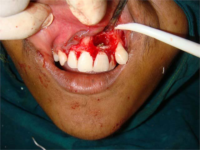
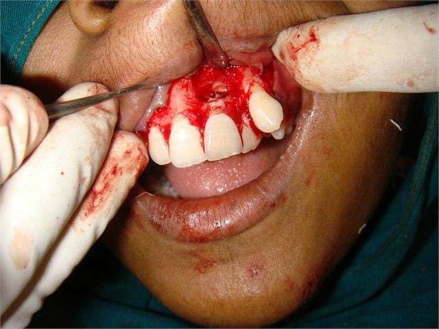
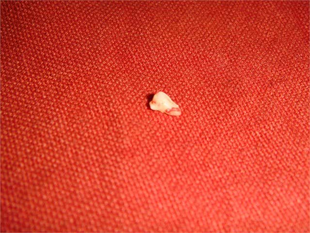
In order to avoid further damage to root structure in an attempt to find the canal, already created space was filled with MTA by orthograde approach. After administration of local anaesthesia full thickness periosteal flap was reflected and buccal bone was found perforated due to presence of the lesion [Table/Fig-3] . All the granulation tissue was curretted out [Table/Fig-4] and pulp space at fractured end was sealed with MTA by retrograde approach [Table/Fig-5] . Displaced apical segment was removed with the help of an elevator [Table/Fig-6] .
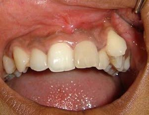
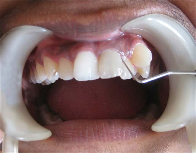
Bony cavity was filled with PRF for rapid healing and suture were placed. Suture were removed after a week and healing was satisfactory [Table/Fig-7,Table/Fig-8] . Follow up radiographs were taken at 6 months, 8 months [Table/Fig-9] and 18 months [Table/Fig-10] interval and it was observed that radiolucency was reducing which is indicative of successful healing; also periodontal probing revealed that tooth mobility was within physiological limit [Table/Fig-11] .
IOPA -apical segment displaced from anatomical position
IOPA - file placed in the space created iatrogenically
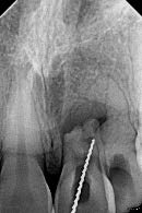
Image showing raised flap
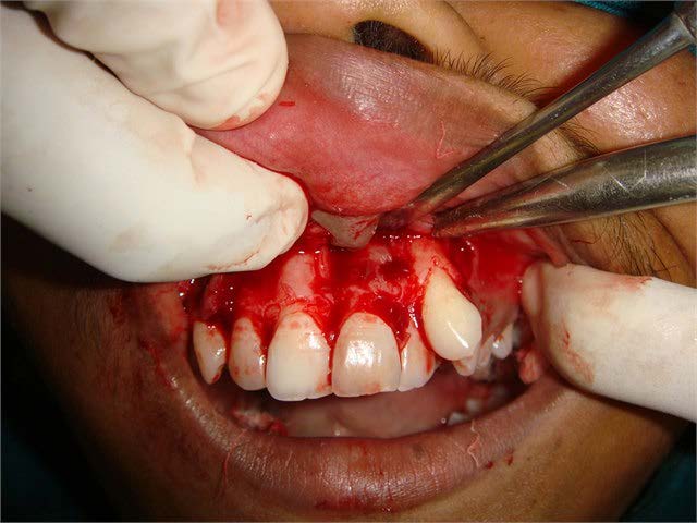
Image showing curettage of the surrounding tissue
Retrograde selaing of MTA
Excised specimen of apical displaced fragment
IOPA - immediate postoperative
IOPA - 8 months follow up
IOPA -18 months follow up
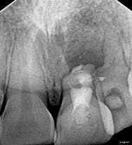
Image showing ‘0 mm’ probing depth
Discussion
Root fractures are relatively uncommon clinical condition with a frequency of 0.5% to 7% in permanent teeth [1]. Although the prognosis of root fractures is generally good [2] histological reaction at fracture line can occur in one of the following 4 types: interposition of calcified tissue (calus formation), interposition of calcified and connective tissue, interposition of connective tissue and by interposition of granulation tissue, caused by an infected or necrotic pulp[1,3]. Though most frequent tissue reaction at the fracture line is interposition of connective tissue, which occurs in almost 65% of cases [4]. Interposition of granulation tissue occurs when the coronal segment loses its vitality and infective products in the coronal pulp cause an inflammatory response.
Complications during healing process of the pulp and periodontium can unfavourably influence the long term prognosis [5]. Pulpal necrosis and lack of healing at the fracture line (interposition of granulation tissue) are the most common complications occurring in almost 20% of the cases [6]. After dental traumas, root resorption, root canal obliteration, interruption in root formation, periapical lesion, and damages to permanent tooth buds may occur as a squeal. Studies have shown high prevalence of root resorption (8-50%) after tooth trauma [1,7].
Clinically teeth may be non symptomatic in the early period of the process and resorption may be seen only in radiographs later on. However, as the process progress, the tooth may become symptomatic and an abscess may develop with sinus formation and increased tooth mobility. Radiographically, radiolucency is seen at the root surface of dentin and adjacent bone. To arrest further resorption, it is critical to control the pulpal bacteria and granulation tissue. In the present case bacterial control was done by root canal treatment and intracanal medicament followed by retro filling and sealing of canal. Granulation tissue was removed surgically. Followup examination shows arrest of resorptive process.
Calcific metamorphosis of pulp space may develop to varying degrees following traumatic injuries to teeth in approximately 20% of cases [8]. Pulpal obliteration is characterized by the apparent loss of the pulp space radiographically and a yellow discoloration of the crown clinically [8]. In the present case calcific metamorphosis and pulpal obliteration was present. As bacteria may harbor even in completely obliterated pulp space, pulp space was sealed coronally as well as at the fractured site.
MTA is biocompatible material with antimicrobial properties and high pH (12.5), which promotes growth of the cementum and formation of bone, resulting regeneration of the PDL around the site of injury [9,10]. In the present case MTA was used to seal the fractured site of the root to promote healing.
Treatment approach for horizontal root fracture is complex and depends on degree of displacement of fracture fragment, patient’s age, stage of root growth, mobility of the coronal fragment, and diastasis of the fragments [11]. Necrotic apical segment should be removed surgically if the remaining coronal root segment is long enough to provide adequate periodontal support. In the present case necrosed apical segment along with granulation tissue was removed surgically.
Platelet-rich fibrin (PRF), a platelet concentrate with growth factors in the form of fibrin membranes was prepared from patient’s own blood and was placed inside the bony cavity to promote healing. PRF has been used in other studies also to promote healing [12]. PRF accelerates wound healing and allows regeneration of tissue thus resulting faster and better healing.
Conclusion
Management of intra alveolar root fracture with complications like calcific metamorphosis, periodontal communication and root resorption is challenging. A tooth with non healing horizontal root fracture with granulation tissue can be treated surgically by removal of necrosed apical segment and granulation tissue followed by PRF placement. Now it is possible to preserve the tooth with the help of newer bioactive materials like MTA and techniques like retro filling, in its healthy state functionally and aesthetically.
[1]. JO Andreasen, FM Andreasen, L Andersson, editors. Textbook and Color Atlas of Traumatic Injuries to the Teeth. Munksgaard, Kopenhagen 2007 :337-71. [Google Scholar]
[2]. JO Andreasen, E Hjørting-Hansen, Intra-alveolar root fractures: radiographic and histologic study of 50 cases.J Oral Surg. 1967 25:414-26. [Google Scholar]
[3]. MK Caliskan, Y Pehlivan, Prognosis of root-fractured permanent incisorsEndod Dent Traumatol 1996 12:129-36. [Google Scholar]
[4]. I Jacobsen, BU Zachrisson, Repair characteristics of root fracture-s in permanent anterior teeth.Scand J Dent Res 1975 83(6):355-34. [Google Scholar]
[5]. JO Andreasen, FM Andreasen, editors. Root fractures. In: Textbook and color atlas of traumatic injuries to the teeth 1994 3rd EdCopenhagenMunksgaard;.:79-314. [Google Scholar]
[6]. M Cvek, G Tsilingaridis, JO Andreasen, Survival of 534 incisors after intra-alveolar root fracture in patients aged 7–17 years.Dental Traumatology 2008 24(4):379-87. [Google Scholar]
[7]. A Majorana, E Bardellini, G Conti, E Keller, S Pasini, Root resorption in dental trauma: 45 cases followed for 5 years.Dent Traumatol 2003 19(5):262-65. [Google Scholar]
[8]. PS McCabe, PMH Dummer, Pulp canal obliteration: an endodontic diagnosis and treatment challenge.International Endodontic Journal 2012 45:177-97. [Google Scholar]
[9]. HW Roberts, JM Toth, DW Berzins, DG Charlton, Mineral trioxide aggregate material used in endodontic treatment: a review of literatureDent Mater 2008 24(20):149-64. [Google Scholar]
[10]. S Agrawal Vineet, Sonali K, Clinical management of severe external root resorption and immature open apex with MTA and calcium hydroxide- A case report.Endodontology. 2013 83(88) [Google Scholar]
[11]. JO Andreasen, FM Andreasen, I Megare, M Cvek, Healing of 400 intra alveolar root fractures: effect of pre-injury and injury factors such as sex, age, stage of root development, fracture type, location of fracture and severity of dislocationDent traumatol 2004 20:192-202. [Google Scholar]
[12]. P Bansal, K Kanyal, V Nikhil, Surgical management of horizontal root fracture with platelet rich fibrin placement: case report of two cases.Dent J of Advance Studies 2014 1(2):28-31. [Google Scholar]