Efficacy of Collagen Particles in Chronic Non Healing Ulcers
Karunakar Reddy Chalimidi1, Yogesh Kumar2, U Anand Kini3
1 Post-graduate, Department of General Surgery, Kasturba Medical College, Manipal university, Mangalore, India.
2 Professor, Department of General Surgery, Kasturba Medical College, Manipal university, Mangalore, India.
3 Associate Professor, Department of General Surgery, Kasturba Medical College, Manipal university, Mangalore, India.
NAME, ADDRESS, E-MAIL ID OF THE CORRESPONDING AUTHOR: Dr. Karunakar Reddy Chalimidi, Room no 109, Falnir Mens PG Hostel, Falnir, Mangalore, India. E-mail : Karunakar_161988@yahoo.co.in
Introduction
Chronic foot ulcers will lead to a significant and prolonged stress to the patients. Pain and discomfort that may be acute or continuous is the usual complaint in chronic non healing ulcers that may even exacerbate with change of the dressings. The end process in any wound healing is wound contracture and scar formation. Collagen plays an important role in this stage of wound healing. Collagen particles were used in chronic non healing ulcer management to prove their efficacy when compared with conventional dressing in a study conducted by us.
Objective
To compare the healing process in non healing ulcers using collagen particles with those of conventional method of dressing (betadine).
Materials and Methods
It was a non randomized, prospective study conducted for a period of October 2012 to October 2014 in hospitals belonging to Kasturba medical college. Non concurrent pre and post comparative study; between collagen group and conventional dressing group. A total of 110 patients with chronic ulcers were included; each group comprising 55 patients.
Results
There was a significant decrease in wound size with a mean difference of 37.29 in experimental group when compared to 14.29 in control group.
Conclusion
Collagen dressing is effective in management of chronic non healing ulcers when compared to conventional betadine dressing. It heals by forming an early granulation tissue and thus reducing the length of hospital stay.
Betadine, Granulation tissue, Wound
Introduction
An ulcer is defined as a break in continuity of covering epithelium, involving skin or mucous membrane and as a result of molecular death. Chronic ulcers of the foot are defined as a gradual breakdown of the epidermal and dermal tissue of the foot lasting for more than 6 weeks. Various factors have been implicated for an ulcer to develop in diabetic patients of which important is peripheral vascular disease which causes decrease in sensation as a result of peripheral neuropathy [1]. Secondary infection in a chronic ulcer is very common that will have a copious amount of discharge that smells foul. Antibiotic treatment for infection, wound dressing, pressure relief (special foot wear, off loading, inserts and casting), debridement and desloughing are the traditional management approaches for diabetic ulcers [2]. Embarrassing dressings, restricted mobility and disability are the other issues that are often a great concern to the patients [3]. Collagens functions as structural proteins of extracellular matrix and are synthesized by the fibroblasts [4,5]. In normal wound healing process special enzymes called matrix metalloproteinases (MMPs) breaks down the malformed and damaged collagen present at the site of wound. So when dressings containing collagen are used, the MMPS are kept busy breaking down that collagen and the healthy collagen that is innate of the body is thus protected, helping in hastening the healing process [6]. There are other modalities of dressing other than conventional betadine dressing in ulcer management like sugar dressing, vacuum assisted closure method. Direct instillation of sugar on the wound will result in low osmolar effect, promoting early granulation tissue formation, reduction in oedema. Hence, bacteriostatic effect is enhanced and thereby hastens the wound healing [7,8]. Knutson and colleagues performed a study on wounds treated with granular sugar with povidone iodine to treat 605 patients with traumatic wounds and burns and observed reduction in healing time by 25% [9]. But there is still a lack in evidence explaining the cellular and molecular interactions between sugar and wound environment, where in our study a molecular basis is explained. Vacuum assisted wound closure is a recent and significant method of wound closure management. This method was first described by Fleishmann in 1993 [10]. This method works on application of controlled negative pressure on wound bed. Gradual traction on living tissues can stimulate and promote regeneration of tissue structures. Morykwas et al., studies showed decrease in the bacterial load in wounds treated with negative pressure therapy [11]. There should be complete hemostasis before application of vacuum dressing and this has to be used in caution in patients with coagulopathies. Also, maceration of skin and pain due to suction make it uncomfortable. Where, in our study such complication was not reported. Pressure maintaining appliance and patient education is also needed.
Dressings containing sterile collagen particles were used in the study and compared with conventional (betadine) dressings. The aim of our study was to evaluate the efficacy of collagen particles, with an objective to compare the rate of healing process using collagen particles with those of conventional methods.
Materials and Methods
The study was a non randomized and prospective comparative study between collagen and betadine dressing group. It included 110 patients with 55 in each group, 55 patients were subjected to collagen dressing and 55 patients to betadine dressing. Comparison between these two groups was done with respect to parameters of wound area, number of debridements, number of dressings and mode of healing. Institutional Ethical committee clearance was taken before starting the study and informed consent was taken from all participants. The study was conducted from October 2012 to October 2014, in Government Wenlock Hospital, Kasturba Medical College and Hospital, Attavar & Ambedkar circle, Mangalore, Manipal University, India.
Inclusion criteria: All non healing ulcers were included like diabetic ulcers, venous ulcers, traumatic ulcers, bedsores, amputation stump ulcers.
Exclusion criteria
Ulcers having exposed bone without granulation tissue.
Ulcers with proven malignancy.
Application methodology
Collagen particles, lyophilized type 1 intact collagen of fish origin, were used. Wound bed was cleaned with normal saline, to the extent that it doesn’t cause any trauma. Measurement of ulcer size and area was done by putting a transparent plastic cover over the ulcer and markings were made along the margin and then the cover was placed on the graph paper and assessed periodically.
Initial debridement was done in case of unhealthy wounds with extensive slough. Later, depending on the amount of dead tissue, periodic debridements were done.
Collagen particles were sprinkled sufficiently to cover the wound surface. In case of tunnelled and undermined wounds, collagen particles were made into paste or a solution with normal saline to ensure that the particles penetrated into the wound. Wound was covered with an absorbent dressing. Frequency of collagen particles application was done initially on daily basis; subsequent dressings were changed every 3-4 days and assessed.
In the study the variables considered were: decrease in wound size, number of debridements required, duration of healing. A comparison was made with that of betadine dressing. Follow up was done for a period of 6 months.
Results
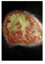
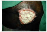
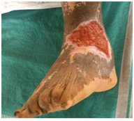
Comparison between these two groups was done in parameters of wound area, number of debridements, number of dressings and mode of healing [Table/Fig-1,2,3,4]. [Table/Fig-5a,5b,5c,6a,6b,6c,7a,7b,7c] depict three different types of ulcers, at their time of presentation and few weeks after collagen particle application.
Comparing the decrease in wound area
| Group | N | Mean | Std. Deviation | Median | Mean Difference | Change (%) | Wilcoxon Signed rank text p-value |
|---|
| Case | Wound initial area | 55 | 82.09 | 67.571 | 67.00 | 37.291 | 45.43 | p=0.000 <0.001,HS |
| Wound final area | 55 | 44.80 | 45.480 | 23.00 |
| Control | Wound initial area | 55 | 61.07 | 51.613 | 36.00 | 14.291 | 23.40 | p=0.000 <0.001,HS |
| Wound final area | 55 | 46.78 | 44.481 | 26.00 |
Wilcoxon signed rank test p-value 0.000, highly significant
(N = number of individuals)
Comparison on number of dressings
| Group | n | Minimum | Maximum | Mean±Std. Deviation | Median | t-test p-value | Mann-whitney test p-value |
|---|
| Cases | 55 | 3 | 18 | 6.96±3.355 | 6.00 | <0.001 | <0.001 |
| Control | 55 | 6 | 35 | 17.89±6.151 | 16.00 | HS | HS |
| Total | 110 | 3 | 35 | 12.43±7.379 | 12.00 | | |
Mann-whitney test p value <0.001, highly significant. (N = no of individuals)
Comparison on number of debridements
| Group | n | Minimum | Maximum | Mean±Std. Deviation | Median | Mannwhitney test p-value |
|---|
| Cases | 55 | 0 | 6 | 2.07±1.260 | 2.00 | .0.035 Sig |
| Control | 55 | 1 | 6 | 2.58±1.357 | 2.00 |
| Total | 110 | 0 | 6 | 2.33±1.328 | 2.00 |
Mann whitney test p-value 0.035; highly significant. (N = no of individuals)
Comparison on mode of healing
| Group | n | Minimum | Maximum | Mean | Std. Deviation | Median | t-test p-value | Mannwhitney test p-value |
|---|
| Cases | 55 | 3 | 18 | 6.96 | 3.355 | 6.00 | .000 HS | .000 HS |
| Control | 55 | 6 | 35 | 17.89 | 6.151 | 16.00 |
| Total | 110 | 3 | 35 | 12.43 | 7.379 | 12.00 |
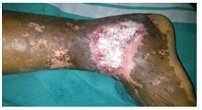
Bed sore at initial presentation
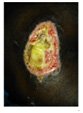
Collagen particles sprinkled over the wound bed
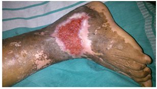
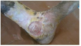
Traumatic ulcer at the time of presentation
After application of collagen particles
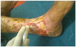
Venous ulcer at the time of presentation
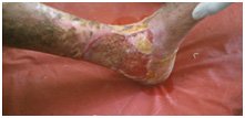
Application of collagen particles
Discussion
Wound healing involves a timely expression of various growth factors that promotes cellular proliferation and migration, collagen deposition and formation of new connective tissue matrix [12,13]. Collagens are Proline-rich proteins that are fibrous with long, stiff, triple stranded helical structure comprising of three α-chains. The major collagen molecules that give tensile strength to skin are heterotrimeric collagen type I, formed by two α 1(I) chains and one α 2(I) chain and homotrimeric collagen type III, formed by three α1(III) chains [14,15]. The role of collagen in improving wound healing is by stimulating fibroblast activity [16].
This study was undertaken with the aim to evaluate the efficacy of collagen particles as a topical wound dressing in patients with chronic non healing ulcers. Donaghue et al., in 75 patients had compared the efficacy of collagen-alginate topical wound dressing with that of regular wound dressing with guaze and normal saline in diabetic foot ulcer patients [17]. There was an 80.6% of reduction in mean wound area, observed in collagen-alginate dressing group compared to 61.1% in guaze-dressing group. In our study a decrease in wound size from initial area to final with mean difference of 37.29 was observed in experimental group when compared to 14.29 in control group, with p-value of (<0.001) that is highly significant. Another study done by Graunlich et al., compared the effectiveness of collagen with that of hydrocolloid in pressure ulcer healing [18]. A total of 65 patients were included, of which 35 were subjected to collagen dressing and 30 to hydrocolloid dressing. Complete ulcer healing was observed in 8 weeks in collagen group (51%) compared to (50%) in control group but the mean healing time was same. In our study complete wound healing was observed in 69% in experimental group as compared to 60% in control group. Above studies were done comparing alginate dressings with guaze dressing and hydrocolloid dressings with collagen, where collagen is shown to be effective in wound healing. As there are not many studies available comparing collagen particle dressing with conventional dressing our study lags in proven data. And comparison with negative pressure wound dressings could not be done as it was applied in wounds with initial wound size being more than our study group. Coming to advantages of collagen dressings over conventional dressings, frequency in change of dressing is reduced causing less discomfort for the patients, no case of allergic response to collagen particles has been reported in our study and application methodology is easier when compared to sugar dressings or vacuum dressings where a negative pressure system is required. Disadvantages are, this is not a cost effective treatment when used for bigger wounds, cannot be applied over infective wounds where debridements at initial setting are needed. The use of new dressing modalities with use of collagen particles can increase the potentials in wound healing but further studies are needed to prove the efficacy.
Limitations
The present study had the following limitations. First is that it was not a randomized study, so similarity among type of ulcers and size of ulcer was not maintained. Second, sample size was very less where we included only 55 patients in each group. Third is the cost factor, as collagen dressing is not very cost effective.
Conclusion
Collagen particles are effective in hastening the healing process by formation of early granulation tissue and wound contraction. So that, the number of debridements and dressings required can be reduced; as supported by the present study. Daily dressings may be uncomfortable for the patient causing pain affecting their social well being. Collagen dressings can be changed once in 2-3 days depending on the wound burden subjecting the patients for less discomfort. A significant decrease in wound size was observed in the experimental group compared to control group. Through our study we could prove that collagen dressing is better in comparison with conventional dressings considering early granulation tissue formation and reduced hospital stay.
Wilcoxon signed rank test p-value 0.000, highly significant(N = number of individuals)
Mann-whitney test p value <0.001, highly significant. (N = no of individuals)
Mann whitney test p-value 0.035; highly significant. (N = no of individuals)
[1]. Pudner R, Wound management: The management of patients with a leg ulcerJournal of Community Nursing 1998 12(3):26-33. [Google Scholar]
[2]. Peter A, Blume, DPM, Jodi Walters, et al. Comparison of Negative Pressure Wound Therapy Using Vacuum-Assisted Closure with Advanced Moist Therapy in the Treatment of Diabetic Foot Ulcers. A multicenter randomized controlled trialDiabetes Care 2008 31(4):631-36. [Google Scholar]
[3]. Bauling PC, A review of the impact of dressings on quality of life. In: Suggett A, Cherry G, Mani R, Eaglstein W, edsEvidence-based wound care: proceedings of a conference sponsored by Smith and Nephew held in New York, UK on 17th November 1997 1998 LondonRoyal society of Medicine Press:39-42. [Google Scholar]
[4]. Heino J, The collagen family members as cell adhesion proteinsBioessays 2007 29:1001-10. [Google Scholar]
[5]. Myllyharju J, Kivirikko KI, Collagens, modifying enzymes and their mutations in humans, flies and wormsTrends in Genetics 2004 20:33-43. [Google Scholar]
[6]. Barbul A, Etron DT, Wound healing. In, Charles brunicardi F.(ed)Schwart’z principles of surgery 2010 9th editionNew YorkMc Graw Hill:209-31. [Google Scholar]
[7]. Chirife J, Herszage L, Joseph A, Kohn ES, In vitro study of bacterial growth inhibition in concentrated sugar solutions: microbiological basis for the use of sugar in treating infected woundsAntimicrob Agents Chemother 1983 23(5):766-73. [Google Scholar]
[8]. Chirife J, Scarmato G, Herszage L, Scientific basis for use of granulated sugar in treatment of infected woundsLancet 1982 1(8271):560-61. [Google Scholar]
[9]. Knutson RA, Merbitz LA, Creekmore MA, Snipes HG, Use of sugar and povidone-iodine to enhance wound healing: five year's experienceSouth Med J 1981 74(11):1329-35. [Google Scholar]
[10]. Cohen I, Diegelmann R, Yager D, Wound care and wound healing 1999 7th ed edNew YorkMcGraw-Hill [Google Scholar]
[11]. Morykwas MJ, Simpson J, Punger K, Argenta A, Kremers L, Argenta J, Vacuum-assisted closure: state of basic research and physiologic foundationPlast Reconstr Surg 2006 117(suppl 7):121S-126S. [Google Scholar]
[12]. Singer AJ, Clark RA, Cutaneous wound healingN Engl J Med 1999 341:738-46. [Google Scholar]
[13]. Writte MB, Barbul A, General principles of wound healingSurg Clin North Am 1997 77:509-28. [Google Scholar]
[14]. Stadelmann WK, Digenis AG, Tobin GR, Physiology and healing dyanamics of chronic cutaneous woundsAm J Surg 1998 176:26S-38S. [Google Scholar]
[15]. Schofiel JD, Uitto J, Prockop DJ, Formation of Interchain Disulfide bonds and Helical Structure During Biosynthesis of Pro collagen by Embryonic Tendon CellsBiochemistry 1974 13:1801-06. [Google Scholar]
[16]. Jiwa F, Diabetes in the 1990s - an overviewStat Bull Metrop Insur Co 1997 78(1):2-8. [Google Scholar]
[17]. Donaghue VM, Chrzan JS, Rosenblum BI, Evaluation of a collagen-alginate topical wound dressing in the management of diabetic foot ulcersAdv Wound Care 1998 11:114-19. [Google Scholar]
[18]. Graunlich JF, Healing pressure ulcers with collagen or hydrocolloid, a Randomized control trialJournal of American Geriatrics society 2003 51(2):147-54. [Google Scholar]