Management of A Rare Case of Communicating Internal-External Inflammatory Resorption
Suraj Arora1, Gurdeep Singh Gill2, Priyanka Saluja3, Vikas Setia4
1 Reader, Department of Conservative Dentistry and Endoodntics, JCD Dental College, Sirsa, Haryana, India.
2 Senior Lecturer, Department of Conservative Dentistry and Endoodntics, JCD Dental College, Sirsa, Haryana, India.
3 Lecturer, Department of Conservative Dentistry and Endoodntics, JCD Dental College, Sirsa, Haryana, India.
4 Assistant Professor, Department of Paediatric Dentistry, Adesh Institute of Dental Sciences and Research, Bathinda, Punjab, India.
NAME, ADDRESS, E-MAIL ID OF THE CORRESPONDING AUTHOR: Dr. Gurdeep Singh Gill, Senior Lecturer, Department of Conservative Dentistry & Endodontics, JCD Dental College, Sirsa, Haryana, India. E-mail : gsg1985@gmail.com
The present case describes the successful management of a rare case of communicating internal-external resorption in which both internal and external resorption seem to develop independent of each other. The case report highlights the importance of correct diagnosis and need of revision of classification system of resorptive defects.
Classification, Endodontics, Mineral trioxide aggregate, Tooth resorption
Case Report
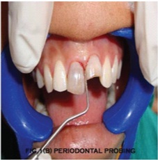
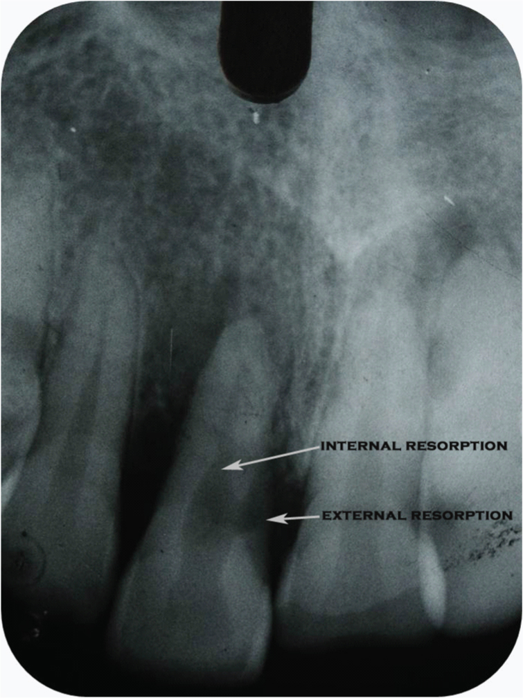
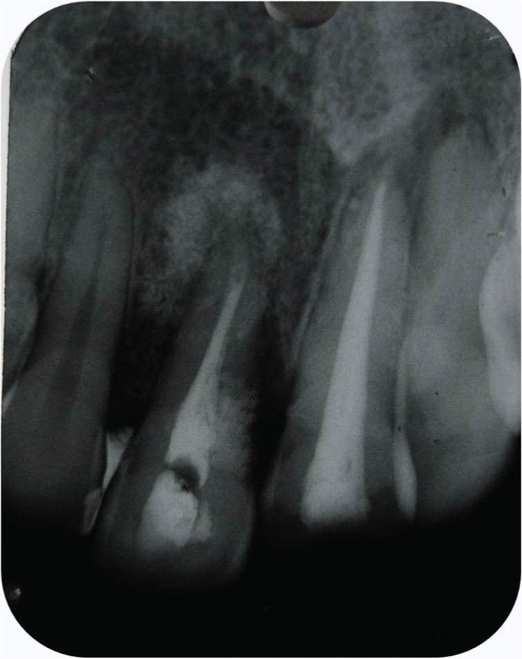
A 28-year-old female patient reported to the Department of Conservative Dentistry and Endodontics with complaint of pus discharge since last 6 months and broken front tooth. Patient had trauma some 3-4 years back. Clinical examination revealed a discoloured 11 (FDI system) and fractured 21 with sinus openings in the apical area of both teeth [Table/Fig-1]. Both teeth were tender on percussion and 11 was grade 2 mobile. On probing pockets were noted on 11(10 mm buccal, 5mm mesial, 7mm distal) and 21(5mm mesial and distal) [Table/Fig-2]. Overall patient had poor oral hygiene. Radiographic examination [Table/Fig-3] revealed widened periodontal ligament and periapical radiolucency w.r.t. both 11 and 21. Internal and external resorption was present w.r.t. 11. Based on clinical and radiographic examination it was tentatively diagnosed as a case of communicating internal-external inflammatory resorption with apical periodontitis w.r.t. 11 and apical periodontitis w.r.t. 21.
Pre-operative photograph of the patient

Diagnostic radiograph showing internal and external resorption
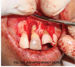
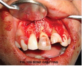
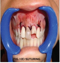
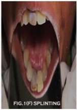
After proper isolation, access opening was done, working length was taken and cleaning and shaping of the root canal was done w.r.t no. 11 & 21. Calcium hydroxide as intracanal medicament was placed in the canal. After 1 week surgery was performed. On flap elevation a large J-shaped bony defect [Table/Fig-4] was found w.r.t. 11 with buccal plate completely denuded. Trichloroacetic (TCA) acid was applied topically (with a small cotton pellet with slowly increasing pressure) on the resorptive defect to control the haemorrhage. Afterthorough curettage of the granulation tissue from the resorptive defect and apical part, retrograde filling of root canal was done. External resorptive defect was filled with MTA (Pro Root MTA-Dentsply Tulsa Dental) [Table/Fig-5]. An artificial bone graft (Ossifi, Equinox medical technologies, Holland) was placed to cover the bony defect [Table/Fig-6] and suturing of flap was done [Table/Fig-7]. After one week sutures were removed. Temporary crown was placed on 21 and teeth were splinted with Ribbond for 3 weeks [Table/Fig-8,9].
BJ-shaped bony defect visible after incision
Immediate post-operative radiograph
Bone graft placed over the defect
Teeth splinting with Ribbond
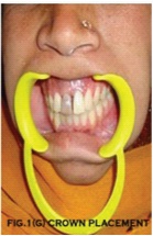
Post-operative photograph
Discussion
Root resorption is a complex process and is defined as the loss of hard dental tissue (i.e. cementum and dentin) as a result of odontoclastic action [1]. Resorptive defects can be challenging to diagnose and incorrect diagnosis can lead to improper treatment. Present case was a diagnostic dilemma as internal resorption was evident from the diagnostic radiograph, but cause of external root resorption presented a challenge. It could have been a sequel of internal root resorption perforating the root. But owing to history of trauma and taking in consideration the periodontal condition of tooth it could well have been developed independently of the internal resorption defect i.e. a true combined endo-perio lesion. Authors failed to find a diagnostic term for such lesion and classified it as per Lindskog’s classification [2] as Communicating Internal-External inflammatory resorption but with independent causes for both.
The selection of proper treatment in resorption cases is related to control of stimulation factors i.e. control of intrapulpal infection, removal of granulation tissue and filling of resorptive defect [3]. In the present case root canal treatment was done to control the intrapulpal infection, granulation tissue was removed with thorough curettage and MTA was used to fill the external resorptive defect. J-shaped bony defect in the case was found to be of Class F category of surgical classification by Kim & Kratchman [4]. Such defects have the poorest prognosis and require concurrent bone grafting and membrane barrier techniques. Splinting was done for stabilization of tooth during healing phase after bone grafting and to reduce patient discomfort.
Conclusion
A newer classification system or update of the older system is needed to include cases such as the present one in the classification system to aid in the proper management of such lesions.
[1]. Patel S, Pitt Ford T, Is the resorption external or internal?Dental Update 2007 34:218-29. [Google Scholar]
[2]. Heithersay GS, Management of tooth resorptionAustralian Dental Journal Supplement 2007 52(1 Suppl):S105-S121. [Google Scholar]
[3]. Fuss Z, Tsesis I, Lin S, Root resorption-diagnosis, classification and treatment choices based on stimulation factorsDental Traumatology 2003 19:175-82. [Google Scholar]
[4]. Kim S, Kratchman S, Modern endodontic surgery concepts and practice: a reviewJ Endod 2006 32:601 [Google Scholar]