Signet Ring Dermatofibroma, a Rare and Confusing Variant
Asmaa Gaber Abdou1, Nancy Youssef Asaad2
1Faculty of Medicine, Pathology Department, Menofiya University, Shebein Elkom, Egypt.
2Faculty of Medicine, Pathology Department, Menofiya University, Shebein Elkom, Egypt.
NAME, ADDRESS, E-MAIL ID OF THE CORRESPONDING AUTHOR: Dr. Asmaa Gaber Abdou, Faculty of Medicine, Department of Pathology, Menofiya University, Shebein Elkom, Egypt.
E-mail: Asmaa_elsaidy@yahoo.com
Dermatofibroma is a common cutaneous benign fibrohistiocytic tumour, which is usually diagnosed without difficulty. In this report we demonstrated a signet ring variant of dermatofibroma as a rare variant of this common neoplasm together with the possible differential diagnosis.The presence of signet cells in cutaneous neoplasm does not necessarily means malignancy. Signet ring dermatofibroma is a rare variant eliciting differential diagnostic problems which can be solved by careful histopathological examination, searching for the classic areas and the help of immunohistochemistry.
Benign fibrous histiocytoma, CD68, Skin
Case Report
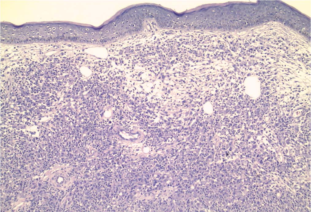
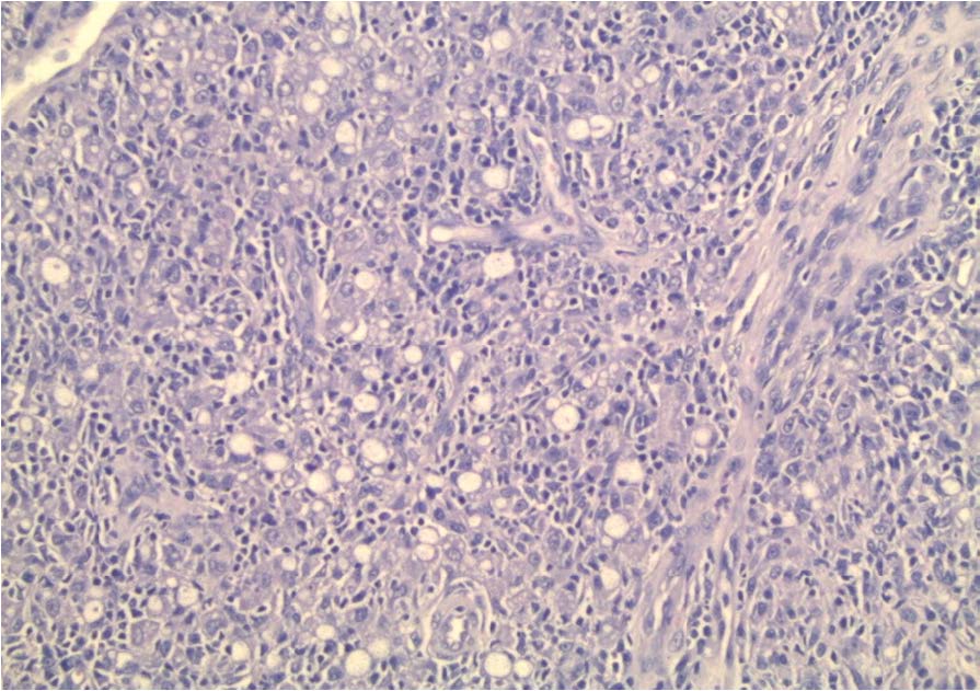
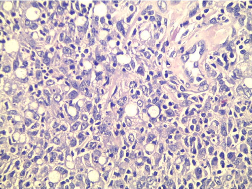
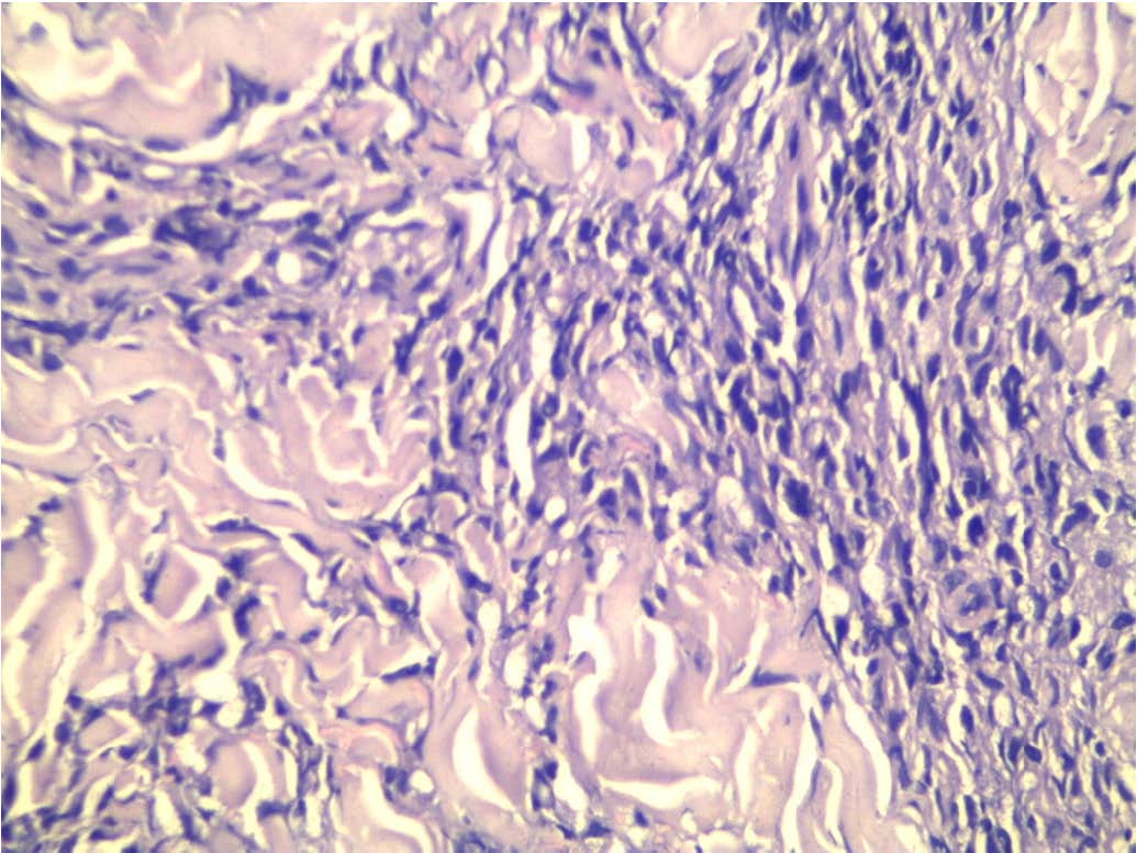
A 35-year-old female presented with painless right leg nodule since 6 months in Surgery Department, Faculty of Medicine, Menofiya University. The excised nodule was covered by skin and measured 2 x 1.5 cm with whitish discolouration. The nodule was examined histopathologically and showed dermal infiltrate formed of sheets of epithelioid cells with pale pink cytoplasm [Table/Fig-1].The infiltrate showed wide spread unmistakable signet ring formation [Table/Fig-2,3]. The infiltrate was admixed with inflammatory cells such as plasma cells [Table/Fig-3]. Other areas showed fibroblastic proliferation and collagen deposition Table/Fig-4]. The infiltrate was separated by a clear zone from the overlying epidermis and did not show atypia, mitoses or necrosis [Table/Fig-1].The infiltrate was diffusely positive for CD68 [Table/Fig-5] and negative for CK, LCA and HMB-45.
Discussion
Dermatofibroma is a common benign fibrohistiocytic dermal skin tumour, which is usually not difficult to diagnose. There are several histological variants of dermatofibroma that sometimes pose diagnostic problems in their recognition. Some variants should be recognized because of their potential liability for recurrence after excision such as cellular and aneurysmal fibrous histiocytomas [1]. The current case presents a rare variant of dermatofibroma characterized by the presence of signet ring cells. This variant was called signet ring cell dermatofibroma according to Garrido-Ruiz et al., [1] who described the only case harbouring this feature in English literature and they explained the signet ring morphology by degenerative process. Their case was a 16-year-old man presented with a painless, slowly growing, 1.5-cm nodule on the lateral side of his right leg. By comparison, our case is a female 35 years who presented with a right leg nodule. Special stains could help in recognizing the type of accumulation inside the cytoplasm of signet ring cells. For example, Alcian blue stains mucin while PAS stains glycogen. The histopathological features of the current case were against the diagnosis of malignancy because of absence of marked atypia, necrosis and mitoses, however exclusion of metastatic adenocarcinoma, melanoma and lymphoma was mandatory by CK, LCA and HMB-45, respectively. Beside the great help of immuno staining, histopathological features could also aid in distinction. Metastatic signet ring adenocarcinoma usually shows nests and cords of malignant cells invading dermis and subcutaneous fat and these cells usually exhibits mild degree of anaplasia. Regarding melanoma, the malignant cells are usually epithelioid to spindle with high degree of anaplasia and show melanin formation in most of the cases. Cutaneous melanoma [2], squamous cell carcinoma [3,4], basal cell carcinoma [4] and T cell lymphoma [5] may show signet ring formation. The presence of collagen, admixture with other inflammatory cells such as plasma cells and the diffuse positivity for CD68 were very helpful in identifying the fibrohistiocytic nature of the lesion.
Dermal infiltrate separated from overlying epidermis by grenz zone (Haematoxylin and eosin staining X100)
The infiltrate is formed of epithelioid cells with pale pink cytoplasm and many of them showed signet ring configuration (Haematoxylin and eosin staining X200)
High power view of the previous slide demonstrating signet ring admixed with inflammatory cells such as plasma cells (Haematoxylin and eosin staining X400)
Fibroblastic proliferation and collagen deposition (Haematoxylin and eosin staining X400)
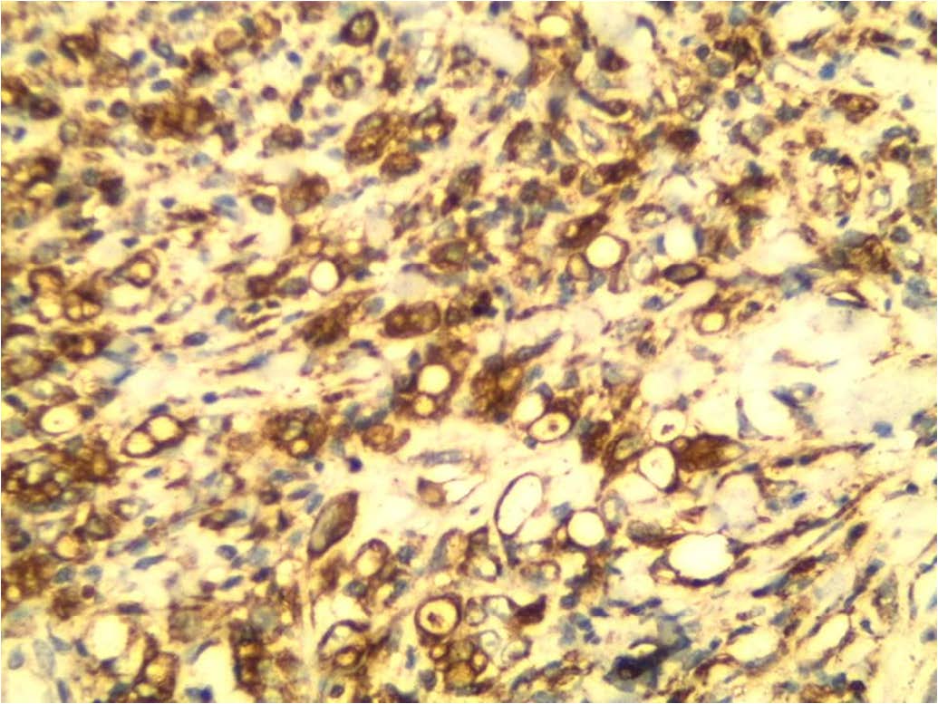
Diffuse cytoplasmic staining for CD68 in the dermal infiltrate including those showing signet ring morphology (Immunohistochemical staining X200)
Conclusion
The presence of signet cells in cutaneous neoplasm does not necessarily means malignancy. Signet ring dermatofibroma is a rare variant eliciting differential diagnostic problems which can be solved by careful histopathological examination, searching for the classic areas and the help of immunohistochemistry.
[1]. MC Garrido-Ruiz, R Carrillo, AB Enguita, MC Rodriguez Peralto, Signet-Ring Cell DermatofibromaAm J Dermatopathol 2009 31:84-87. [Google Scholar]
[2]. L Kocovski, S Alowami, Signet-ring cell melanoma: a potential diagnostic pitfallAm J Dermatopathol 2014 36(12):985-88. [Google Scholar]
[3]. K Nakajima, T Kaneko, T Aizu, H Nakano, Y Matsuzaki, D Sawamura, Signet-ring cutaneous squamous cell carcinoma arising on the back of the finger Case Rep Dermatol 2013 5:215-18. [Google Scholar]
[4]. DN Lortscher, EK Satter, LS Romero, Signetring-like cells: no longer a “signature” of glandular differentiationDermatol Online J 2012 18:3 [Google Scholar]
[5]. Massone C, El-Shabrawi-Caelen L, Kerl H, Cerroni L, The morphologic spectrum of primary cutaneous anaplastic large T-celllymphoma: a histopathologic study on 66 biopsy specimens from 47 patients with report of rare variantsJ Cutan Pathol 2008 35:46-33. [Google Scholar]