Erasmus Syndrome in a 42-Year-Old Male: A Rare Case Report
Subrata Chakrabarti1, Koushik Pan2
1 Post Graduate Trainee, Department of General Medicine, IPGMER, Kolkata, India.
2 Post Graduate Trainee, Department of General Medicine, IPGMER, Kolkata, India.
NAME, ADDRESS, E-MAIL ID OF THE CORRESPONDING AUTHOR: Dr. Subrata Chakrabarti, Doctor’s Hostel, AJC Bose Road, Kolkata-700020, India.
E-mail: subratachakrabarti2011@gmail.com
Erasmus syndrome is a rare entity in which systemic sclerosis develops following exposure to silica with or without silicosis. Few case reports are available in literature. We report here a case of Erasmus syndrome in a 42-year-old manual labourer. The patient presented with arthralgia, Raynoud’s phenomenon, skin tightening and microstomia along with features of Interstitial Lung Disease (ILD) and pulmonary arterial hypertension. Evidence of Interstitial Lung Disease (ILD) with mediastinal lymphadenopathy as well as pulmonary arterial hypertension with vascular reactivity was found in appropriate investigations. Serological markers of systemic sclerosis were strongly positive. After a diagnosis of Erasmus syndrome was made, a combination of drugs including Prednisone, Cyclophosphamide and Nifedipine was instituted this led to moderate improvement in his symptoms over 6 months.
Interstitial lung disease, Pulmonary arterial hypertension, Silicosis, Systemic sclerosis
Case Report
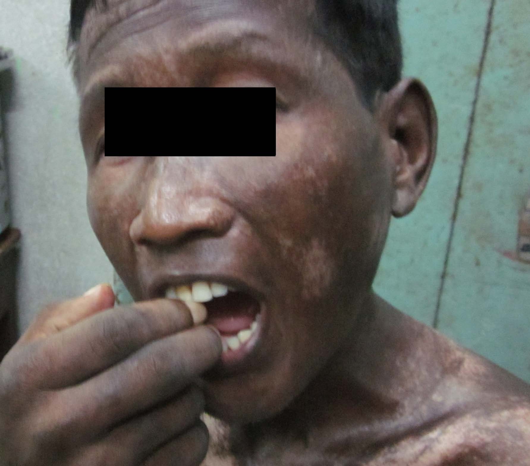
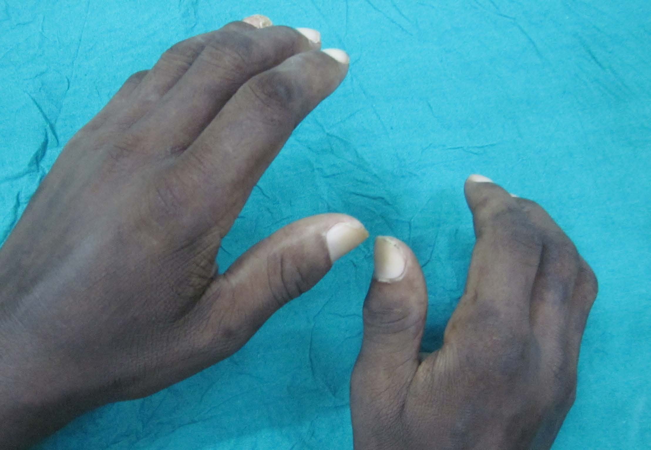
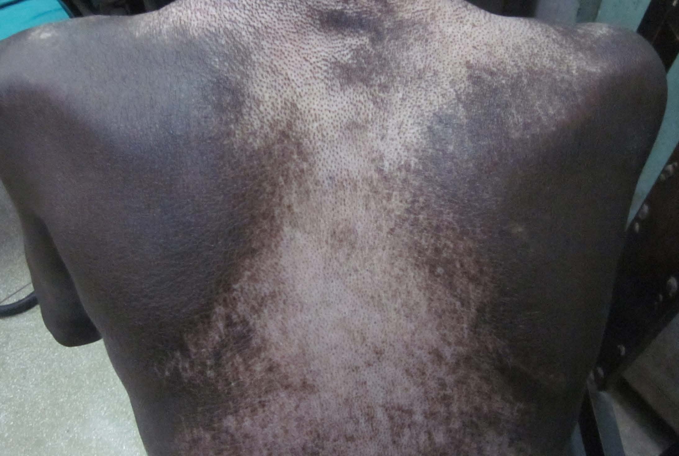
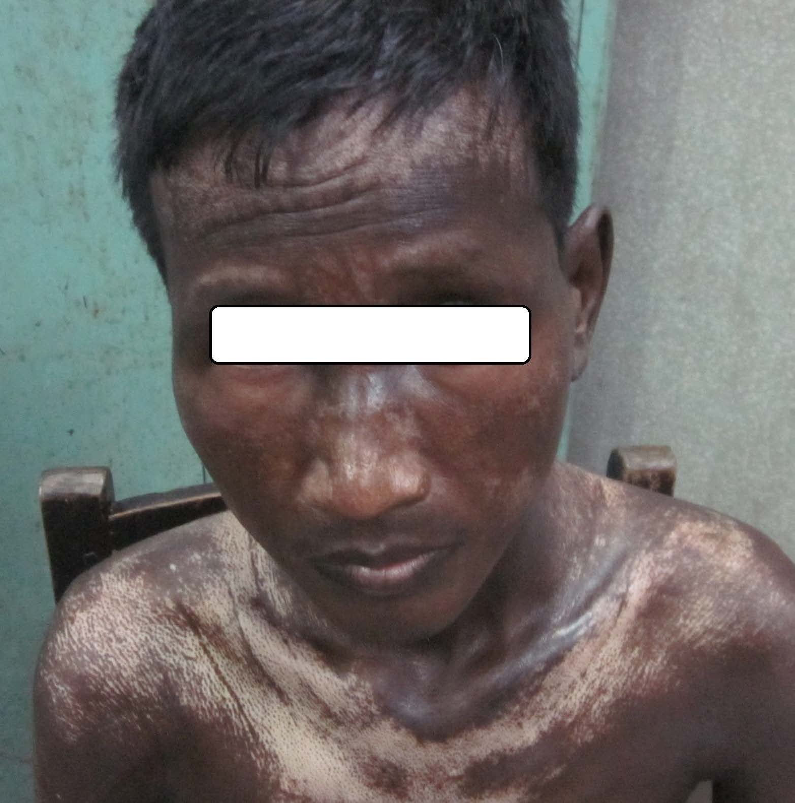
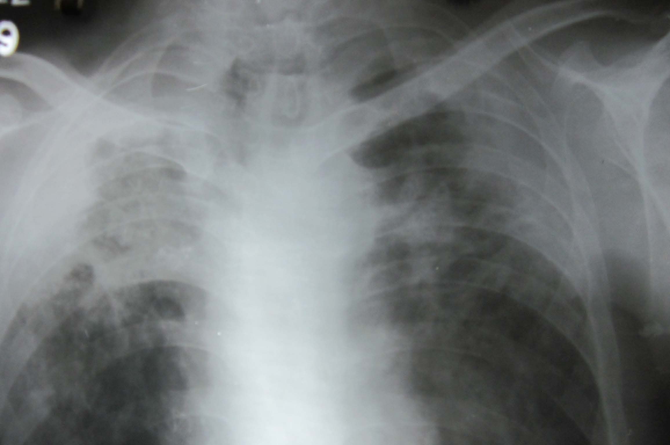
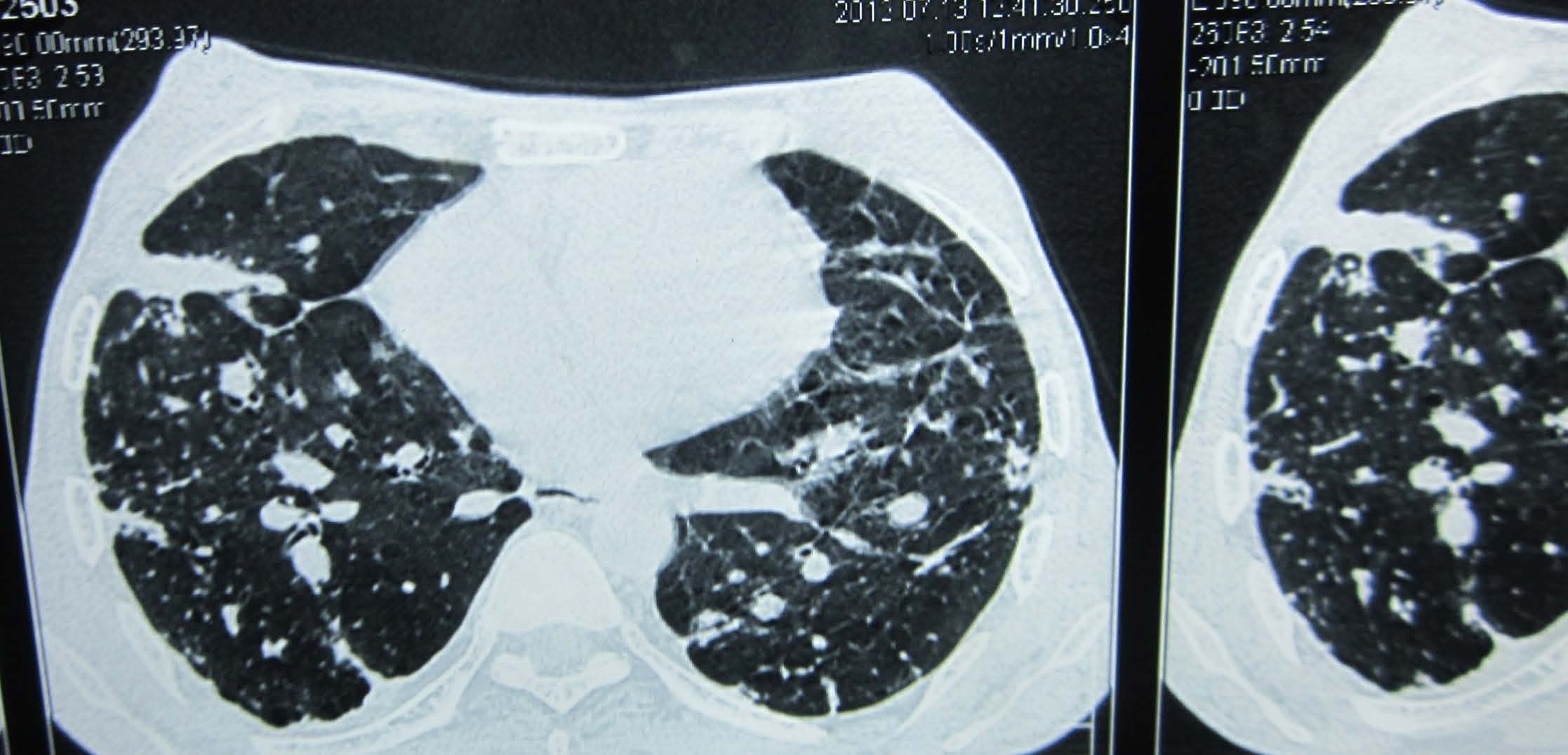
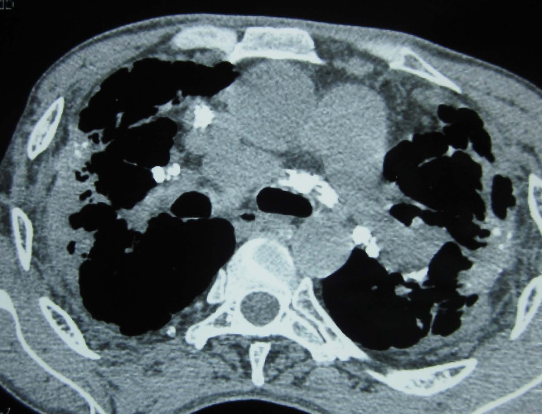
A 42-year-old non-diabetic and non-hypertensive male patient presented to the General medicine Out-patient Department of SSKM Hospital with a history of progressively worsening shortness of breath for last 4 months and low-grade irregular fever with persistent dry cough for 1 month. He had developed progressive skin tightening mainly over the face and extremities for last 8 months and suffered from episodic bluish discolouration of fingers on exposure to cold suggestive of Raynaud’s phenomenon for the last 6 months. He also had arthralgia mainly involving the small joints of the upper limb (metacarpo-phalangeal and inter phalangeal joints) for last 1 year but no dysphagia was present. Occupational history revealed that he worked as a stone crusher for nearly 12 years in a stone quarry and he was forced to leave that job 7 years ago. Neither past history of pulmonary tuberculosis nor any history of any chemotherapeutic drug administration chronic drug intake in the past was present. No family history of similar complaints was noted. On examination, there was pallor, loss of wrinkling over the forehead, microstomia [Table/Fig-1], skin fixity over the hands leading to apparent clawing [Table/Fig-2], skin thickening and areas of irregular hyperpigmentation and depigmentation of skin on the arms and back suggestive of salt pepper dermopathy [Table/Fig-3,4]. However, clubbing and calcinosis cutis was not noted. Respiratory system examination revealed bilateral end-inspiratory coarse crepitations in bilateral upper and mid-zones and breath sounds were decreased in the right infraclavicular area infrascapular area. Cardiological evaluation showed normal S1 and loud P2 with no added sounds and normal heart rate and rhythm. No organomegaly or free fluid was noted in abdominal examination. Complete blood count , urinalysis, liver and renal function tests and serum electrolytes were within normal limits except normocytic normochromic anemia (Hb-8.8 g/dl) and hypoalbuminemia (2.2 g/dl). HIV, HBV and HCV serology was negative. Bilateral upper zone opacities (right>left) was noted in Chest X-ray [Table/Fig-5]. High resolution Computed Tomography (HRCT) of the thorax revealed diffuse fibrosis, nodular lesions, occasional ground glass changes involving mainly the upper and middle lobes suggestive of Interstitial Lung Disease (ILD) along with mediastinal lymphadenopathy [Table/Fig-6,7]. Pulmonary function test revealed severe restrictive defect. ANA by Hep-2 method was positive in high titres (1:640); nuclear speckled pattern and Anti Scl-70 antibody was also strongly positive. Echocardiogram showed left ventricular ejection fraction of 58% and raised pulmonary artery systolic pressure (62 mm Hg). Vascular reactivity test for pulmonary arterial hypertension (PHT) was positive. Considering the presence of tell-tale clinical manifestations like arthralgia, Raynoud’s phenomenon, skin tightening over the face and extremities, microstomia, salt pepper dermopathy, Interstitial Lung Disease (ILD) with mediastinal lymphadenopathy, significant pulmonary arterial hypertension with vascular reactivity along with unequivocal serological markers of Systemic sclerosis, a clinic-pathological diagnosis of Systemic sclerosis was safely made.
Showing presence of microstomia. Skin tightness of face is also evident
Showing skin tightening leading to apparent clawing
Showing ‘salt pepper dermopathy’ on the back
Showing ‘salt pepper dermopathy’ on the chest and shoulders
Showing mainly right upper lobe fibrosis; comparatively mild left upper lobe fibrotic change is also seen
Showing fibrosis, nodules and ground glass changes suggestive of ILD in HRCT Thorax pulmonary window
Showing mediastinal lymphadenopathy in HRCT Thorax mediastinal window
Again considering the significant occupational exposure to silica (for 12 y) and absence of any chronic drugs like bleomycin, methysergide, cyclophosphamide, etc. (known to induce lung fibrosis) or any family history of similar ailments, a final etiological diagnosis of Erasmus syndrome (diffuse cutaneous systemic sclerosis associated with silica exposure) was made and no alternative possibility was entertained. Treatment was made with Prednisone (40 mg/day based on body weight 38 kg, to be continued lifelong) and cyclophosphamide (1 g/month pulse dosage for 6 months) for ILD and Nifedipine (started with 20 mg/day and increased up to 120 mg/day over 6 months and advised to continue lifelong) for PHT and Raynoud’s phenomenon. Advice was given to strictly avoid cold exposure and attend regular follow-up in OPD. He was found to have significant improvement in skin tightening, arthralgia, Raynaud’s phenomenon and cough although marginal improvement in dyspnoea over treatment for 6 months during follow-up visit.
Discussion
Systemic sclerosis is an autoimmune disease with vascular changes and inflammation and degeneration with diffuse tissue fibrosis involving skin, lung, kidney, heart, gastrointestinal tract, and synovium [1]. Although pathology has been elucidated in recent studies, exact pathogenesis is still enigmatic. Environmental and occupational exposures have been implicated in some studies, namely vinyl chloride, epoxy benzene, organic solvent, silica [2].
Silicosis is probably the most common form of pneumoconiosis, caused by the inhalation of mineral dust containing silica. Silicosis is an inflammatory disease of the lung characterized by irreversible lung fibrosis which develops from prolonged pulmonary inhalation and retention of crystalline silica and immune reaction mounted by the body to this extraneous chemical. It mainly appears as an occupational hazard in persons involved in stone-quarrying, mining and sand blasting [3]. Silicosis is often associated with the development of other diseases, such as pulmonary tuberculosis, lung carcinoma and less commonly, autoimmune diseases like systemic sclerosis (SSc), rheumatoid arthritis and systemic lupus erythematosus [4]. Exposure to silica is associated with abnormalities of the humoral and cellular immunity, hypergammaglobulinemia and alterations in T-helper and T-suppressor lymphocytes; antinuclear antibody and rheumatoid factor often become positive [5]. Based on these immunological aberrance, silica exposure has been incriminated as a cause of progressive systemic sclerosis.
The association of previous exposure to silica and later development of systemic sclerosis was first analysed by Erasmus in 1957 [6] although credit for first observation of this association goes to Bramwel in 1914 [7]. This entity is rarely reported from India. Khanna et al., reported a case of Erasmus syndrome in a 53-year-old stone-cutter who initially manifested with arthralgia, Raynoud’s phenomenon, skin tightening and later on developed dyspnoea and dysphagia after two years of initial symptom onset [8]. Recently, Ganguly et al., reported a similar case in a 26-year-old manual labourer who had dyspnoea, haemoptysis, arthralgia, Raynoud’s phenomenon for 5 y before coming to medical attention [9]. The index patient was a 42-year-old stone quarry worker who presented to the OPD due to progressively worsening shortness of breath for last four months and low-grade irregular fever with persistent dry cough for one month but detailed enquiry revealed that he was suffering from arthralgia, Raynoud’s phenomenon, skin tightening for almost one year. However, he had no dysphagia. The index case is notable for the fact that the patient required medical attention within one year of onset of symptoms.
The postulated mechanism is that pulmonary macrophages on exposure to silica produce lymphokines which may stimulate collagen production directly or induce other cells like monocyte or mast cells to release factors that in turn induce chronic inflammation and fibroblastic proliferation and collagen biosynthesis in the lungs [5]. Alterations in soluble interleukin-2 (IL-2) receptors may also play a role [10,11]. The connective tissue disease following exposure to silica occurs usually 15 y after the initial exposure. The index patient was continually exposed to silica dust for 12 y and systemic sclerosis developed after nearly 19 y. An accelerated form has been noticed in some patients in whom silica induced systemic sclerosis develops after a period of short intense exposure (5-10 y). A predominance of male sex has been noted in some studies although further studies are warranted [12]. HRCT image findings include linear ground glass opacities, honeycombing and small sub-pleural nodules which are inseparable from idiopathic SSc [13]. In fact, features of silica associated systemic sclerosis are also clinically, serologically, immunologically and prognostically indistinguishable from idiopathic scleroderma though the former expresses a higher prevalence of pulmonary involvement and anti Scl-70 antibody [14].
Conclusion
An underlying systemic sclerosis which contributes to dyspnoea by multiple mechanisms- pulmonary fibrosis, pulmonary artery hypertension and localised thoracic skin disease, must be kept in mind whenever the degree of dyspnoea cannot be explained by extent of silicosis induced lung fibrosis alone and especially if additional clinical pointers are present.
[1]. Mora GF, Systemic Sclerosis: environmental factorsJ Rheumatol 2009 36(11):2383-96. [Google Scholar]
[2]. Mayes MD, Lacey JV Jr, Beebe-Dimmer J, Gillespie BW, Cooper B, Laing TJ, Prevalence, incidence, survival, and disease characteristics of systemic sclerosis in a large US populationArthritis Rheum 2003 48(8):2246-55. [Google Scholar]
[3]. Smith V, Vanthuyne M, Vander Cruyssen B, Van Praet J, Vermeiren F, Smets H, Over-representation of construction-related occupations in male patients with systemic sclerosisAnn Rheum Dis 2008 67:1448-50. [Google Scholar]
[4]. Koeger AC, Lang T, Alcaix D, Milleron B, Rozenberg S, Chaibi P, Silica - associated connective tissue disease. A study of 24 casesMedicine (Baltimore) 1995 74(5):221-37. [Google Scholar]
[5]. Zaghi G, Koga F, Nisihara RM, Skare TL, Handar A, Utiyama SR, Autoantibodies in silicosis patients and in silica-exposed individualsRheumatol Int 2010 30(8):1071-75. [Google Scholar]
[6]. Erasmus LD, Scleroderma in Gold-Miners on the Witwatersrand with Particular Reference to Pulmonary ManifestationsS Afr J Lab Clin Med 1957 3(3):209-31. [Google Scholar]
[7]. Bramwell B, Diffuse scleroderma: its frequency; its occurrence in stonemasons, its treatment by fibrinolysin, elevation of temperature do to fibrinolysin injectionsEdinburgh Med J 1914 12:387-401. [Google Scholar]
[8]. Khanna N, D’Souza P, Sud A, Pandhi RK, Systemic sclerosis in a stone cutterIndian J Dermatol Venereol Leprol 1997 63(2):111-13. [Google Scholar]
[9]. Ganguly J, Kumar A, Samanta SK, Mitra R, Kundu S, Erasmus Syndrome: A Case Report of Silicosis-induced Scleroderma in a 26-year-old MaleOman Med J 2013 28(5) [Google Scholar]
[10]. Hayashi H, Maeda M, Murakami S, Kumagai N, Chen Y, Hatayama T, Soluble interleukin-2 receptor as an indicator of immunological disturbance found in silicosis patientsInt J Immunopathol Pharmacol 2009 22(1):53-62. [Google Scholar]
[11]. Kahaleh MB, LeRoy EC, Interleukin-2 in scleroderma: correlation of serum level with extent of skin involvement and disease durationAnn Intern Med 1989 110(6):446-50. [Google Scholar]
[12]. McCormic ZD, Khuder SS, Aryal BK, Ames AL, Khuder SA, Occupational silica exposure as a risk factor for scleroderma: a meta-analysisInt Arch Occup Environ Health 2010 83(7):763-69. [Google Scholar]
[13]. Cointrel C, Tillie-Leblond I, Lamblin C, Furon D, Tonnel AB, Wallaert B, Erasmus syndrome: clinical, tomographic, respiratory function and bronchoalveolar lavage characteristicsRev Mal Respir 1997 14(1):21-6. [Google Scholar]
[14]. Rustin MH, Bull HA, Ziegler V, Silica-associated systemic sclerosis is clinically, serologically and immunologically indistinguishable from idiopathic systemic sclerosisBr J Dermatol 1990 123:725-34. [Google Scholar]