Dentigerous Cyst Associated with Adenomatoid Odontogenic Tumour
Sumit Majumdar1, Divya Uppala2, Ayyagari Kameswara Rao3, Sunil Talasila4, Mahesh Babu5
1 Head of the Department, Department of Oral Pathology and Microbiology, GITAM Dental College and Hospital, Rushikonda, Visakhapatnam, India.
2 Senior Lecturer, Department of Oral Pathology and Microbiology, GITAM Dental College and Hospital, Rushikonda, Visakhapatnam, India.
3 Post Graduate Student, Department of Oral Pathology and Microbiology, GITAM Dental College and Hospital, Rushikonda, Visakhapatnam, India.
4 Reader, Department of Oral and Maxillofacial Surgery, GITAM Dental College and Hospital, Rushikonda, Visakhapatnam, India.
5 Reader, Department of Oral Pathology and Microbiology, GITAM Dental College and Hospital, Rushikonda, Visakhapatnam, India.
NAME, ADDRESS, E-MAIL ID OF THE CORRESPONDING AUTHOR: Dr. Ayyagari Kameswara Rao, GITAM Dental College and Hospital, Rushikonda, Visakhapatnam-530045, India. E-mail : calmua@gmail.com
Adenomatoid odontogenic tumour (AOT), a tumour composed of odontogenic epithelium, is an uncommon tumour of odontogenic origin that accounts for only 2.2- 7.1% of all odontogenic tumours. Very few cases of AOT associated with Dentigerous cyst (DC) have been reported till date, most cases are in females and have a striking tendency to occur in the anterior maxilla. The present case is that of a 14-year-old female who revealed a large radiolucent lesion associated with the crown of an unerupted canine located in the left maxillary anterior region. The microscopic examination revealed the presence of AOT in the fibrous capsule of a DC. In this paper, we describe the importance of grossing, sectioning and complete examination of the slide to diagnose such hybrid lesions.
Grossing, Hybrid, Odontogenic tumor
Case Report
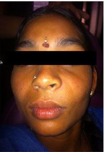
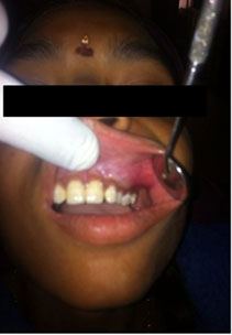
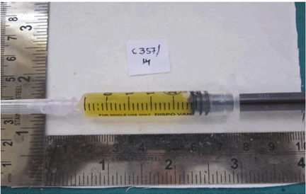
A 14-year-old female patient reported to the GITAM Dental College and Hospital with a chief complaint of painless swelling over left front side of upper jaw causing disfigurement. On examination of the patient, she had a diffuse extraoral swelling measuring approximately 2×2 cm extending from left infraorbital region superiorly to left corner of mouth, obliterating the nasolabial fold [Table/Fig-1]. There was no paraesthesia over infraorbital region. Intraoral examination revealed a soft fluctuant swelling extending from left upper lateral incisor to second premolar region, obliterating the labial vestibule. The left deciduous canine was retained and the permanent canine was missing [Table/Fig-2]. Mucosa overlying the swelling was normal. On aspiration, straw-coloured fluid was obtained [Table/Fig-3].
Demonstrating extraoral swelling obliterating the nasolabial fold
Demonstrating intraoral swelling obliterating the labial vestibule with retained deciduous canine
Demonstrating aspirated straw-coloured fluid
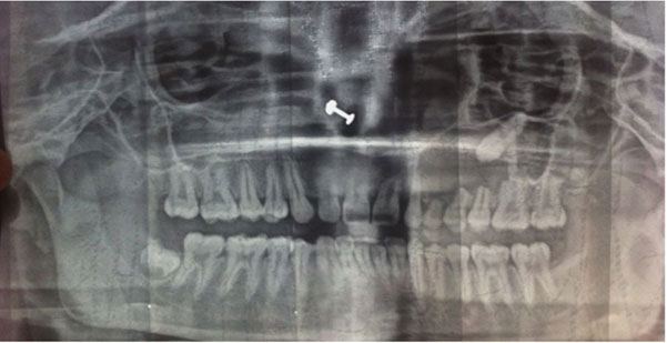
Orthopantomogram revealed a well-defined, unilocular corticated radiolucency extending from the apical region of left maxillary lateral incisor to the mesial root of left maxillary first molar measuring 3×3 cms in size with an impacted permanent canine and the resorption of roots of left maxillary 1st premolar and 2nd premolars [Table/Fig-4].
Orthopantomogram demonstrating well-defined, unilocular radiolucency with an impacted permanent canine
On basis of clinical and radiographical findings, the clinical diagnosis of dentigerous cyst was done and differential diagnosis of unicystic ameloblastoma and AOT were considered. Electric pulp vitality test showed delayed response to maxillary lateral incisor and premolars and no response to deciduous canine. The patient underwent surgery under general anesthesia. A mucoperiosteal flap was raised from the left incisor to premolar region in the labial vestibule. The buccal cortex was resorbed completely with thin shells of bone in between. The lining of the cystic lesion was carefully separated from the mucoperiosteum and the lesion was enucleated along with the impacted canine. The wound was irrigated with saline and betadine. Homeostasis was achieved. Wound was sutured with 3-0 vicryl. Healing was uneventful and patient is under follow up.
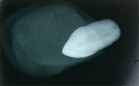
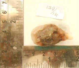
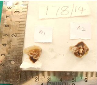
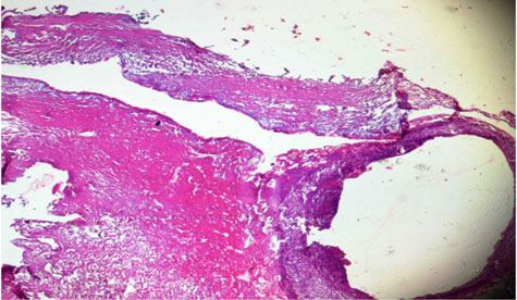
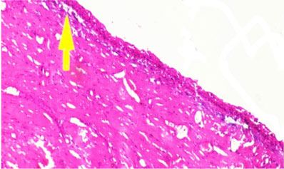
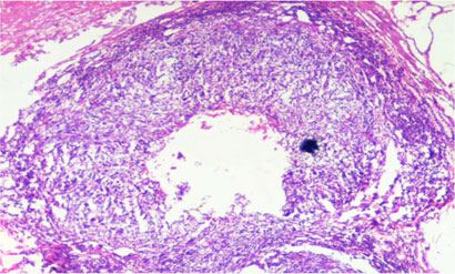
Macroscopic features: On gross examination, the specimen was greyish white in colour, measured 3.5×2×2 cms along with the impacted permanent canine. Radiograph of the gross specimen revealed an impacted canine within the soft tissue extending beyond cement-enamel junction (CEJ) and without any calcifications [Table/Fig-5]. The specimen was sectioned into two bits and the cut specimen showed both cystic space and solid tissue [Table/Fig-6,7,8]. The hematoxylin and eosin stained soft tissue section showed non keratinized cystic lining epithelium in association with fibro-vascular connective tissue and in areas of connective tissue capsule, solid masses of cells were seen in a scant fibrous stroma arranged in ductal pattern [Table/Fig-9]. The cystic epithelial lining epithelium was 2-4 layered, lined by cuboidal cells [Table/Fig-10]. The connective capsule had solid masses of cells in a scant fibrous stroma arranged in ductal pattern [Table/Fig-11]. Spindle shaped odontogenic epithelial cells were arranged in rosette pattern with few tubular or duct like structures were seen with a central space [Table/Fig-12].
Radiograph of the gross specimen demonstrating an impacted canine within the soft tissue without any calcifications
Gross specimen demonstrating soft tissue extending beyond CEJ of an impacted canine
Gross specimen demonstrating both cystic space and solid tissue along with an impacted canine
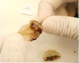
gross specimen which was sectioned into two bits demonstrating both cystic space and solid tissue
The hematoxylin and eosin stained soft tissue section demonstrating non keratinized cystic lining epithelium in association with fibro-vascular connective tissue and in areas of connective tissue capsule, solid masses of cells in a scant fibrous stroma arranged in ductal pattern.(4× view)
Demonstrating 2-4 layered cuboidal cells in the cystic epithelial lining epithelium
Demonstrating solid masses of cells in a scant fibrous stroma arranged in ductal pattern
Demonstrating spindle shaped odontogenic epithelial cells arranged in rosette pattern with few tubular or duct like structures with a central space
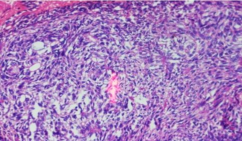
With the above findings, the lesion was diagnosed as dentigerous cyst associated with adenomatoid odontogenic tumour.
Discussion
AOT is uncommon, slow growing tumour accounting only for 2.2- 7.1% of all odontogenic tumours [1,2]. Most incidence of AOT is in females and has a striking tendency to occur in the anterior maxilla [3]. This tumour can also referred as 2/3rd tumour as 2/3rd of cases occurs in maxilla, 2/3rd in young females, 2/3rd are associated with unerupted teeth and 2/3rd are associated with impacted canines [4].
Philipsen et al., categorized AOT into three variants (follicular, extrafollicular, and peripheral). The ‘follicular type’ has a central lesion associated with an impacted tooth and is the most common type accounting for 73% of cases. The ‘extrafollicular type’ has a central lesion and is not associated with the tooth, accounts for 24% of cases. The ‘peripheral type’ contributes to 4.4% of cases and is extraosseous in origin [3].
The follicular variant of AOT is usually clinically misdiagnosed as DC as both will have a unilocular, well-defined radiolucency surrounding the crown of an impacted canine. Aspiration reveals straw coloured fluid which helps in differentiating DC from the solid tumour clinically and grossly follicular type of AOT sometimes extends apically beyond the cement enamel junction (CEJ) [5], while DC is attached to the tooth at the cervical region [6]. Cut surface of the AOT gross specimen shows a solid mass or a partly cystic space [2] in contrast to cystic space of DC. In this case, aspiration revealed straw coloured fluid thinking in the way of DC, on gross examination the soft tissue extended beyond the CEJ with a positive finding towards AOT, but the cut section showed both the solid and cystic areas which made us to consider a hybrid variant. DC is an odontogenic cyst develops by expansion of follicle in an unerupted tooth [6]. Benign odontogenic epithelial neoplasms like ameloblastoma [7], AOT [1,2,6,8–12] and malignant neoplasms like squamous cell carcinoma [13]. The mucoepidermoid carcinoma [14] can arise from the epithelial lining of DC present case is of particular interest as AOT is found in association with the fibrous capsule of DC and such hybrid variant was not discussed in the previous classifications.
In addition to the anterior maxilla, AOT has been reported in other areas of the jaws, such as in the sinus, in the posterior maxillary regions, and in the mandibular anterior regions [1] suggesting dental laminar remnants may likely represent the progenitor cells for this benign odontogenic tumour. The histogenesis of AOT is still controversial with various theories being proposed ranging from fully formed enamel organ, dental lamina and/or its remnants to odontogenic cysts [5]. Envelopmental theory hypothesis proposes that AOT grows next to or into a nearby dental follicle while forming a cystic space [15]. So, AOT in the present case might have developed from the dental laminar remnants along with DC at the time of cyst expansion.
Very few cases have been reported that arise in association with DC. A systematic search of the English language medical literature revealed only 19 such cases and 14 cases occurred in the maxillary region of which 11 cases are associated with impacted canine, 8 cases occurred in females of second decade. The clinical characteristics of these 19 cases along with the current case are summarized in the [Table/Fig-13].
Reported cases of AOT arising from DC
| S.No | Reference | Age/Sex | Race | Year | Site | Features |
|---|
| 1. | Valderrama [16] | 16/f | Philippino | 1988 | Maxilla | Unilocular radiolucency, surrounding tooth 14 crown |
| 2. | Warter et al., [17] | 8/m | Nigerian | 1990 | Maxilla sinus | Unilocular radiolucency, surrounding tooth 13 crown |
| 3. | Tajima et al., [18] | 15/m | Japanese | 1992 | Maxilla | A well-defined radiopaque mass and crown of unerupted 28 |
| 4. | Garcia-Pola et al., [19] | 12m | Spanish | 1998 | Maxilla | Unilocular radiolucency, surrounding tooth 23 |
| 5. | Bravo et al., [20] | 14/f | Not stated | 2005 | Maxilla | Unilocular radiolucency, surrounding tooth 23 crown |
| 6. | Nonaka et al., [21] | 13/f | Brazil | 2007 | Maxilla | Unilocular radiolucency with few radiopaque areas 23 and 24 |
| 7. | Chen et al., [22] | 15/m | Chineese | 2007 | Maxilla | Impacted 23 |
| 8. | Sandhu et al., [23] | 25/f | Indian | 2010 | Maxilla | Impacted 13 |
| 9. | J Baby John, Reena Rachel John [24] | 38/f | Indian | 2010 | Maxilla | Impacted 27 |
| 10. | Khot and Vibhakar [25] | 17/f | Indian | 2011 | Maxilla | Impacted 33 |
| 11. | Zama Moosvi [26] | 13/f | Indian | 2011 | Mandible | Impacted 32 |
| 12. | Anita Dnyanoba Munde et al., [1] | 20/f | Indian | 2013 | Mandible | Impacted 33 |
| 13. | Vikramjeet singh et al., [2] | 15/f | Indian | 2012 | Maxilla | Impacted 13 |
| 14. | Anshita Agarwal et al., [8] | 15/f | Indian | 2012 | Maxilla | Impacted 23 |
| 15. | Sushruth Nayak et al., [9] | 32/m | Indian | 2012 | Mandible | Impacted 43 |
| 16. | Latti BR, Kalburge JV [6] | 15/f | Indian | 2013 | Mandible | Impacted 33 |
| 17. | Harish Saluja et al., [10] | 18/F | Indian | 2013 | Mandible | Impacted 43. |
| 18. | Shivesh Acharya [11] | 14/F | Indian | 2014 | Maxilla | Impacted 13 |
| 19. | Ludmila De Faro Valverde et al., [12] | 17/F | Unknown | 2014 | Maxilla | Impacted 23 |
| 20. | Present case | 14/f | Indian | 2014 | Maxilla | Impacted 23 |
AOT and DC are both benign, encapsulated lesions. Conservative surgical enucleation or curettage is the treatment of choice. The prognosis is good as all the patients are under follow up and no recurrence was reported till to date.
Conclusion
This case highlights the importance of clinical examination, grossing and meticulous histopathological examination to diagnose such rare variants and if such cases are reported the exact pathogenesis of AOT can be revealed in near future.
[1]. Anita DM, Ravindra RK, Swapna S, Sunil S, Adenomatoid Odontogenic Tumour of the Mandible arising from a Dentigerous Cyst: A Case Report. Research and ReviewsJournal of Dental Sciences 2014 2(1):87-91. [Google Scholar]
[2]. V Singh, Adenomatoid Odontogenic tumour with Dentigerous cyst: Report of a case with review of literatureContemporary Clinical Dentistry 2012 3(2):S244-47. [Google Scholar]
[3]. Philipsen HP, Reichart PA, Adenomatoid odontogenic tumour: Facts and figuresOral Oncol 1999 35(2):125-31. [Google Scholar]
[4]. Marx RE, Stern D, Oral and Maxillofacial pathology: A rationale for diagnosis and treatment 2003 Hanover ParkQuintessence publishing:609-12. [Google Scholar]
[5]. F Ide, Development and Growth of Adenomatoid Odontogenic Tumour Related to Formation and Eruption of TeethHead and Neck Pathol 2011 5:123-32. [Google Scholar]
[6]. Latti BR, Kalburge JV, Dentigerous cyst associated with Adenomatoid Odontogenic tumour (AOT) A Rare Case Report and Review of LiteratureMedical Science 2013 1(2):44 [Google Scholar]
[7]. Jivan V, Altini M, Meer S, Mohamed F, Adenomatoid Odontogenic tumour (AOT) originating in a unicystic ameloblastoma: A case reportHead and Neck Pathol 2007 1(2):146-49. [Google Scholar]
[8]. A Agarwal, KY Giri, Alam S, The Interrelationship of Adenomatoid Odontogenic Tumour and Dentigerous Cyst: A Report of a Rare Case and Review of the LiteratureCase Reports inPathology 2012 2012Article ID 358609, 4 pages [Google Scholar]
[9]. Nayak Sushruth, Nayak Prachi, Mannae Rakesh Kumar, Mahendra Ashish, An Unusual Case of Adenomatoid Odontogenic Tumour Associated with Dentigerous CystIndian Journal of Dental Education 2012 5(4):233-36. [Google Scholar]
[10]. Saluja H, Kasa V, Gaikwad P, Mahindra U, Dehane V, A rare occurrence of adenomatoid odontogenic tumour arising from cystic lining in the mandible: Review with a case reportJournal of Orofacial Sciences 2013 5(1):50-53. [Google Scholar]
[11]. Acharya S, Goyal A, Rattan V, Vaiphei K, Bhatia SK, Dentigerous Cyst or AdenomatoidOdontogenic Tumour: Clinical Radiological and Histopathological DilemmaCase Reports in Medicine 2014 2014Article ID 514720, 5 pages [Google Scholar]
[12]. Ludmila De Faro Valverde, AdenomatoidOdontogenic Tumour associated with Dentigerous CystCase Report. OOOO 2014 117(2):e137 [Google Scholar]
[13]. Colbert S, Brennan PA, Theaker J, Evans B, Squamous cell carcinoma arising in dentigerous cystsJournal of CranioMaxilloFac Surg 2012 40(8):e355-57. [Google Scholar]
[14]. Spoorthi BR, Predominantly cystic central mucoepidermoid carcinoma developing from a previously diagnosed dentigerous cyst: case report and review of the literatureClinics and Practice 2013 3:e19 [Google Scholar]
[15]. Batra P, AdenomatoidOdontogenicTumour: Review and Case ReportJ Can Dent Assoc 2005 71(4):250-53. [Google Scholar]
[16]. Valderrama L S, Dentigerous cyst with intracystic adenomatoid odontogenic tumor and complex odontomaThe Journal of the Philippine Dental Association 1988 41(3):35-41. [Google Scholar]
[17]. Warter A, George-Diolombi G, Chazal M, Ango A, Melanin in a dentigerous cyst and associated adenomatoid odontogenic tumorCancer 1990 66:786-88. [Google Scholar]
[18]. Tajima Y, Sakamoto E, Yamamoto Y, Odontogenic cyst giving rise to an adenomatoid odontogenic tumor: report of a case with peculiar featuresJournal of Oral and Maxillofacial Surgery 1992 50(2):190-93. [Google Scholar]
[19]. Garcia-Pola VM, Gonzalez Garcia M, Lopez-Arranz JS, Herrero Zapatero A, Adenomatoid odontogenic tumour arising in a dental cyst: Report of unusual caseJ Clin Pediatr Dent 1998 23:55-58. [Google Scholar]
[20]. Bravo M, White D, Miles L, Cotton R, Adenomatoid odontogenic tumor mimicking a dentigerous cystInternational Journal of Pediatric Otorhinolaryngology 2005 69(12):1685-88. [Google Scholar]
[21]. Nonaka C. F. W, De Souza L. B, Quinderé L. B, Adenomatoid odontogenic tumour associated with Dentigerous cyst-unusual case reportBrazilian Journal of Otorhinolaryngology 2007 73(1):135-37. [Google Scholar]
[22]. Chen Adenomatoid odontogenic tumor arising from a Dentigerous cyst—a case reportInternational Journal of Pediatric Otorhinolaryngology 2007 2(4):257-63. [Google Scholar]
[23]. Sandhu Simarpreet V, Narang Ramandeep S, Jawanda Manveen, Rai Sachin, Adenomatoid odontogenic tumor associated with dentigerous cyst of the maxillary antrum: A rare entity- a case ReportJournal of Oral and Maxillofacial Pathology 2010 14(1):24-28. [Google Scholar]
[24]. John J Baby, John Reena Rachel, Adenomatoid odontogenic tumor associated with dentigerous cyst in posterior maxilla: A case report and review of literatureJournal of Oral and Maxillofacial Pathology 2010 14(2):59-62. [Google Scholar]
[25]. Khot K, Vibhakar P. A, Case report: mural adenomatoid odontogenic tumor in the mandible—a rare caseInternational Journal of Oral and Maxillofacial Pathology 2011 2(2):35-39. [Google Scholar]
[26]. Moosvi Zama, Taayar S Amsavardani, Kumar G.S, Neoplastic potential of odontogenic cystsContemp Clin Dent 2011 2(2):106-09. [Google Scholar]