Carotid Artery Calcification and Sailolith in Partially Edentulous Patient: An Accidental Finding on Panoramic Radiograph
Bader K Alzarea1
1 Assistant Professor, College of dentistry, AlJouf University, Skaka, AlJouf, Kingdom Saudi Arabia.
NAME, ADDRESS, E-MAIL ID OF THE CORRESPONDING AUTHOR: Dr. Bader K Alzarea, Assistant Professor, College of dentistry, AljJouf University, Skaka, Aljouf, Kingdom Saudi Arabia.
E-mail: bkzarea@ju.edu.sa
Calcified carotid artery atheromas, Calcified body, Orthopantomograph, Risk factors
Orthopantomograms are very useful in detecting the pathologies affecting the teeth, development disorders of jaws, foreign bodies, fractures and other hard tissue variations [1]. Calcifications of various structures located in the head and neck region are a relatively common and are detected on routine panoramic radiographs. Three cases with unusual calcification seen incidentally during radiographic examination are reported here.
Case 1
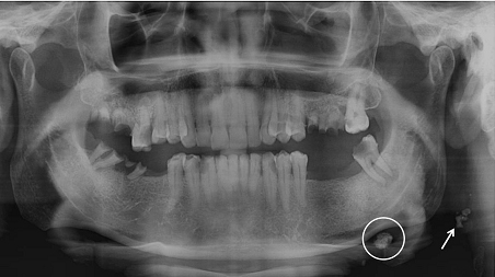
A 59-year-old male patient reported to us for replacing his missing teeth and removal of his broken teeth. Patient gave a previous history of the extraction of few mandibular teeth bilaterally. Panoramic radiograph was made for the patient who revealed missing mandibular right first molar and left first and second molars. Multiple grossly carious teeth and root stumps were also observed in both the arches. A well defined, solitary nodular radiopaque structure was seen inferior to the left mandibular border, near the angle. Apart from this, multiple well defined punctuate radiopacities were seen at the level of the lower margin of the cervical vertebra on the left side [Table/Fig-1]. Based on the radiographic location and appearance of the lesion on the radiograph, provisional diagnosis of the left submandibular gland sialolothiasis and carotid artery calcification was made.
OPG showing left submandibular sialolith (circle) and left carotid artery calcification (arrows)
Case 2
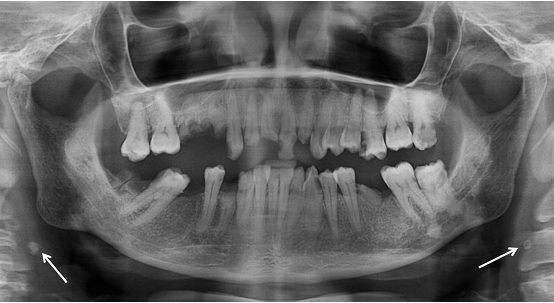
A 56-year-old male patient reported to us for replacing his missing teeth and restoration of carious teeth. Patient has undergone extraction of his mandibular teeth due to caries and pain. Panoramic radiograph was made for the patient which revealed missing mandibular right molar and multiple carious teeth and root stumps. Also, well defined, bilateral nodular radiopacities were seen at the level of the lower margin of the third and the fourth cervical vertebra (C3 & C4) [Table/Fig-2]. Based on the radiographic location and appearance of the lesion on the radiograph, provisional diagnosis of the coronary artery calcification was made.
OPG showing bilateral nodular radopacities (arrows)
Case 3
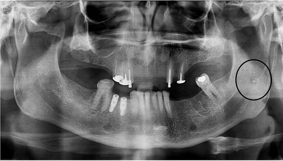
A 49-year-old male patient reported to us for replacing his missing teeth. Patient has undergone extraction of his teeth due to caries and periodontitis. A panoramic radiograph was made, which revealed multiple missing teeth and previously placed implants. Apart from this multiple punctuate radiopacites were seen on the superior portion of the left side of mandibular ramus. These radiopacites were discrete and measured around 2 mm in diameter [Table/Fig-3]. Considering the radiographic location and appearance of the lesion on an orthopantomograph, diagnosis of the tonsillolith was made.
OPG revealing punctuate radiopacities over left mandibular ramus (circle)
Orthopantomograms proved as an important diagnostic tool in detecting the various pathologies affecting the teeth and jaws [1]. Dentists should be knowledgeable regarding the interpretation and identification about the various calcified structures seen on the orthopantomograph.
Differential diagnosis of calcification seen on panoramic radiographs: Calcifications seen on the panoramic radiogragraphs are usually the soft tissue structures of the head and neck region which undergo physiological or pathological mineralization. Arthur Friedlander et al., described carotid artery calcifications as nodular and discrete radiopaque masses in the soft tissues of the neck at or above the level of the third cervical vertebra or the intervertebral space between third and fourth cervical vertebra [2]. These calcifications may also be noted as linear, punctate or sometimes as irregular non-homogenous radiopacities [3]. Calcifications that can be seen in the same location include phleboliths, and calcified acne or lymph nodes. Phleboliths are usually multiple, laminated with a radiopaque centre giving an “onion-like” appearance and are spherical in shape [4]. On panoramic radiographs calcified lymph nodes are noted as distinct, irregularly shaped opacities described as “cauliflower-like” in appearance [5]. Tonsilloliths appear as multiple small discrete opaque bodies superimposed on the anterior border of the oropharyngeal airway space and sometimes extending over the mandible. Calcified triticeous cartilage can be seen on a panoramic radiograph as oval, with a smooth and well defined borders which are corticated. They are located within the pharyngeal air space adjacent to the superior portion of C4. On panoramic radiology, calcified stylohyoid chain appears as an elongated bony projection that continues from the styloid process extending towards the lesser cornu of the hyoid bone. Sialoliths of parotid glands are seen on a panoramic film superimposed over the ramus of the mandible. Submandibular sialoliths are clearly seen if they are located inferior located inferior to the lower border of mandible [6].
Dental practitioners should be aware about the various calcified structures seen on the panoramic radiographs, especially those which are associated with systemic illnesses. If the dentists are aware of these conditions, they can timely refer the patients to the respective specialists those patients who may present pathologies described here.
[1]. Sharan A, Madjar D, Correlation between maxillary sinus floor topography and related root position of posterior teeth using panoramic and cross-sectional computed tomography imagingOral Surg Oral Med Oral Pathol Oral Radiol Endod 2006 102(3):375-81. [Google Scholar]
[2]. Friedlander AH, Lande A, Panoramic radiographic identification of carotid arterial plaquesOral Surg Oral Med Oral Pathol 1981 52:102-04. [Google Scholar]
[3]. Altug HA, Buyuksoy V, Okcu KM, Dogan N, Hemangiomas of the head and neck with phleboliths: clinical features, diagnostic imaging, and treatment of 3 casesOral Surg Oral Med Oral Pathol Oral Radiol Endod 2007 103:e60-4. [Google Scholar]
[4]. Kato H, Ota Y, Sasaki M, Arai T, Sekido Y, Tsukinoki K, A phlebolith in the anterior portion of the masseter muscleTokai J Exp Clin Med 2012, 20 37(1):25-29. [Google Scholar]
[5]. Lee JS, Kang BC, Screening panoramic radiographs in a group of patients visiting a Health Promotion CenterKorean J Oral Maxillofac Radiol 2005 35:199-202. [Google Scholar]
[6]. Monsour PA, Romaniuk K, Hutchings RD, Soft tissue calcification in the differential diagnosis of opacities superimposed over the mandible by dental panoramic radiographyAust Dent J199136(2):94-101. [Google Scholar]