The smear layer is a surface film of 1 to 2 μm thickness containing dentin debris, residual pulp tissue and bacteria that remains on the dentinal walls following mechanical instrumentation of the root canal [1]. The physical presence of smear layer and debris especially in the apical portion of the root canal is of clinical relevance as microorganisms contained within it along with unfavourable local host factors at the periapex could be a reason behind failed root canal procedures. This layer may interfere with the action of irrigants, prevent sealer adaptation to the canal walls and allow penetration of irritants into the periapical tissues [2].
Rotary nickel titanium instruments (RNT) represent a relatively new approach to rapid and simplified canal preparation with a standardized uniform taper [3]. But effective cleansing of the entire root canal system is still challenging as every available file system generates a smear layer and more so in the apical thirds where the cleaning efficiency is limited [4]. In general, the flute as well as cross sectional design of RNT files plays an important role in the cleaning efficiency of these instruments [5]. During the past few years RNT instruments with advanced blade designs have been developed to improve cleaning efficiency during root canal preparation. The ProTaper file system has been one of the most frequently used and widely recommended RNT system [6–8]. The ProTaper cross-sectional design resembles that of a reamer, with three machined cutting edges and convex core [8].
Recently two new RNT instruments with different cross sectional design and manufacturing methods have been marketed as Mtwo files and Twisted Files. Unlike ProTaper, Mtwo files cross-sectional design resembles that of the S-file [9]. The instrument has positive rake angles, no radial lands, progressive blade pitch from tip to shaft and a non-cutting tip. These files have two cutting edges with minimal radial contact providing maximum space for dentin removal [9]. According to the manufacturer, all files in the instrumentation sequence should be used to the full length of the root canal [9,10].
Twisted Files manufactured by SybronEndo (Orange, CA, USA), is a recently introduced rotary file system. These files have a triangular cross section with constant tapers. These files are manufactured by twisting a triangular piece of nickel titanium (NiTi) wire and adding three new design features; R-phase heat treatment, twisting of the metal wire and a special surface conditioning [11]. As a result of this novel manufacturing process, Twisted Files are considered superior to file systems made by the traditional grinding method with respect to their flexibility; cyclic fatigue resistance, cutting efficiency, and their ability maintain the original canal shape with minimal transportation [12–15].
Research on cleaning effectiveness of RNT instruments has paid much attention to the comparison of such systems as ProTaper crown down sequence and Mtwo file system single length technique. [6–8,9,10,16] On the contrary, little information exists about the performance of Twisted Files in terms of their cleaning efficiency in natural teeth.
The purpose of this study was to compare the cleaning efficacy of rotary Twisted, Mtwo and ProTaper file systems by evaluating the presence of smear layer on canal walls after chemo-mechanical preparation using SEM.
Materials and Methods
This study was done In the Department of Conservative Dentistry and Endodontics, Sardar Patel Postgraduate Institute Of Dental And Medical Sciences, Lucknow in collaboration with Birbal Sahni Institute of Paelobotany, Lucknow in the year 2011-12 for a duration of one year and six months.
Selection of Teeth
Sixty extracted human single rooted teeth indicated for orthodontic extraction or for periodontal reasons were selected with the approval of the ethics and research committee of the institute. All the patients were informed regarding the procedure and consent was obtained. After extraction the teeth were stored in 0.2% thymol solution at room temperature. Each root was radiographed in bucco-lingual and mesio-distal projections to exclude multiple canals and apical foramina. The inclusion criteria were caries free teeth with mature apices and an intact pulp chamber with moderate curvature (<25o). The degree of curvature was determined using the Schneider method [17].
Canal Preparation
A conventional access cavity was prepared in each tooth with a round diamond point at high speed to allow direct access to all the root canals. Occlusal surface was flattened so that a standardized reference point will be maintained. Access cavity preparation was made. Pulp extirpation was done with barbed broaches. Patency of the apical foramen was confirmed by inserting a size #15 K-file (Dentsply, Maillefer, Ballaigues, Switzerland) so that the tip was just visible. Individual working lengths was calculated 1mm short of this position. Teeth with apical diameters larger than size #15 were excluded from the study. The root apices were sealed with cold cure acrylic and the teeth were randomly divided into three groups, each containing 20 teeth.
Root Canal Instrumentation
The instrumentation sequence for each rotary instrument was used according to the manufacturer’s instructions at the recommended speeds using a 16:1 gear reduction handpiece powered by a torque controlled electric motor (X-Smart; Dentsply, Maillefer, CA, USA). The root canals were irrigated between each instrument using a 3mL of 3% sodium hypochlorite (NaOCl) solution via a 27 gauge side-vented ProRinse needles (DENTSPLY Tulsa, USA) placed as deep as possible into the canal without resistance. Thus the depth varied depending on the stage of instrumentation. As a lubricant a small amount of Glyde chelator paste (File Prep, Dentsply, Maillefer, Ballaigues, Switzerland) was coated on the flute of every NiTi file and instrumentation was completed. The instrumentation sequences used in the three Groups A, B and C according to the manufacturer’s recommendation has been described tabulated [Table/Fig-1]. Our study design was based on previous published literature by Foschi et al., and Schafer et al., where ProTaper was used as a standard for comparison but included in the experimental group [9,10].
Summary of recommended instrumentation sequence and speed by manufacturer for each group
| RNT FILE | RECOMMENDED SPEED (rpm) | RECOMMENDED TECHNIQUE | RECOMMENDED INSTRUMENTATION PREPARATION SEQUENCE |
|---|
| Group A Twisted File | 500 | Crown down canal preparation using a gentle in-and-out motion in accordance with the manufacturer’s instruction. | Manual glide path up to size 20 hand instrument was created before rotary instrumentation 0.08 taper size 25 instrument 0.06 taper size 25 instrument 0.06 taper size 30 instrument at working length
Once each instrument had negotiated the root canal and rotated freely, it was removed. |
| Group B Mtwo File | 280 | Full working length canal preparation using a gentle in-and-out motion in accordance with the manufacturer’s instruction | 0.04 taper size 10 instrument at working length (WL) 0.05 taper size 15 instrument at WL 0.06 taper size 20 instrument at WL 0.06 taper size 25 instrument at WL 0.05 taper size 30 instrument at WL
Once each instrument had negotiated the root canal and rotated freely, it was removed. |
| Group C ProTaper File | 250 | Crown down canal preparation using a gentle in-and-out motion in accordance with the manufacturer’s instruction. | SX instrument at two third of working length (WL) S1 instrument at WL-1mm.Taper 0.02-0.11. Size 17 S2 instrument at WL-1mm. Taper 0.04-0115. Size 20 F1 instrument at WL-1mm. Taper 0.055-0.07. Size 20 F2 instrument at WL-1mm. Taper 0.055-0.08. Size 25 F3 instrument at WL-1mm. Taper 0.05-0.09. Size 30
Once each instrument had negotiated the root canal and rotated freely, it was removed. |
Group A - Twisted File (SybronEndo, Orange, CA, USA):
Group B - MTwo(VDW, Munich, Germany):
Group C - ProTaper (Dentsply Maillefer, Ballaigues, Switzerland):
SEM Preparation
All root canal preparations were completed by one operator. The canals were dried with absorbent paper points, and orifices protected with a cotton pellet. Using carborundum disc, the crowns were removed at the cemento-enamel junction, and deep longitudinal grooves were cut on the buccal and palatal surfaces of the roots, without penetrating the canals. To avoid contamination of the canals with smear during the separation process, the tooth was split with chisel and mallet. Samples were dehydrated using a series of graded ethanol solutions (70%, 80%, 90%, and 100%). After assembly on coded stubs the specimens were prepared in a vacuum chamber and sputter coated with a 300Å Gold-Palladium layer and viewed under SEM. Dentinal wall of the cervical, middle and apical thirds of root canals was observed at magnification of 2000X for the presence or absence of smear layer and visualization of the entrance to the dentinal tubules. Photomicrographs (2000X) of those areas representative of the predominant condition on each of the thirds were taken. The amount of smear layer on the canal walls was rated for each photomicrograph.
Scoring and Evaluation
A 4-score system was used to evaluate the cleaning of root canal walls as described [18].
0 = No smear layer/all tubules clean and open;
1 = Slight superficial smear layer/tubule openings visible, but some contain debris plug or soft tissue remnants;
2 = Moderate smear layer/some tubules open and others closed;
3 = Heavy smear layer and most/all tubule opening obscured.
The SEM evaluations were performed by a second examiner who was blind with respect to all experimental groups but trained with reference to the scoring system of SEM evaluations. The final result of the smear layer analysis was obtained for each specimen on the screen. The raw data was recorded and analysed statistically
Statistical Analysis
Continuous data were summarized as Mean ± SD. Groups were compared by two way (Groups x Sites) analysis of variance (ANOVA) using general linear models (GLM) and the significance of mean difference within and between the groups was done by Tukey HSD (honestly significance difference) post-hoc test after ascertaining the normality by Shapiro-Wilk test and the homogeneity of variance by Levene’s test. A two-sided (α=2) p<0.05 was considered statistically significant. All analyses were performed on STATISTICA (window version 6.0).
Results
Bar graphs show the result for the absence or presence of smear layer among the three groups and the three canal areas (coronal, middle and apical thirds) [Table/Fig -2]. None of the three instruments used produced completely clean root canal walls in the apical portion. For all groups maximum smear layer was observed at the apical thirds as compared to the middle and coronal thirds (p<0.01) [Table/Fig-2,3]. The use Mtwo instruments resulted in overall significantly less smear layer formation in the coronal, middle and apical thirds (p < 0.05) compared to canals prepared with Twisted and ProTaper instruments [Table/Fig-4a-c,5].
Mean score comparison of smear layer between the three groups at coronal, middle and apical thirds
ns p>0.05 or ***p<0.001

Smear layer presence scores (Mean ± SD) of three groups at three root sites
| Instruments | Coronal | Middle | Apical |
|---|
| GROUP A |
| Twisted | 0.35 ± 0.49 | 1.25 ± 0.79 | 2.40 ± 0.50 |
| (n=20) | (0-1) | (0-3) | (2-3) |
| GROUP B |
| Mtwo | 0.25 ± 0.44 | 0.90 ± 0.72 | 1.65 ± 0.52 |
| (n=20) | (0-1) | (0-2) | (1-3) |
| GROUP C |
| ProTaper | 0.40 ± 0.50 | 1.40 ± 0.75 | 2.60 ± 0.50 |
| (n=20) | (0-1) | (0-3) | (2-3) |
Numbers in parenthesis indicates the range (min-max)
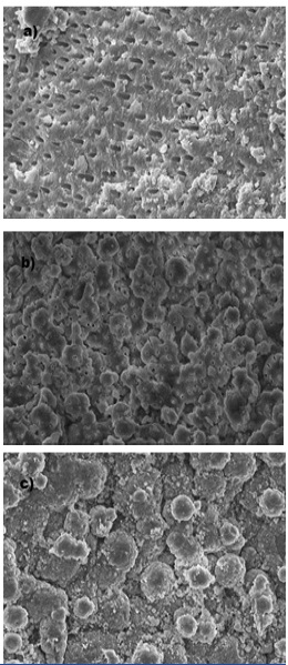
Scanning electron micrograph of canal wall after preparation with rotary NiTi instruments in the apical thirds
(a) Mtwo group: Slight smear layer with mostly open dentinal tubules (score 1 magnification 2000x)
(b) Twisted File group: Smear layer with some open dentinal tubules (score 2 magnification 2000x)
(c) Pro Taper group: Canal wall covered with agglomerations of thick smear layer with no open dentinal tubules (score 3 magnification 2000x)
Summary of score for smear layer for each group
| Instrument | Coronal third* | Middle third** | Apical third*** | Total |
|---|
| Score | Score | Score | Score |
|---|
| 0 | 1 | 2 | 3 | 0 | 1 | 2 | 3 | 0 | 1 | 2 | 3 | 0 | 1 | 2 | 3 |
| GROUP A | | | | | | | | | | | | | | | | |
| Twisted | 13 | 7 | 0 | 0 | 3 | 10 | 6 | 1 | 0 | 0 | 12 | 8 | 16 | 17 | 18 | 9 |
| GROUP B | | | | | | | | | | | | | | | | |
| Mtwo | 15 | 5 | 0 | 0 | 6 | 10 | 4 | 0 | 1 | 14 | 5 | 0 | 22 | 29 | 9 | 0 |
| GROUP C | | | | | | | | | | | | | | | | |
| ProTaper | 12 | 8 | 0 | 0 | 2 | 9 | 8 | 1 | 0 | 0 | 7 | 13 | 14 | 17 | 15 | 14 |
p- values; **p<0.05 or ***p<0.001- as compared to *Coronal
When compared to Twisted Files and ProTaper, Mtwo left significantly less smear in the apical thirds of the root canal (p<0.001) [Table/Fig–2,6].
Mean Smear layer comparison between the three groups at three root sites
| Comparisons | Coronal | Middle | Apical |
|---|
| Protaper vs. Twisted | 1.000 | 0.997 | 0.979 |
| Protaper vs. Mtwo | 0.997 | 0.161 | p<0.001 |
| Twisted vs. Mtwo | 1.000 | 0.638 | 0.002 |
p- values; p<0.05 or p<0.001
Twisted files showed lesser overall scores in the coronal, middle and apical portions compared to ProTaper but was not statistically significant (p>0.05) [Table/Fig 4b,5]. Twisted files produced cleaner canal wall scores in the apical third compared to ProTaper however, the result was not statistically significant (p>0.05) [Table/Fig-2,6].
ProTaper files produced statistically significant (p<0.05) maximum smear layer scores overall in the coronal, middle and apical portions [Table/Fig-4c,5]. ProTaper files resulted in significantly more smear layer (p <0.001) in the apical thirds of the canal compared Mtwo instrument [Table/Fig- 2,6].
Discussion
The ability to effectively clean the endodontic space relies on both instrumentation and irrigation. With all three systems partially uninstrumented areas were found in all canal sections. The present study confirms previous observations by that cleanliness decreased from the coronal to the apical part of the root canal for all three RNT files [19,20]. This could be attributed to sufficient access to mechanical flushing with root canal irrigants in this area. In addition, the effectiveness of irrigants is also reduced closer to the apex [21]. The use of antibacterial irrigants has been recommended in combination with chelating agents in order to effectively remove inorganic and organic components of the smear layer [22].
Previous SEM studies have investigated the cleaning efficacy of RNT instruments using NaOCl alone to avoid any influence of different irrigation solutions [6,10,23]. These studies reported the presence of a greater amount of smear layer in the apical third of the canals compared to the middle and coronal thirds highlighting the importance of an irrigation protocol that renders the canal free of debris and smear layer and improves the overall efficiency of the instruments [24]. In our study, we have incorporated a chemo-mechanical regimen of NaOCl and chelating agent that is most commonly used in a clinical setting so that the results obtained could be extrapolated to have a clinical significance.
In the present study, the cleaning efficacy of Twisted, Mtwo and ProTaper rotary instruments were examined on the basis of a numerical evaluation scheme for presence/absence of smear layer, in the coronal, the middle, and the apical portions of the canals. The cleaning efficiency of the three instruments was evaluated using a standard irrigation combination of NaOCl and EDTA containing chelating agent routinely used in clinical situations.
ProTaper systems were chosen as a standard for comparison in this study due to their popularity and published research evaluating these systems. Mtwo rotary systems was used as comparison for the results obtained with Twisted Files because their cleaning superior effectiveness has been investigated in recent studies [13,14,21,25]. However, very few studies exist evaluating the cleaning ability of Twisted Files in curved canals [26,27].
In general, the use of Mtwo files showed significantly less smear layer formation in all three canal areas compared to Twisted and ProTaper instruments (p<0.001) [Table/Fig-2,3]. This difference in cleanliness between RNT files could be attributed to their cross sectional design. Mtwo files are characterised by a positive rake angle with two sharp cutting edges. The smaller cross sectional area increases its flexibility and greater chip space allows increased debris clearance [9]. Also, an increasing helical pitch from tip to shaft reduces the transportation and accumulation of debris towards the apex [10]. In the apical third of the canals, instrumentation with Mtwo resulted in significantly less smear layer formation compared to Twisted and ProTaper instruments (p<0.001) [Table/Fig-2,6]. This result is consistent with previous reports that showed Mtwo to be superior in cleaning efficacy compared to ProTaper files [9,10,16].
Twisted Files are made by twisting NiTi metal. The manufacturer’s claim that Twisted Files are superior to file systems manufactured by the traditional grinding method [11]. Twisted Files by design have a triangular cross-section that enhances flexibility and generates less friction inside the canal walls due to a lack of peripheral lands. It has a variable pitch that minimizes the “screw-in” effect, allows debris to be effectively channelled out of the canal due to flute widths and flute depths that become accentuated toward the handle. In spite of its novel manufacturing process and unique flute design, our study concluded that the cleaning efficacy of Twisted Files seemed to be inferior to Mtwo files in the apical thirds [Table/Fig-2,3]. Clinically, this finding may be more important than the significant difference between the three RNT instruments in the amount of smear layer remaining in the coronal and middle parts of the canals because the microorganisms which remain in the apical portion of root canal have been considered the main cause of root canal treatment failure. When comparing with ProTaper group, Twisted Files produced cleaner canal wall scores in the apical third. However, the result was not statistically significant (p>0.05) [Table/Fig-2,6]. This result is in agreement with Kadhom et al., who noted no significant difference between Twisted Files and ProTaper rotary in the amount of debris removal in the apical thirds of oval shaped root canals [27]. Our results also support claims by Li et al., that moderately curved canals prepared by Twisted Files and ProTaper rotary showed no statistical difference in debris and smear layer scores in the apical and middle canals, but presented significance in coronal canals with Twisted Files group possessing lower scores [26].
ProTaper specimen group showed the presence of maximum remaining smear layer compared to Twisted and Mtwo groups [Table/Fig-2,3]. In our study, the SEM evaluations of the ProTaper group displayed a dense, thick, and non homogeneous smear layer with almost no dentinal tubule openings seen on the canal walls. ProTaper files have a triangular convex cross-section with three sharp cutting edges that contribute to more aggressive cutting. The relatively small chip space could be responsible for less debris removal which means more smear [8].
A key part of root canal cleanliness also depends on the type of instrumentation technique used. The RNT crown-down preparation technique is very effective and allows predictable shaping in significantly less time [28]. It creates a smooth funnel form shape allows deeper penetration of needles and irrigating solutions during early phases of instrumentation [29]. All three instruments produced an almost smear free dentine surface in the coronal and middle thirds. However, the apical third of the canals were less clean regardless of the instrument used. Our findings show that the rotary file systems used in conjunction with NaOCl solution and chelator paste which was both used with each new file did not effectively help in removing the smear layer. This difference was particularly pronounced in the apical portion of the canal. At the apical part, smear layer covered the root canal walls in a large number of the specimens instrumented with Twisted and ProTaper rotary files. This was probably due to the fact that for these two rotary instruments, the crown-down preparation as recommended by the manufacturer was done until resistance was felt. Hence, deeper placement of the needle slowly improved as the instrumentation progressed towards the apex. However, final flushing of the root canal occurred only after the irrigating needle reached as deep as 1mm short of the working length following complete crown-down preparation of the apical third of the canals. In comparison, Mtwo instrumentation was completed to the full working length according to the manufacturer’s instruction. This operational sequence allowed the irrigating solutions to effectively reach and remain at the end of the prepared canal at all times during rotary preparation. A more extensive tissue- chemicals contact was achieved which may have contributed to a more effective debridement in this group. Our SEM analysis showed that instruments and instrumentation procedures used in the Mtwo group gave the most favourable results in attempting to remove the smear layer from the apical thirds of the root canal walls.
All canals were prepared to the same apical diameter equivalent to size 30 (Mtwo and Twisted # 30, ProTaper F3) and a similar taper of 8% for the purpose of standardisation and simulation of a clinical situation.
There is still an ongoing debate regarding the optimum size of apical root canal enlargement. It has been suggested that the minimal apical size of a prepared canal should be ISO # 30 to allow proper chemo-mechanical cleansing with deeper penetration of the irrigating needles and solutions towards the apex thereby improving quality of treatment outcome [29].
Conclusion
Within the limitations of this study it may be concluded that none of the three canal preparation instruments used; Twisted, Mtwo and ProTaper, left completely clear root canal walls. Mtwo rotary files removed smear layer more effectively than Twisted and ProTaper files in the apical portion of the canal. There was no significant difference in canal cleanliness between Twisted and ProTaper rotary instruments in the apical thirds of the root canal. However, further studies using different methodologies are warranted to establish the cleaning efficacy of RNT files used in this study design.
Numbers in parenthesis indicates the range (min-max)
p- values; **p<0.05 or ***p<0.001- as compared to *Coronal
p- values; p<0.05 or p<0.001