Bilobed Lipoma of Submandibular Region: An Unusual Presentation
Vineet Kumar1, Sanjay Rastogi2, Roy Thomas3, Himanshu Pratap Singh4, Rupshikha Choudhury5
1Professor, Department of Orthodontics, SBB-Dental College, Masuri, Ghaziabad, UP, India.
2Associate Professor, Department of Oral and Maxillofacial Surgery, IDS-Dental College, Bareilly, UP, India.
3Professor and Consultant, Oral and Maxillofacial Surgery, Private Practice, Kerala, India.
4Senior Lecturer, Department of Oral and Maxillofacial Surgery, IDS-Dental College, Bareilly, UP, India.
5P.G. Student, Department of Oral and Maxillofacial Surgery, ITS-Dental College, Murad Nagar, Ghaziabad, UP, India.
NAME, ADDRESS, E-MAIL ID OF THE CORRESPONDING AUTHOR: Dr. Sanjay Rastogi, Z-23, Ashiyana Phase II, Moradabad-244001, UP, India.
E-mail: docos79@gmail.com
Hemangiopericytoma, Lipomatous hemangiopericytoma, Solitary fibrous tumour
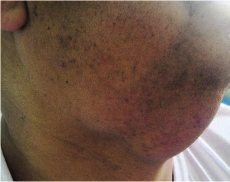
A 55-year old male patient presented to the Department of Oral and Maxillofacial Surgery with a swelling of the right submandibular region since four years [Table/Fig-1]. Swelling was painless asymptomatic and gradually increasing in size. On palpation swelling was firm with well-defined margins approximately (7x4 cm), swelling was mobile not fixed to overlying skin and no sign of inflammation. Patient was systemically normal. A tentative diagnosis was formed based on clinical examination which includes non-specific enlargement of right submadibular gland, lipoma, and lymphoma.
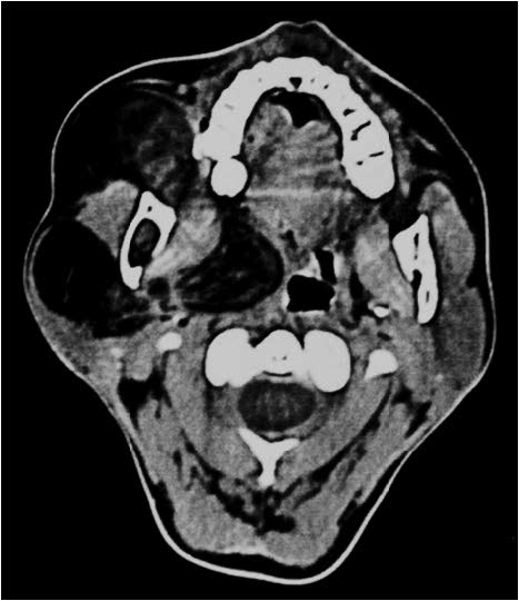
Contrast CT of right submandibular region showed a well circumscribed bilobed mass with mild extension into the parapharynegal space [Table/Fig-2] which was suggestive of benign lipomatous condition. A decision was made to surgically explore the lesion.
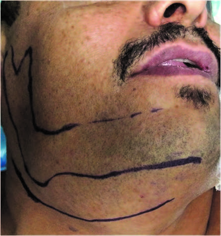
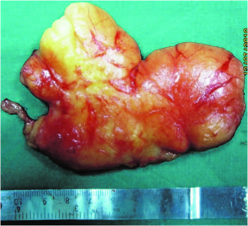
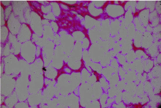
After obtaining pre anaesthetic clearance, patient was admitted for excision of mass under GA [Table/Fig-3]. A submandibular incision was given to create access to soft tissue mass blunt dissection was done preserving marginal mandibular branch of facial nerve and a yellowish bilobed soft encapsulated mass was excised [Table/Fig-4,5]. Postoperative recovery was uneventful. Histological examination under 5 x magnifications showed sheets of mature adipocytes and lobules of adipose tissue separated from the surface epithelium by fibrous connective tissue stroma. The adipocytes were loosely arranged in large areas which showed presence of empty cytoplasm and small nuclei. Tumour cells were arranged in lobules. These lobules were separated from each other by fibrovascular connective tissue septae [Table/Fig-6].
The presence of lipoma in the submandibuar region is extremely rare and it accounts for only 1-2% of all lipomas of the body. Angiolipomas and infiltrating lipomas are rarely found in the oral cavity [1] .
There have been reports of deep intra muscular lipoma in the submandibular region by Adachi et al., [2] . Pusiol e t al., reported an oncocyticsialo lipoma of submandibular gland [3] .
Diagnostic imaging techniques such as ultrasonograpghy, MRI and CT help to differentiate lipomas from other soft tissue lesion. And helps to identify the nature and exact location of lesion. However, the soft tissue characterization with ultrasonography is less specific than CT or MRI. When the mass is difficult to identify on ultrasonogram, CT or MRI is necessary. On CT scan it shows a high density from 83 to 143 Hounsfield units with well or poorly defined margins depending on the capsule [4].
Clinical picture of lipoma in right submandibular region
Contrast CT scan of the right submandibular region revealing the bilobed Tumour mass with mild extension into the parapharyngeal space
Outline of submandibular incision along with marking of inferior border of the mandible in order to save the marginal mandibular nerve during dissection
Intraoperative view of bilobed lesion separated from surrounding
structures
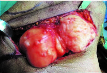
Completely excised lesion along with parapharyngeal extension
Depicting the mature adipocytes and fat lobules separated by fibrous septae
[1]. N Buric, D Krasic, M Visnjic, V Katic, Intraoseous mandibular lipoma: a case report and review of the literatureJ Oral Maxillofac Surg 2001 59:1367-71. [Google Scholar]
[2]. P Adachi, SP Kaba, AM Soubhia, EH Shinohara, Intermuscularlipoma of the submandibular spaceIndian journal of dental research 2011 22(6):871-72. [Google Scholar]
[3]. T Pusiol, I Franceschetti, M Scialpi, I Piscioli, Oncocyticsialolipoma of the submandibular gland with sebaceous differentiation: a new pathological entityIndian journal of pathology and microbiology 2009 52(3):379-82. [Google Scholar]
[4]. Cristalli Maria Paola, Monaca Gerardo La, Giannone Nicola, Francesco Nunzio, Testa Lucio Lo, Russo Lorenzo Lo, Muzio Hefferren, Lipoma in the Soft Tissues of the Floor of the Mouth: A Case Report Susanna AnnibaliThe Open Otorhinolaryngology Journal 2009 3:11-13. [Google Scholar]