Inverse Bell’s Phenomenon: Rare Ophthalmic Finding Following Ptosis Surgery
Satish Shitole1, Tapan Jakkal2, Bhasakar Khaire3
1Assistant Professor, Department of Ophthalmology, Government Medical College, Aurangabad, Maharashtra, India.
2Assistant Professor, Department of Ophthalmology, Government Medical College, Aurangabad, Maharashtra, India.
3Professor and HOD, Department of Ophthalmology, Government Medical College, Aurangabad, Maharashtra, India.
NAME, ADDRESS, E-MAIL ID OF THE CORRESPONDING AUTHOR: Dr. Satish Shitole, 45/1792.Apna Ghar hsg, Vartak Nagar, Thane (West), Maharashtra-400606, India.
E-mail: drsatish.shitole@gmail.com
Bell’s phenomenon is protective reflex in which the globe is turned upwards and slightly outwards during the eyelid closure to avoid corneal exposure. In Inverse Bell’s phenomenon, the eye moves downward instead of upward, this may be seen in the normal population, patients with Bell’s palsy or following conjunctival scarring. We hereby present the unusual complication of transient inversion of Bell’s phenomenon following extensive levator resection surgery performed for congenital ptosis. A 24-year-old male was undergone ptosis correction surgery. On postoperative day two, ocular examination revealed down rolling of eye ball during eyelid closure. It underwent spontaneous resolution within four weeks without any corneal complication. The patients were given frequent lubricating eye drops during this period and advised frequent follow-up for early diagnosis of corneal complication.
Here we highlight an inverse Bell’s phenomenon following levator resection surgery, its possible mechanism and risk of corneal complication.
Corneal complication, Levator resection surgery, Variation in bells phenomenon
Case Report
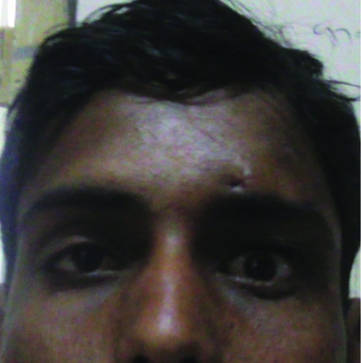
A 24-year-old male patient visited to our ophthalmology department with chief complaints of bilateral drooping of eyelids. Patient was a known case of bilateral congenital ptosis and had undergone surgery for same complaints eight years ago. Old surgical records revealed that it was frontalis sling with fascia lata surgery done under local anaesthesia at another institute. On ophthalmic evaluation, visual acuity (unaided) of both eyes was 6/6. The margin reflex distance (MRD 1) of the upper lid from corneal light reflex was 1 mm for right eye and 0 mm for left eye. Levator muscle function was assessed by measuring the excursion of the upper eye lid from extreme down gaze to up gaze and found to be 12 mm for right eye and 14 mm for left eye. There was no evidence of marcus gunn phenomenon. The Bell’s phenomenon was good and corneal sensations were normal. He had simple moderate bilateral ptosis with good levator function. Extensive levator resection surgery was planned of 12 mm for both Surgeryeyes for him in view of under correction and asymmetry of eye lids under local anaesthesia. Procedure was uneventful with satisfactory correction of ptosis [Table/Fig-1].
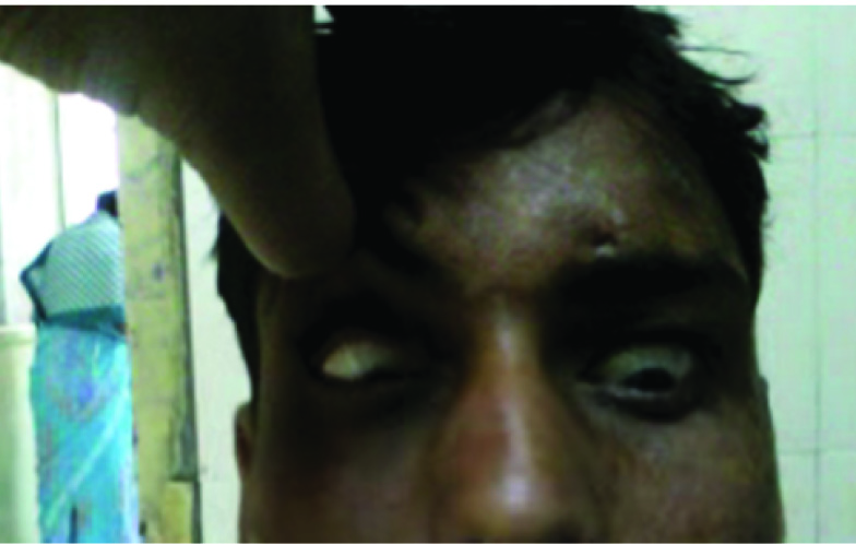
On postoperative day two downward rolling of eye ball (Inverse bells phenomenon) and mild lid oedema was observed on ocular examination [Table/Fig-2] [video-1]. Rest of the ocular examination was normal. Patient was prescribed lubricating eye drops and eye ointment to prevent exposure keratitis and frequent follow up for early detection of corneal complications like corneal erosion, exposure keratitis etc. On follow up visits both spontaneous resolution of Inverse bells phenomenon and lid oedema was noted at fourth week without any corneal complication.
Discussion
Exact mechanism underlying bells phenomenon is unknown. Possible explanation is the reciprocal muscle activity of superior rectus, levator palpebrae and orbicularis oculi with innervation from Trigemino-oculomotor nucleus [1] . The variation in bells phenomenon (reduction or inversion) following eye lid surgery can be seen in residual ptosis, ptosis with myopathy and congenital complicated ptosis [2] . Patients with residual ptosis undergoing repeat surgery are more prone for variation in bells phenomenon and both these factors were present in our case [2]. Inversion of bells phenomenon is known but unusual variation of bells phenomenon following levator resection surgery for congenital ptosis [3]. It can also be seen in normal population, patients with Bell’s palsy or following conjunctival scarring [4].
Hence, preoperative documentation of bells phenomenon is necessary in every case posted for ptosis surgery and also postoperative examination to look for variations in bells phenomenon following ptosis correction surgery.
There are two possible mechanisms for inverse bell phenomenon in early postoperative period. During extensive and repeated resection of levator palpebrae muscle the superior rectus muscle and oculomotor nerve can get injured. This may alter the Trigeminooculomotor projection leading to abnormal eye ball movement [5]. In such cases paradoxical eye ball movement may remain for many years [5]. Secondly severe oedema and hyperemia of superior fornix in post-operative period may affect the relationship between eyelid and superior rectus muscle. In such case eye ball movement can revert back to normal within few weeks after resolution of eyelid oedema and it usually requires no treatment.
In our case the inverse bell phenomenon subsided without any treatment and following resolution of eye lid oedema. Hence, it was probably due to soft tissue oedema in postoperative period than aberrant connections of nervous system. Similar finding were reported by Betharia et al., [2] and Kyung et al., [6] where they observed spontaneous resolution of phenomenon.
When inverse bells phenomenon develops in postoperative period, cornea is at risk of developing exposure keratitis. Hence, copious use of eye lubricant along with frequent ophthalmic checkup is recommended for early diagnosis of corneal complication. In our case, it was managed successfully as he did not get any corneal complication [6].
Postoperative day 1 photograph showing correction of ptosis

Photograph showing downward rolling of eye balls following lid closure (inverse bell’s phenomenon)

Conclusion
Preoperative and postoperative examination of eye ball for bells eye phenomenon is necessary. Patients with congenital ptosis, residual ptosis and requiring repeat surgery for ptosis correction are more prone to development of inverse bell phenomenon. Post-operative soft tissue oedema or aberrant connections of nervous system is the possible mechanism for it. Once inverse bell phenomenon develops copious lubricating eye drops and frequent examination of eye is required until it get resolved.
[1]. C Evinger, KA Manning, Pattern of extraocular muscle activation during reflex blinkingExp brain res 1993 92:502-06. [Google Scholar]
[2]. SM Betharia, BR Kalra, Observations on Bell’s phenomenon after levator surgeryIndian J Ophthalmol 1985 33:109-11. [Google Scholar]
[3]. SM Betharia, V Sharma, Inverse Bell’s Phenomenon observed following levator resection for blepharoptosisGraefes Arch Clin Ophthalmol 2006 244:868-70. [Google Scholar]
[4]. JS Gupta, A Chatterjee, Inverse Bell’s Phenomenon as a protective mechanismAm J Ophthalmol 1965 59:931-33. [Google Scholar]
[5]. EG Buckley, FD Ellis, Posttraumatic abducens to oculomotor nerve misdirectionJ AAPOS 2005 9:12-16. [Google Scholar]
[6]. SN Kyung, WY Suk, Two cases of inverse bell’s phenomenon following levator resection: contemplation of the mechanismEuropean Journal of Ophthalmology 2009 19(2):285-87. [Google Scholar]