Tetra-Phocomelia: A Rarest of Rare Case
Anil Kumar Shukla1, S.C. Sanjay2, L. Krishna3, N. Krishnappa4
1 Professor, Department of Radiodiagnosis, Kempegowda Institute of Medical Sciences, Bangalore, India.
2 Associate Professor, Department of Radiodiagnosis, Kempegowda Institute of Medical Sciences, Bangalore, India.
3 Former Professor & HOD, Department of Obstretic & Gynaecology, Kempegowda Institute of Medical Sciences, Bangalore, India.
4 Professor & HOD, Department of Radiodiagnosis, Kempegowda Institute of Medical Sciences, Bangalore, India.
NAME, ADDRESS, E-MAIL ID OF THE CORRESPONDING AUTHOR: Dr. Anil Kumar Shukla, 77, Himagiri Apartment, Flat 4C, 15 Cross, 4 Main, Malleswaram, Bangalore-560 055, India.
E-mail: shookla2007@yahoo.co.in
We present a rarest of rare case of Tetra-Phocomelia evaluated by antenatal Ultrasonography. It is a condition seen in 0.62 per 100,000 live births. An ultrasonogram was done at 18 wk of pregnancy to assess the fetus and after termination gross specimen was evaluated and X-ray infantograms were done to confirm the findings. The case showed classic Tetra-Phocomelia with limbs like flippers of a seal. Our findings make it rarest of rare as only few cases have been so far reported.
Flippers, Limbs, Phocomelia, Ultrasonogram
Case Report
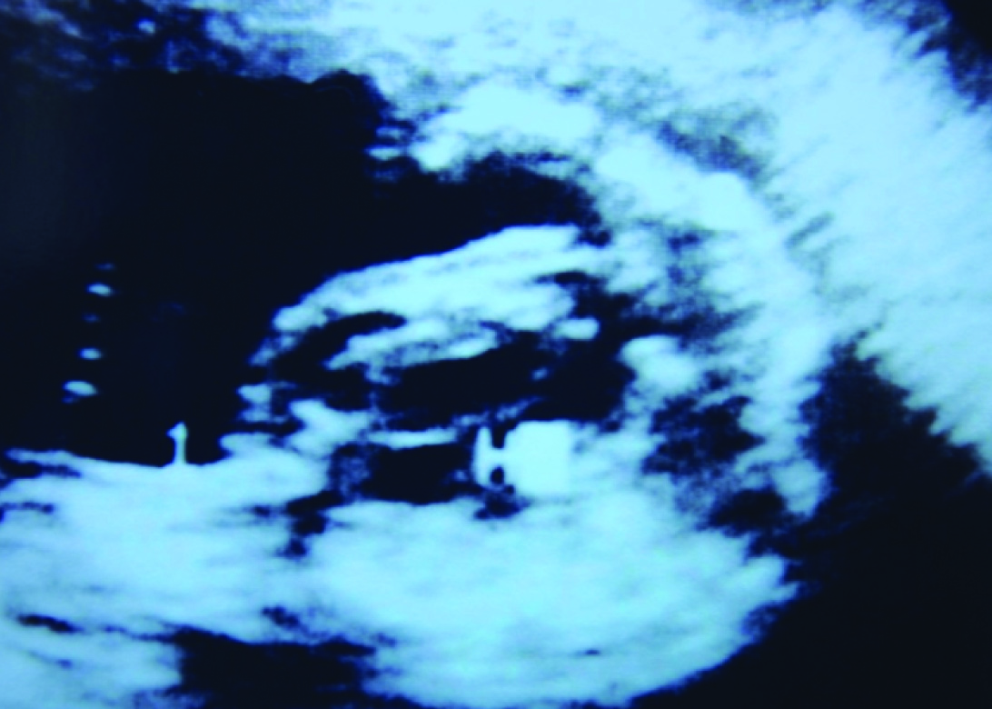
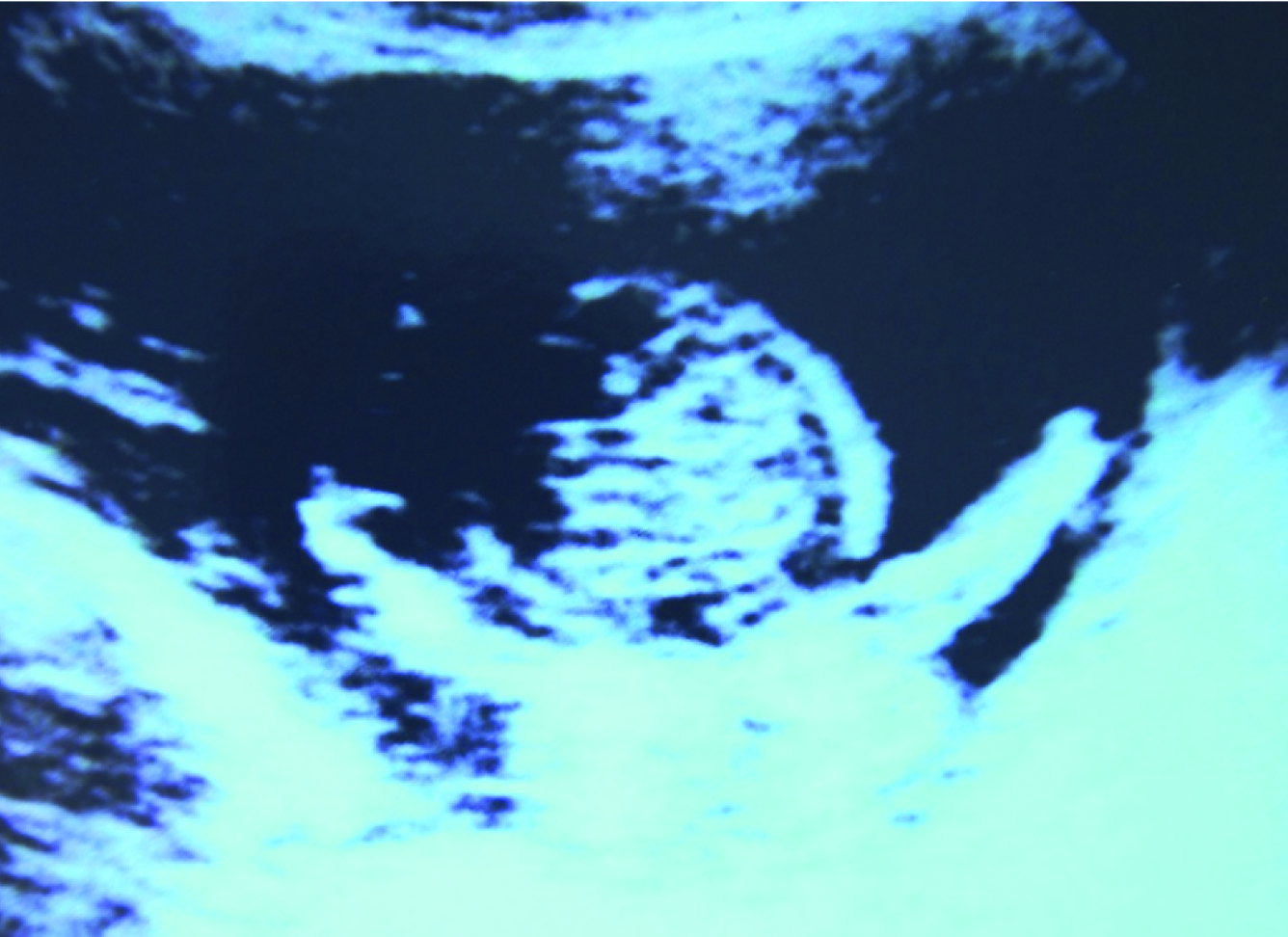
An 18-year-old primigravida came to our department for routine checkup. An ultrasound scan was conducted to evaluate for fetal anomalies. Fetus was found to be of about 18 weeks with various skeletal anomalies. Mainly we saw absence of long bones in upper or lower limbs with hands and feet arising directly from the shoulders and hips [Table/Fig-1,2]. The pelvic girdle was not visible. Spine showed absent distal sacrum. Shoulders did not show full scapulae. There were multiple facial abnormalities like big nose with two nostrils, eyes were slightly widely separated and hypoplastic jaw.
Ultrasound image of fetal upper limbs and thorax showing hands like flippers
Ultrasound image of fetal lower limbs and abdomen showing lower limbs directly arising from pelvis
Fetal heart and brain did not show any abnormality. Umbilical cord and the placenta were normal. It was decided to end the pregnancy with parental consent after explaining to them about defects.
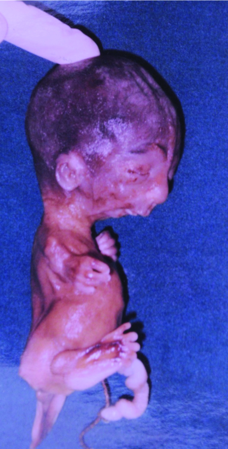
Delivery was induced and it was still birth. On gross evaluation baby showed limbs, as seen antenatal with hands showing four fingers on both sides and legs with four toes on right and five on left sides [Table/Fig-3,4,5]. The facial features also corroborated with the USG findings.
(3) Post delivery baby’s gross specimen from the front showing all four limbs as flippers; (4) Post delivery baby’s gross specimen from the left showing hypoplastic mandible, hands and feet

Post delivery baby’s gross specimen from the right showing fingers and toes
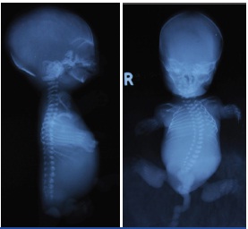
An x-ray infantogram was performed in both anterioposterior and lateral views [Table/Fig-6,7]. The skeletal X-ray and pictures confirmed our antenatal findings. There were 10 ribs on either side. Spine showed absent distal sacral segments. Biochemical evaluation of the mother was normal however due to cost factor genetic mapping of fetus was not done.
(6) X-ray infantogram lateral view showing hypoplastic mandible and hardly any limb bones’ (7) X-ray infantogram AP view showing bones of hands and feet
Discussion
Fetal limb defects are known to occur although are included in rare category. Amelia is a condition in which one arm or leg is absent. It is also known as phocomelia, this term was used first time in 1836 by Etienne Geoffroy Saint Hilaire. When all the four limbs are affected is known as “Tetra-Phocomelia”. “Tetra” = Four, “Phoco” = Seal and “Melos” = Limb. In this condition hands and feet are only parts of limbs from shoulder or pelvis like seal flippers [1–3].
The true phocomelia is seen in 0.62 per 100,000 births, rarest of rare defect is known to be seen in much less proportion [1]. Various causes have been attributed to the disease like some drugs, substance abuse, radiation, diabetes, genetic, etc [4–6].
Use of some drugs during pregnancy like Thalidomide, substance abuse like cocaine or alcohol, gestational diabetes, use of X-ray radiation, fetal chromosomal anomalies like trisomy 18 or genetic inheritance is considered to be the cause [6,7]. Fetal limbs start developing at about 24 to 36 days of fetal life [1]. Upper limbs by 12 and lower by 14 wk are formed [8]. If formation process is disturbed at this early age can lead to severe phocomelia. About 25 % of limb defects are Amelia, in 60% cases a single limb is involved [9]. Mechanical disruption or vascular disturbances at the time of limb development is also considered to be the cause of deformities [1,8,9]. Our case had distinctive features of deficiency of rays in hands and feet [Table/Fig-6,7]. However, in some cases talipes equinovarus was observed [6].
The present patient did not have other associated anomalies like cleft lips, encephalocele, hydrocephalous, renal or cardiac malformations [6,7]. The patient had micrognathia and mild hyperteliorism, some of cases have shown to have short, wide neck, small ears besides other anomalies [6]. A normal umbilical cord, however, a single umbilical artery is also seen. The patient had normal kidneys and urinary bladder, sometime renal agenesis is also observed [4].
Fetal ultrasound is practiced all over the world for last three-four decades. Most of the limb defects can be seen or suspected at time of nuchal translucency evaluation. Their close observation further at around 16-18 wk can give us correct view about limb deformity. Ultrasonography should be meticulously performed and should be cautiously interpreted. It is very rare these days however still there are instances when it is not used [2]. Present case fulfilled all the criteria described for Tetra-Phocomelia.
Conclusion
Based on our observations and experience, antenatal ultrasono- graphy remains the hallmark for detecting all fetal anomalies including that of limbs. No other tests are required to confirm this diagnosis. Such cases are isolated or sporadic due to certain genetic inheritances.
[1]. Bermejo-Sanchez E, Cuevas L, Amar E, Bianca S, Bianchi F, Botto L, Phocomelia: A worldwide descriptive epidemiologic study in a large series of cases from the International Clearinghouse for Birth Defects Surveillance and Research, and overview of the literatureAm J Med Genet 2011 157(4):305-20. [Google Scholar]
[2]. Keypour F, Naghi I, Roberts Behnam B, SC Phocomelia Syndrome (Pseudothalidomide Syndrome): A Case ReportJournal of Family & Reproductive Health 2013 7(1):45-47. [Google Scholar]
[3]. Rao R, Rao N, Tetra-Phocomelia-A CASE REPORTIndian Journal of Medical Case Reports [Internet] 2012 1(2-3):20-2.available from: http://www.cibtech.org/jcr.htm 2012 Vol. 1 (2-3) Jul.-Sept. & Oct.- Dec., pp.20-22/Rao and Rao [Google Scholar]
[4]. Kashiwagi M, Chaoui R, Stallmach T, Hurlimann S, Lauper U, Hesbisch Gl, Fetal bilateral renal agenesis, phocomelia and single umbilical artery associated with cocaine abuse in early pregnancyBirth Defect Res. A 2003 67(11):951-52. [Google Scholar]
[5]. Weiss S, Foreshortened Phantom Limbs and PhomeliaJAMA 1963 183(12):1053 [Google Scholar]
[6]. Jinghui Hu, Weijie Du, Xiangming Fang, Xinyan Wang, A case of Trisomy 18 Presenting with Severe Facial Abnormality and PhocomeliaInt J Hum Genet 2012 12(4):325-27. [Google Scholar]
[7]. Subhani M, Akangire G, Kulkarni A, Wilson G, Al-Awadi/ Raas-Rothschild/Schinzel(AARRS)phocomelia syndrome: case report and developmental field analysisAmerican Journal of Medical genetics Part A 2009 149A(7):1494-98. [Google Scholar]
[8]. Pujarii D, Imran SS, Hogade J BMS, Phocomelia - a Case ReportAnatomica Karnataka 2012 6(3):86-89. [Google Scholar]
[9]. Fundukian L, The Gale encyclopedia of genetic disorders 2010 Farmington Hills, MichGale [Google Scholar]